. 2015 Jul;1(4):381-394.e7.
doi: 10.1016/j.jcmgh.2015.05.001.

Mutations in Plasmalemma Vesicle Associated Protein Result in Sieving Protein-Losing Enteropathy Characterized by Hypoproteinemia, Hypoalbuminemia, and Hypertriglyceridemia

Affiliations

Mutations in Plasmalemma Vesicle Associated Protein Result in Sieving Protein-Losing Enteropathy Characterized by Hypoproteinemia, Hypoalbuminemia, and Hypertriglyceridemia

Abdul Elkadri et al. Cell Mol Gastroenterol Hepatol. 2015 Jul.

Abstract

Background & aims methods: Severe intestinal diseases observed in very young children are often the result of monogenic defects. We used whole exome sequencing (WES) to examine the genetic cause in a patient with a distinct severe form of protein losing enteropathy (PLE) characterized by hypoproteinemia, hypoalbuminemia, and hypertriglyceridemia.

Methods: WES was performed at the Centre for Applied Genomics, Hospital for Sick Children, Toronto, Canada. Exome library preparation was performed using the Ion Torrent AmpliSeq RDY Exome Kit. Functional studies were carried out based on the identified mutation.

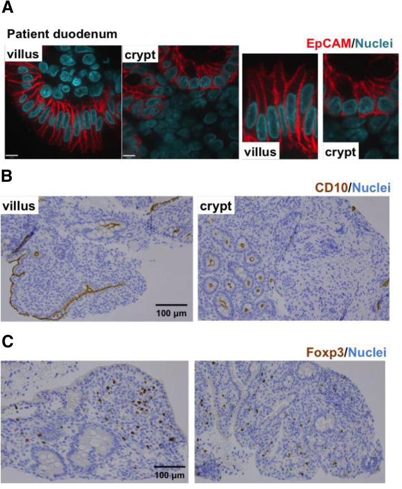
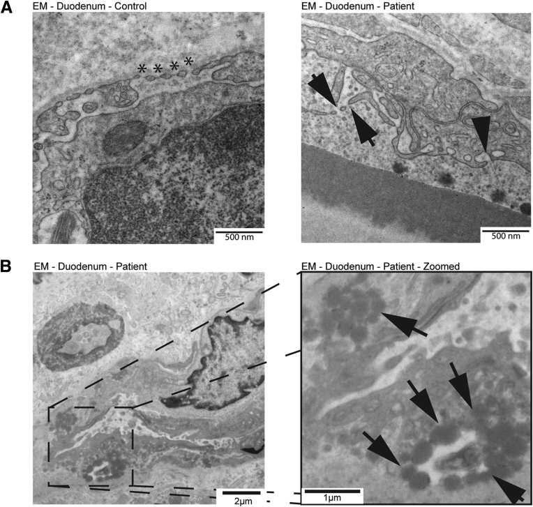
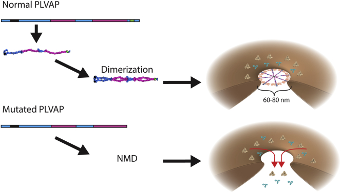
Results: Using whole exome sequencing we identified a homozygous nonsense mutation (1072C>T; p.Arg358*) in the PLVAP (plasmalemma vesicle associated protein) gene in an infant from consanguineous parents who died at five months of age of severe protein losing enteropathy. Functional studies determined that the mutated PLVAP mRNA and protein were not expressed in the patient biopsy tissues, presumably secondary to nonsense-mediated mRNA decay. Pathological analysis showed that the loss of PLVAP resulted in disruption of endothelial fenestrated diaphragms.

Conclusions: PLVAP p.Arg358* mutation resulted in loss of PLVAP expression with subsequent deletion of the diaphragms of endothelial fenestrae leading to plasma protein extravasation, protein-losing enteropathy and ultimately death.

Keywords: IBD; PLE; PLVAP; Protein losing enteropathy; VEOIBD; and hypertriglyceridemia; endothelium; fenestrae; hypoalbuminemia; hypoproteinemia; monogenic diseases; very early onset IBD.


Conflict of interest statement

The authors have no potential conflicts including financial, professional, or personal that are relevant to the manuscript.

Figures

Supplementary Figure 1
Immunohistochemistry staining for epithelial cell adhesion molecule (EpCAM, A), CD10 (B), and Foxp3 (C) in duodenal biopsy samples from the patient. Normal expression and localization of EpCAM, CD10, and Foxp3 in the duodenum. Scale bars: (A) 8 μm; (B, C) 100 μm.
Supplementary Figure 2
(A) Immunofluorescence analysis of tight junctions (ZO-1) and adherens junctions (β-catenin) in the duodenum of control patient, disease control patient, and the study patient. A congenital tufting enteropathy (CTE) case presenting as protein-losing enteropathy (PLE) served as a proper disease control for the small intestine. Alexa 568 red stains ZO-1, Alexa 488 β-catenin, and Hoechst nuclei in blue. ZO-1 localizes subapical and β-catenin basolateral in villus as well as crypt enterocytes of control, CTE, and study patient. Scale bar: 10 μm. (B) Immunofluorescence analysis of tight junctions (ZO-1) and adherens junctions (β-catenin) in the colon of control, disease control, and study patient. A patient with inflammatory bowel disease (IBD) with active disease (inflamed areas in the colon) served as the disease control for the colon. Alexa 568 red stains ZO-1, Alexa 488 β-catenin, and Hoechst nuclei in blue. ZO-1 localizes subapical and β-catenin basolateral in villus as well as crypt enterocytes of the control patient, the IBD case, and the study patient. Scale bar: 10 μm. (C) Immunofluorescence analysis of vessels in the duodenum of the control patient, the disease control patient, and the study patient. A CTE case presenting as PLE served as a proper disease control for the small intestine. Alexa 568 red stains CD31 (vessel marker), Alexa 488 PLVAP, and Hoechst nuclei in blue. CD31 marks the vessel walls and colocalizes with PLVAP in the control and CTE. In the patient the vessels are stained by CD31, but in the absence of PLVAP. Scale bar: 10 μm. (D) Immunofluorescence analysis of vessels in the colon of control, disease control, and the patient. A patient with IBD with active disease (inflamed areas in the colon) served as a disease control for the colon. Alexa 568 red stains CD31 (vessel marker), Alexa 488 PLVAP, and Hoechst nuclei in blue. CD31 marks the vessel walls and colocalizes with PLVAP in the control and IBD cases. In the study patient, the vessels are stained by CD31, but in the absence of PLVAP. Scale bar: 10 μm.
Figure 1
Figure 1
Low-power light and electron microscopy showing edema and absence of intestinal structural defects in PLVAP p.R358* mutation. (A) H&E and PAS stain of the duodenum from a control patient (left) and the PLVAP p.R358* patient (right). H&E and PAS stains show prominent interstitial edema in the duodenum (shown by the pale interstitium in the villus area). Scale bars: 200 μm. (B) Transmission electron microscopy analysis of the duodenum from a control patient (left) and the PLVAP p.R358* patient (right) showing no abnormalities within the ultrastructure of the enterocyte epithelium. Scale bars: 2 μm.
Figure 2
Figure 2
Identification of a PLVAP mutation in an infant with severe protein-losing enteropathy. (A) Pedigree. (B) Whole-exome sequencing variant filtration algorithm showing the variants identified in the patient and parents (listed in Supplementary Table 2). (C) Domain, gene, and mRNA view. Gray arrows show glycosylation sites. The identified mutation (red), coil-coiled regions (pink), transmembrane regions (black), and a proline-rich region (green) are noted.
Figure 3
Figure 3
PLVAP p.R358* mutation causing loss of the diaphragms of endothelial fenestrae and caveolae and severely decreased levels of mRNA and protein. (A) Electron micrographs of small intestinal biopsies from control patient (left panel) and PLVAP p.R358* patient (right panel). Fenestrae and caveolae of the mutant endothelial cells do not have diaphragms (arrows) whereas diaphragms are readily present in normal endothelial cells (*). Scale bars: 500 nm. (B) Electron micrographs of PLVAP p.R358* patient duodenum biopsy, showing extracellular lipid droplets (black arrows). The image on the right is a higher magnification of the indicated area in the left micrograph. Scale bars: left, 2 μm; right, 1 μm.
Figure 4
Figure 4
PLVAP p.R358* mutation causing loss of the diaphragms of endothelial fenestrae and caveolae and severely decreased levels of mRNA and protein. (A) Detection of human PLVAP mRNA (blue-green staining) using RNAscope probes in duodenal biopsies from a normal control patient (left), a congenital tufting enteropathy (CTE) patient (middle), and out PLVAP p.R358* patient (right). The general architecture of the tissue is revealed by H&E counterstain. Scale bars: 100 μm. (B–D) Nuclei were stained with Hoechst (blue). The disease controls used are microvillous inclusion disease (MVID) in B, CTE in E, and active colonic inflammatory bowel disease (IBD) in F. Scale bars (B–D): 100 μm. (B) Multiplex immunofluorescence with anti-PLVAP (clone PAL-E)-Alexa 647 (purple) and anti-CD31 (yellow) on methanol-fixed frozen sections of duodenal biopsies from normal control patient (left), control MVD patient (middle), and PLVAP p.R358* patient (right). Hoechst stains nuclei in blue. (C, D) Multiplex immunofluorescence with anti-PLVAP (clone 174/2) (green) and anti-CD31 (red) on formalin-fixed, paraffin-embedded sections from normal control patient (left), control disease patient (middle), and PLVAP p.R358* patient (right). Sections were obtained from duodenum (C) and colon (D) biopsies.
Figure 5
Figure 5
Functional studies of mutated forms of PLVAP. (A) Schematic constructs of various truncation mutations generated. CC, coiled coil domain; 3xHA, trio tandem of HA epitopes; N-Glyc, N-glycosylation; PRR, proline rich region; TM, transmembrane region. Downward orange lines mark the position of cysteines. (B) PLVAP R358* forms homodimers detected by the anti-PLVAP mAb clone PAL-E. Immunoblotting analysis of 10% (top) or 4%–20% (middle and bottom) SDS-PAGE resolved lysates of Ea.hy926 cells transfected with empty vector (EV) or with human PLVAP full length-3xHA (FL), human PLVAP 1-389-3xHA (389), human PLVAP 1-357-3xHA (357-3xHA), or human PLVAP R358* truncation constructs. The membranes have been probed with three available anti-human PLVAP antibodies recognizing different epitopes: PAL-E, mouse anti-human PLVAP mAb clone PAL-E (top panel); mouse anti-human PLVAP mAb clone 174/2 (top panel); and anti-hPV1C, chicken anti-human PV1C pAb. Ea.hy926 are devoid of endogenous PLVAP, but bands of expected molecular weight were readily detected by all three antibodies in PLVAP FL-3xHA transfected cell lysates in reducing (r, ∼63 kDa) and nonreducing (n, ∼126 kDa) conditions (corresponding to PLVAP FL-3xHA monomers or homodimers, respectively). The only antibody that recognized PLVAP R358* and PLVAP 357-3xHA was PAL-E. (C) PLVAP R358* is N-glycosylated. Immunoblotting with PAL-E (top) or anti-HA mAb (bottom) of total cellular proteins from Ea.hy926 endothelial cells expressing PLVAP truncation constructs treated (+) or not (−) with PNGase F to remove N-glycans. The proteins were resolved by nonreducing 8% and reducing 4%–20% SDS-PAGE, respectively. Controls consisted of nontransfected (NT) or empty vector (EV) transfected Ea.hy926 cells. The PAL-E antibody recognized bands of an appropriate size corresponding to the glycosylated and deglycosylated forms of PLVAP FL-3xHA and -389-3XHA and R358* truncations constructs. (D) Immunoblotting with mouse anti-HA mAb of total cellular proteins from endothelial cells expressing PLVAP truncation constructs treated (+) or not (−) with PNGase F to remove N-glycans resolved by reducing 8% SDS-PAGE. A ∼15 kDA drop was detected in 357-3xHA upon PNGase F treatment (+).
Figure 6
Figure 6
PLVAP R358* trafficked to the plasma membrane of endothelial cells. (A) Confocal microscopy demonstrating the expression of PLVAP R358* on the surface of live endothelial cells. Live transfected cells were labeled with PAL-E mAb followed by goat anti-mouse IgG-AlexaFluor 647 (red). The cells were transfected with bicistronic vectors encoding for full-length PLVAP-3xHA (FL), PLVAP 389-3xHA (1-389), PLVAP R358* (R358*), or empty vector (EV). Transfected cells were detected by hrGFP fluorescence (green). Scale bars: 50 μm. (B) Flow cytometric analysis of Ea.hy962 cells transfected with full-length PLVAP (FL-HA) (pink trace), PLVAP R358* (R358*) (orange trace), and PLVAP 357-3xHA (357 HA) (green trace). Controls consisted of nontransfected Ea.hy962 cells (black trace) or Ea.hy962 cells transfected with empty vector (EV) (grey trace). PLVAP constructs were expressed from bicistronic vectors also encoding for hrGFP as described in Materials and Methods. Left: Gating on humanized Renilla green fluorescent protein (hrGFP)-positive cells. Right: PAL-E mAb signal in hrGFP-positive cells. (C) Flow cytometric analysis of Ea.hy962 cells transfected with full-length PLVAP (FL-3xHA), PLVAP R358* (R358*), and PLVAP 357*-3xHA (1–357 HA) (green trace). Controls consisted of nontransfected Ea.hy962 cells (black trace) or Ea.hy962 cells transfected with EV (grey trace). PLVAP constructs were expressed from bicistronic vectors also encoding for hrGFP. Cells were labeled with either PAL-E mAb (two left columns) or with anti-HA mAb (two right columns). Left: Gating on hrGFP-positive cells. Right: Gating on PAL-E or anti-HA mAb signal in hrGFP-positive cells.
Figure 7
Figure 7
Proposed effect of PLVAP R358* mutation on endothelial fenestrae diaphragms. Expression of PLVAP leads to the formation of diaphragms in fenestrae (top). PLVAP R358* mutation results in the degradation of the vast majority of mRNA possibly via nonsense mediated decay (NMD), resulting in loss of PLVAP protein and failure in fenestrae diaphragm formation (bottom). Plasma proteins such as albumin (yellow) and immunoglobulins (blue) are subsequently lost due to absence of fenestrae.

References

    1. Braamskamp M.J., Dolman K.M., Tabbers M.M. Clinical practice: protein-losing enteropathy in children. Eur J Pediatr. 2010;169:1179–1185. - PMC - PubMed
    1. Umar S.B., DiBaise J.K. Protein-losing enteropathy: case illustrations and clinical review. Am J Gastroenterol. 2010;105:43–49. - PubMed
    1. Uhlig H.H., Schwerd T., Koletzko S. The diagnostic approach to monogenic very early onset inflammatory bowel disease. Gastroenterology. 2014;147:990–1007.e3. - PMC - PubMed
    1. Avitzur Y., Guo C., Mastropaolo L.A. Mutations in tetratricopeptide repeat domain 7A result in a severe form of very early onset inflammatory bowel disease. Gastroenterology. 2014;146:1028–1039. - PMC - PubMed
    1. Stan R.V., Tse D., Deharvengt S.J. The diaphragms of fenestrated endothelia: gatekeepers of vascular permeability and blood composition. Dev Cell. 2012;23:1203–1218. - PMC - PubMed

LinkOut - more resources