. 2015 Jul;56(8):4725-33.
doi: 10.1167/iovs.15-16924.

Targeting Caspase-12 to Preserve Vision in Mice With Inherited Retinal Degeneration

Affiliations

Targeting Caspase-12 to Preserve Vision in Mice With Inherited Retinal Degeneration

Yogesh Bhootada et al. Invest Ophthalmol Vis Sci. 2015 Jul.

Abstract

Purpose: The unfolded protein response is known to contribute to the inherited retinal pathology observed in T17M rhodopsin (T17M) mice. Recently it has been demonstrated that the endoplasmic reticulum stress-associated caspase-12 is activated during progression of retinal degeneration in different animal models. Therefore, we wanted to explore the role of caspase-12 in the mechanism of retinopathy in T17M mice and determine if inhibiting apoptosis in this way is a viable approach for halting retinal degeneration.

Methods: One, two-, and three-month-old C57BL6/J, caspase-12-/-, T17M, and T17M caspase-12-/- mice were analyzed by scotopic ERG, spectral-domain optical coherence tomography (SD-OCT), histology, quantitative (q)RT-PCR, and Western blot of retinal RNA and protein extracts. Calpain and caspase-3/7 activity assays were measured in postnatal (P) day 30 retinal extracts.

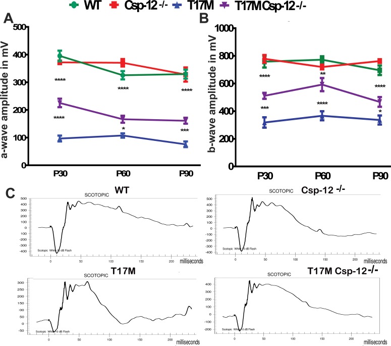
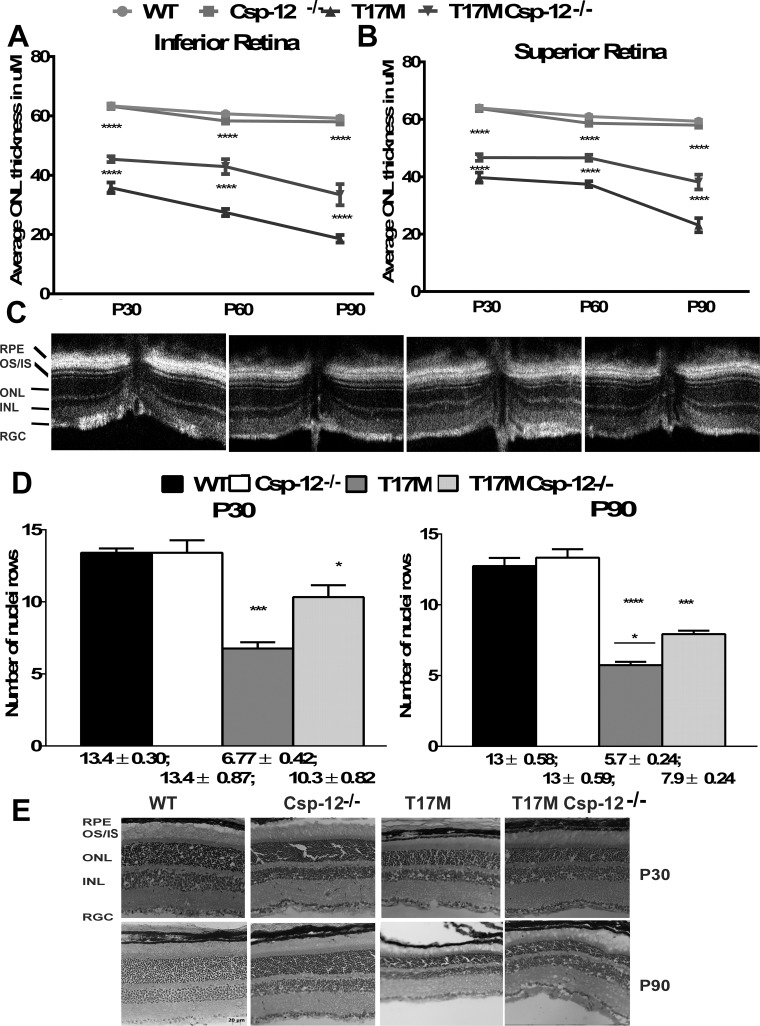
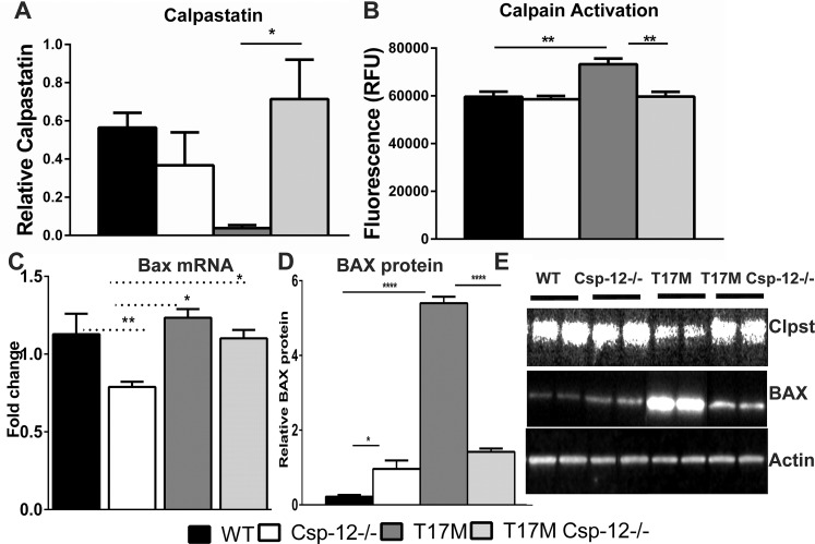
Results: Caspase-12 ablation significantly prevented a decline in the a- and b-wave ERG amplitudes in T17M mice during three months, increasing the amplitudes from 232% to 212% and from 160% to 138%, respectively, as compared to T17M retinas. The SD-OCT results and photoreceptor row counts demonstrated preservation of retinal structural integrity and postponed photoreceptor cell death. The delay in photoreceptor cell death was due to significant decreases in the activity of caspase-3/7 and calpain, which correlated with an increase in calpastatin expression.

Conclusions: We validated caspase-12 as a therapeutic target, ablation of which significantly protects T17M photoreceptors from deterioration. Although the inhibition of apoptotic activity alone was not sufficient to rescue T17M photoreceptors, in combination with other nonapoptotic targets, caspase-12 could be used to treat inherited retinopathy.


Figures

Figure 1
Figure 1
Expression and activation of Csp-12 in the T17M retina during ADRP progression. We used four animals per group in this experiment and analyzed retinal protein and RNA extracts at early time points from P12 to P25. (A) Starting at P18, we observed a 6-fold (P < 0.01) increase in Csp-12 mRNA in T17M mice compared with wt. At P21 and P25, Csp-12 gene expression was upregulated between 4.5- and 1.6-fold. (B) At P21, we detected a 3-fold (P < 0.001) increase in Csp-12 activation in T17M retinas, suggesting that in addition to the UPR activation detailed in previous studies,, ADRP photoreceptors also experience activation of Csp-12. One-way ANOVA was used to calculate differences. Data are shown as mean ± SEM (*P < 0.05, **P < 0.01).
Figure 2
Figure 2
Lack of Csp-12 protects T17M retinas from degeneration, as measured by scotopic ERG responses at 10 dB. We analyzed four groups of animals (n = 6). (A) Using two-way ANOVA, we demonstrated that a-wave amplitudes of the scotopic ERG were significantly increased in T17M Csp-12−/− mice at 1 month of age and that the value of a-wave amplitudes was 225 μV ± 15 vs. 97.0 μV ± 11 in T17M. These data represent a 2.3-fold difference in a-wave amplitudes between T17M and T17M Csp-12−/− mice. At 2 and 3 months, the difference between T17M and T17M Csp-12−/− groups was also dramatic, but somewhat diminished due to a decrease in T17M Csp-12−/− a-wave amplitudes. For example, we registered 166.0 μV ± 13 vs. 108.0 μV ± 7.8 and 161.0 μV ± 11 vs. 76.0 μV ± 10.0 in T17M Csp-12−/− versus T17M at 2 and 3 months, respectively. (B) Interestingly, b-wave scotopic ERG amplitudes were also elevated in T17M Csp-12−/− retinas. However, the magnitude of this elevation was less dramatic than for a-wave over the 3 examined months. Thus, 1-month-old T17M Csp-12−/− mice demonstrated 511.0 μV ± 22.0 vs. 318.0 μV ± 37.0 in T17M. In the second month, the b-wave amplitudes in the T17M Csp-12−/− mice were also increased by 1.6-fold as compared to T17M. We registered the b-wave amplitude of 592.2 μV ± 22.0 in T17M Csp-12 vs. 366.8 μV ± 32.0 in T17M. By 3 months of age the stable preservation of b-wave amplitude observed during the first 2 months changed, and the ratio of T17M Csp-12 to T17M amplitudes dropped from 1.6- to 1.38-fold (Supplementary Table S1). This would suggest that the bipolar cells are less sensitive to Csp-12 ablation in degenerating retinas. The differences between all groups were statistically significant. (C) Images of the scotopic ERG amplitudes registered at 10 dB or 25 cd*s/m2 in four groups of animals. Data are shown as mean ± SEM (*P < 0.05, **P < 0.01, ***P < 0.001, ****P < 0.0001).
Figure 3
Figure 3
The preservation of retinal structure and prevention of photoreceptor cell death in T17M Csp-12−/− retinas. We analyzed four groups of animals (n = 6). (A) Two-way ANOVA analysis of data from the inferior retina revealed that this region was better preserved than the superior region, with increases in the average ONL thicknesses over a period of 3 months of 1.26-, 1.56-, and 1.8-fold. These data also pointed to the fact that despite experiencing the same molecular mechanisms responsible for retinal denegation, activation of prodeath cascades in the inferior and superior regions might occur to different degrees, suggesting that the therapeutic benefit from Csp-12 ablation could also be different. (B) Interestingly, the data demonstrated a steady preservation of the average ONL thickness in T17M Csp-12−/− superior retinas from 1.2 to 1.5 over a period of 3 months, while T17M mice experienced a continuous decline in the average ONL thickness. (C) Spectral-domain OCT representative images of wt, Csp-12−/−, T17M, and T17M Csp-12−/− retinas. (D) Histological analyses of wt, Csp-12−/−, T17M, and T17M Csp-12−/− cryostat-sectioned retinas stained with hematoxylin and eosin (H&E). Results from photoreceptor row counts in control and experimental mouse retinas at 1 and 3 months of age show preservation of photoreceptor cell death in T17M Csp-12−/− mice. Results from histological analysis of H&E-stained cryostat sections demonstrated partial preservation of T17M retinas from photoreceptor cell death. For example, 1-month-old T17M Csp-12−/− had 10.3 ± 0.82 rows, and this number was significantly (1.5-fold) higher when compared to T17M retinas (6.7 ± 0.82). However, by 3 months of age, the number of photoreceptor rows in T17M Csp-12−/− had already declined to 7.9 ± 0.24 rows, and preservation was only 1.38 times higher as compared to T17M mice (5.7 ± 0.24). Two-way ANOVA with multiple comparison analysis demonstrated differences in all three groups of animals at 1 and 3 months of age. (E) Representative images of 1- and 3-month-old wt, Csp-12, T17M, and T17M Csp-12−/− retinas stained with H&E. RGC, retinal ganglion cell layer; INL, inner nuclear layer; ONL, outer nuclear layer; IS, inner segments; OS, outer segments. Scale bar: 20 μm. Data are shown as mean ± SEM (*P < 0.05, ***P < 0.001, ****P < 0.0001).
Figure 4
Figure 4
Ca2+ sensor protein expression in T17M Csp-12−/− retinas. Western blot analysis of retinal protein extracts isolated from four animals in each group at P30. Statistical analysis was performed by using one-way ANOVA. (A) We found that the total calpain activity was increased in T17M retinas by 1.25-fold as compared to wt. Csp-12 ablation, however, dramatically decreased the levels of active calpains in these retinas and reduced them to wt levels. (B) In agreement with downregulation of calpain activity in T17M Csp-12−/− mice, we found that the calpain inhibitor calpastatin was upregulated by 4.5-fold in ADRP retinas that were deficient in the ER stress–associated Csp-12. This points to a link between calpain activation and the calpain inhibitor. Both of these results were in agreement with downregulation of BAX in T17M Csp-12−/− retinas. Although the Bax mRNA levels in the two ADRP groups were not significantly different (C), BAX protein production was significantly elevated in T17M retinas and was dramatically decreased (3.8-fold) in T17M Csp-12 mice (D), suggesting less inhibition of the antiapoptotic BCL-2 protein and, consequently, stronger antiapoptotic activity undermining ADRP progression in T17M Csp-12−/− retinas. (E) Representative images of Western blots treated with calpastatin-, BAX-, and actin-specific antibodies. Data are shown as mean ± SEM (*P < 0.05, **P < 0.01, ****P < 0.0001).
Figure 5
Figure 5
Csp-12 ablation in T17M retinas results in decreased levels of apoptosis. Four animals in each group were analyzed using one-way ANOVA. (A) In the current study we confirmed that the level of TRAF2 was 12-fold higher in T17M retinas, implying that the apoptotic IRE1-TRAF2-cJNK pathway could be upregulated in these retinas. Ablation of Csp-12, however, dramatically cut TRAF2 protein expression to levels comparable to wt. (B) Interestingly, downregulation of TRAF2 in T17M Csp-12−/− retinas occurred concomitantly with a decline in Csp-3/7 activity in T17M Csp-12−/− retinas. However, the level of active caspases was still 50% higher as compared to that in wt retinas. (C) Reduction of Csp-3 was confirmed by Western blot analysis. Csp-3 activation was found to be 16.9-fold higher in T17M retinas than in wt retinas. Meantime, ablation of Csp-12 in ADRP retinas led to diminished Csp-3 activity. The observed increase in Csp-3 activity in T17M Csp-12 retinas reached 9.9-fold as compared to wt retinas. These results, along with the observed TRAF2 downregulation, indicate that a reduction in apoptotic signaling in ADRP retinas resulted in partial protection of photoreceptors from functional loss and cell death. (D) NF-κB activity (p65) was measured in all four groups of animals. A greater than 3-fold elevation was found in T17M retinas as compared to wt retinas. A slight decline in NF-κB expression was observed in T17M Csp-12 mice as compared to T17M retinas, which was not statistically different when compared by one-way ANOVA but was significant when compared by t-test (P = 0.009). Despite its 20% downregulation, the level of NF-κB in T17M Csp-12 mice was still 2.4-fold higher than in wt retinas. (E) Representative images of Western blots treated with TRAF2-, NF-κB, Csp-3, and actin-specific (internal control) antibodies. Data are shown as mean ± SEM (*P < 0.05, **P < 0.01, ***P < 0.001, ****P < 0.0001).

References

    1. Li T,, Sandberg MA,, Pawlyk BS,, et al. Effect of vitamin A supplementation on rhodopsin mutants threonine-17 → methionine and proline-347 → serine in transgenic mice and in cell cultures. Proc Natl Acad Sci U S A. 1998; 95: 11933–11938. - PMC - PubMed
    1. Kunte MM,, Choudhury S,, Manheim JF,, et al. ER stress is involved in T17M rhodopsin-induced retinal degeneration. Invest Ophthalmol Vis Sci. 2012; 53: 3792–3800. - PMC - PubMed
    1. Rana T,, Shinde VM,, Starr CR,, et al. An activated unfolded protein response promotes retinal degeneration and triggers an inflammatory response in the mouse retina. Cell Death Dis. 2014; 5: e1578. - PMC - PubMed
    1. Nashine S,, Bhootada Y,, Lewin AS,, Gorbatyuk M. Ablation of C/EBP homologous protein does not protect T17M RHO mice from retinal degeneration. PLoS One. 2013; 8: e63205. - PMC - PubMed
    1. Choudhury S,, Nashine S,, Bhootada Y,, et al. Modulation of the rate of retinal degeneration in T17M RHO mice by reprogramming the unfolded protein response. Adv Exp Med Biol. 2014; 801: 455–462. - PMC - PubMed

Publication types

MeSH terms