. 2016 Jul;26(4):452-64.
doi: 10.1111/bpa.12292. Epub 2015 Sep 22.

A New Targeted Model of Experimental Autoimmune Encephalomyelitis in the Common Marmoset

Affiliations

A New Targeted Model of Experimental Autoimmune Encephalomyelitis in the Common Marmoset

Ruth Martha Stassart et al. Brain Pathol. 2016 Jul.

Abstract

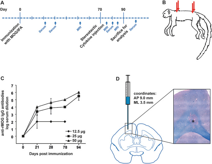
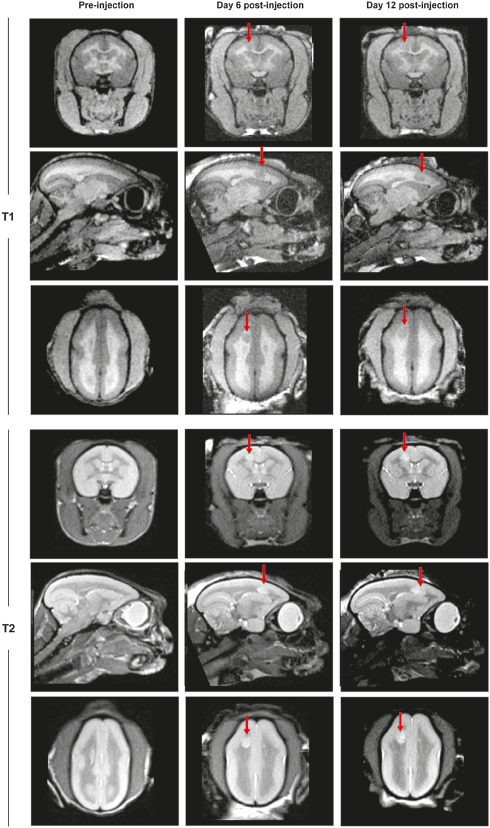
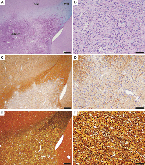
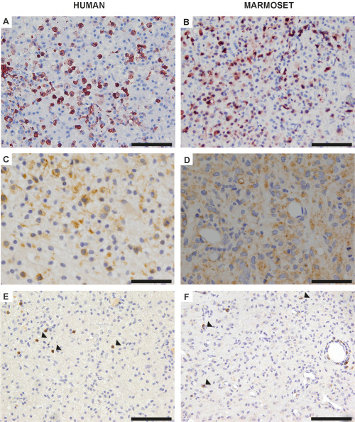
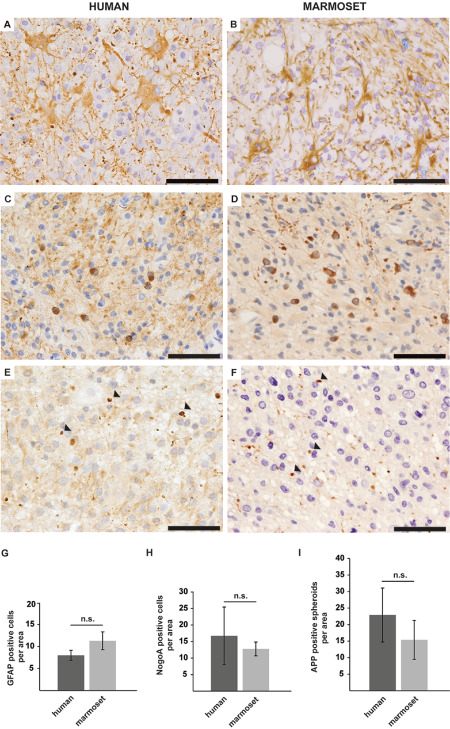
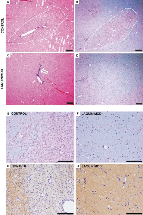
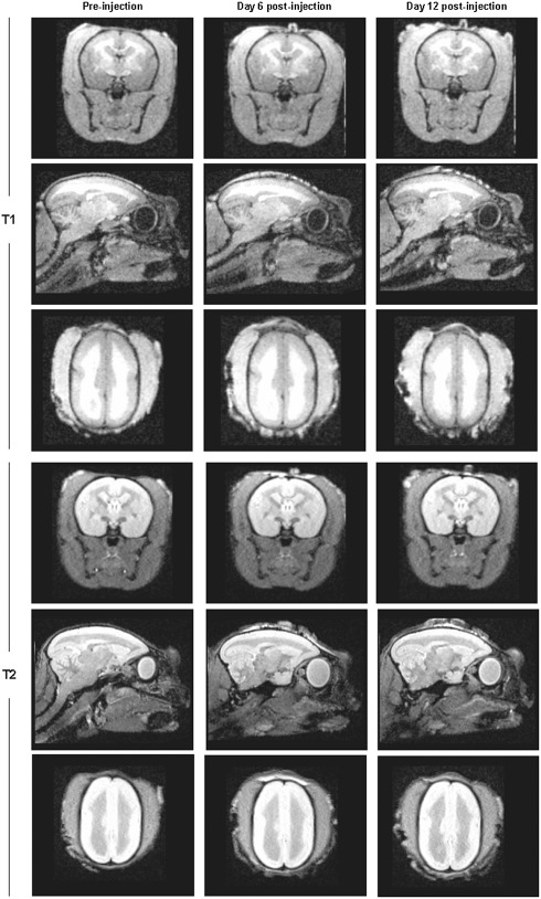
Multiple sclerosis (MS) is the most common cause for sustained disability in young adults, yet treatment options remain very limited. Although numerous therapeutic approaches have been effective in rodent models of experimental autoimmune encephalomyelitis (EAE), only few proved to be beneficial in patients with MS. Hence, there is a strong need for more predictive animal models. Within the past decade, EAE in the common marmoset evolved as a potent, alternative model for MS, with immunological and pathological features resembling more closely the human disease. However, an often very rapid and severe disease course hampers its implementation for systematic testing of new treatment strategies. We here developed a new focal model of EAE in the common marmoset, induced by myelin oligodendrocyte glycoprotein (MOG) immunization and stereotactic injections of proinflammatory cytokines. At the injection site of cytokines, confluent inflammatory demyelinating lesions developed that strongly resembled human MS lesions. In a proof-of-principle treatment study with the immunomodulatory compound laquinimod, we demonstrate that targeted EAE in marmosets provides a promising and valid tool for preclinical experimental treatment trials in MS research.

Keywords: common marmoset; demyelination; experimental autoimmune encephalomyelitis; immunomodulation; laquinimod; multiple sclerosis.


Conflict of interest statement

The authors have nothing to disclose and no conflict of interest to report.

Figures

Figure 1
Figure 1
A. Schematic overview demonstrating the experimental strategy for the induction of focal experimental autoimmune encephalomyelitis (EAE) in the common marmoset. B. Immunization with subclinical dosages of recombinant myelin oligodendrocyte glycoprotein (rMOG) was performed subcutaneously at four injection sites. C. Marmosets were immunized subclinically with different dosages of rMOG (12.5, 25 and 50 μg) and serum concentrations of anti‐MOG immunoglobulin G (IgG) antibodies were determined at 21, 28, 78 and 94 days post immunization by enzyme‐linked immunosorbent assay (ELISA). The applied rMOG concentrations correlated with the IgG titers, which reached similar levels in animals immunized with 25 or 50 μg, respectively. Mean, SD; IgG antibodies shown as log of serum dilution. N, numbers are given within the Materials and Methods section. D. Proinflammatory cytokines were injected intracerebrally with the help of a stereotactic instrument at the coordinates AP (anteroposterior) 9.0 mm and ML (mediolateral) 3.5 mm, resulting in lesions of the corpus callosum and adjacent grey matter, as indicated on the schematic marmoset brain cross section. The light microscopic image represents a demyelinating lesion in the corpus callosum (see asterisk) as visualized by Luxol fast blue–periodic acid Schiff (LFB‐PAS) staining, which marks myelin in blue.
Figure 2
Figure 2
Representative T1‐ and T2‐weighted magnetic resonance imaging (MRI) images of a marmoset brain in coronal, sagittal and transverse planes. Images were performed at three different time points to allow the analysis of lesion development. The first imaging was performed after immunization but before cytokine injection (Pre‐injection), followed by MRI at day 6 and 12 post lesion induction by cytokine injections. Lesions are indicated by arrows.
Figure 3
Figure 3
Representative light microscopic images depict a demyelinating lesion of the corpus callosum and neighboring gray matter of focal experimental autoimmune encephalomyelitis (fEAE) in marmosets (20 days post lesion induction by the injection of cytokines). A. A low magnification overview demonstrates the demyelinated area (LESION) and the transition to the normal appearing white matter (WM) and gray matter (GM). Luxol fast blue–periodic acid Schiff (LFB‐PAS) staining, scale bar 200 μm. B. In a higher magnification, complete demyelination and a strongly increased cellularity becomes apparent. LFB‐PAS staining, scale bar 50 μm. C. Overview of a lesion stained with an antibody against myelin basic protein (MBP) demonstrates a sharp border between the demyelinated area and the adjacent white and gray matter. Scale bar 200 μm. D. MBP immunohistochemistry confirms complete demyelination within the lesion area at a higher magnification. Scale bar: 50 μm. E. A Bielschowsky silver impregnation was performed to visualize axonal density within the lesion. Scale bar 200 μm. F. Note that axons are distended but remain relatively preserved in the lesion. Bielschowsky silver impregnation. Scale bar 50 μm.
Figure 4
Figure 4
A. Representative image of an early active human multiple sclerosis (MS) plaque, which reveals a prominent infiltration of early active macrophages as determined by myeloid‐related protein 14 (MRP14) immunohistochemistry. Scale bar 100 μm. B. The infiltration of numerous MRP14‐positive macrophages also characterizes demyelinating lesions of focal experimental autoimmune encephalomyelitis (fEAE) in marmosets. Scale bar 100 μm. C. Macrophages in human MS lesions demonstrate intracytoplasmic myelin oligodendrocyte glycoprotein (MOG)‐positive degradation products as revealed by immunohistochemistry. Scale bar 100 μm. D. MOG‐positive myelin degradation products are also detected within macrophages of demyelinating lesions in the common marmoset. Scale bar 100 μm. E. T‐cells (arrowheads) are homogeneously distributed within lesions of human MS. CD3 immunohistochemistry; scale bar 100 μm. F. Likewise, a comparable, diffuse T‐cell infiltration (arrowheads) with a perivascular accentuation is detected in fEAE lesions. CD3 immunohistochemistry, scale bar 100 μm.
Figure 5
Figure 5
A. Glial fibrillary acidic protein (GFAP) immunohistochemistry reveals a reactive astrocytosis in human multiple sclerosis (MS) lesions. Scale bar 50 μm. B. A prominent astrogliosis is as well a feature of focal experimental autoimmune encephalomyelitis (fEAE) lesions in marmosets. GFAP immunohistochemistry. Scale bar 50 μm. C. Numerous NOGO‐A positive oligodendrocytes are detected within demyelinated areas of human MS plaques. Scale bar 50 μm. D. Likewise, NOGO‐A positive oligodendrocytes are abundantly detected in lesions of fEAE in marmosets. Scale bar 50 μm. E. Anti‐Alzheimer precursor protein (APP) immunohistochemistry reveals axonal damage in MS lesions, as exemplarily indicated by arrowheads. Scale bar 50 μm. F. Axonal degeneration is likewise observed in fEAE lesion (see arrowheads). APP immunohistochemistry. Scale bar 50 μm. G. The quantification of GFAP‐positive cells per area (0.04 mm2) reveals a similar degree of astrogliosis in human (n = 3) and marmoset (n = 4) lesions. Mean, SD, n.s. = nonsignificant. H. The number of NOGO‐A‐positive oligodendrocytes in human MS lesions (n = 3) varies between individual patients, but is overall comparable to the density of oligodendrocytes in marmoset fEAE (n = 4). Quantification per area (0.04 mm2), mean, SD, n.s. = nonsignificant. I. APP‐positive axonal spheroids are detectable in both, human (n = 3) and marmoset (n = 3) lesions to a similar extent. However, the interindividual variability is much higher in human MS lesions. Quantification per area (0.04 mm2), mean, SD, n.s. = nonsignificant.
Figure 6
Figure 6
A. Representative light microscopic overview of a demyelinating lesion (indicated by white line) in a marmoset with focal experimental autoimmune encephalomyelitis (fEAE). Hematoxylin–eosin (HE) staining, scale bar 200 μm. B. Luxol fast blue–periodic acid Schiff (LFB‐PAS) staining of the same animal as (A) reveals complete demyelination within the lesion. Scale bar 200 μm. C. Representative image of the injection site of cytokines in a marmoset which received a prophylactic treatment of the immunomodulatory drug laquinimod. Note the injection channel, which is marked by Monastral blue (see arrow). Scale bar 200 μm. D. In the respective LFB‐PAS staining of the marmoset described in (C), no lesion formation can be detected. Scale bar 200 μm. E. The lesion edge of a control animal is demonstrated at a higher magnification of the LFB‐PAS staining (see B). The lesion shows complete demyelination and an increased cell number. Scale bar 100 μm. F. In contrast, no signs of demyelination can be detected in laquinimod treated marmosets. Representative image of the injection site of cytokines in a higher magnification. LFB‐PAS staining, scale bar 50 μm. G. myelin basic protein (MBP) immunohistochemistry of a control animal demonstrates MBP‐positive myelin sheaths at the lesion edge (left part of the image) whereas the lesion itself shows complete demyelination (see right part of the image). Scale bar 100 μm. H. In laquinimod‐treated animals no loss of MBP‐positive myelin sheaths can be detected. Representative image of the injection site of cytokines in a higher magnification. MBP immunohistochemistry, scale bar 100 μm.
Figure 7
Figure 7
Representative T1‐ and T2‐weighted magnetic resonance imaging (MRI) images of the brain of a marmoset that received a prophylactic laquinimod treatment. Shown are the coronal, sagittal and transverse planes. Images were performed at three different time points to allow the analysis of lesion development. The first imaging was performed after immunization but before cytokine injection (Pre‐injection), followed by MRI at day 6 and 12 post lesion induction by cytokine injections. Importantly, MRI imaging revealed no lesion formation in marmosets that were treated with laquinimod.

References

    1. Boretius S, Schmelting B, Watanabe T, Merkler D, Tammer R, Czéh B et al (2006) Monitoring of EAE onset and progression in the common marmoset monkey by sequential high‐resolution 3D MRI. NMR Biomed 19:41–49. - PubMed
    1. Brück W, Vollmer T (2013) Multiple sclerosis: oral laquinimod for MS—bringing the brain into focus. Nat Rev Neurol 9:664–665. - PubMed
    1. Brück W, Porada P, Poser S, Rieckmann P, Hanefeld F, Kretzschmar HA, Lassmann H (1995) Monocyte/macrophage differentiation in early multiple sclerosis lesions. Ann Neurol 38:788–796. - PubMed
    1. Brück W, Gold R, Lund BT, Oreja‐Guevara C, Prat A, Spencer CM et al (2013) Therapeutic decisions in multiple sclerosis: moving beyond efficacy. JAMA Neurol 70:1315–1324. - PMC - PubMed
    1. Gardner C, Magliozzi R, Durrenberger PF, Howell OW, Rundle J, Reynolds R (2013) Cortical grey matter demyelination can be induced by elevated pro‐inflammatory cytokines in the subarachnoid space of MOG‐immunized rats. Brain 136:3596–3608. - PubMed

MeSH terms

Substances