. 2015 Sep 15;75(18):3788-99.
doi: 10.1158/0008-5472.CAN-15-0054. Epub 2015 Jul 24.

IL17 Promotes Mammary Tumor Progression by Changing the Behavior of Tumor Cells and Eliciting Tumorigenic Neutrophils Recruitment

Affiliations

IL17 Promotes Mammary Tumor Progression by Changing the Behavior of Tumor Cells and Eliciting Tumorigenic Neutrophils Recruitment

Luciana Benevides et al. Cancer Res. .

Abstract

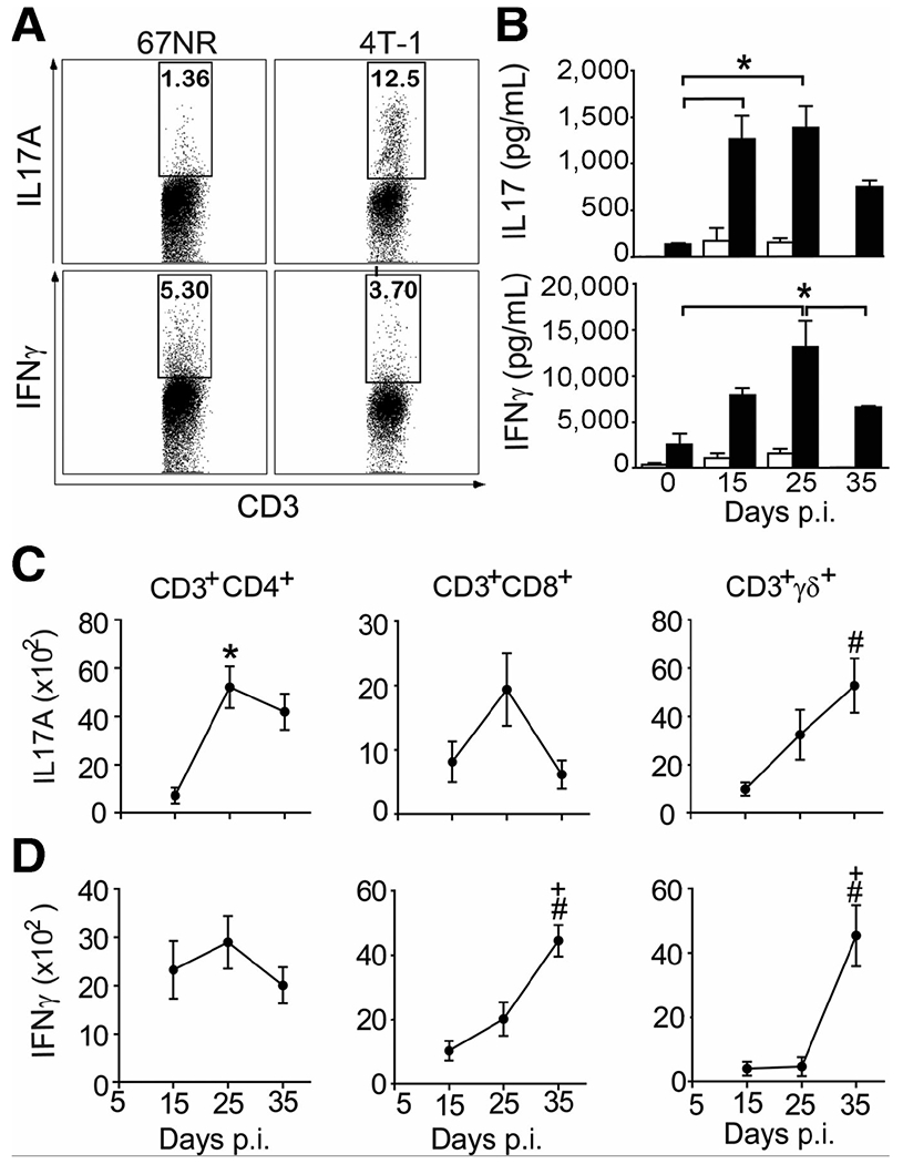
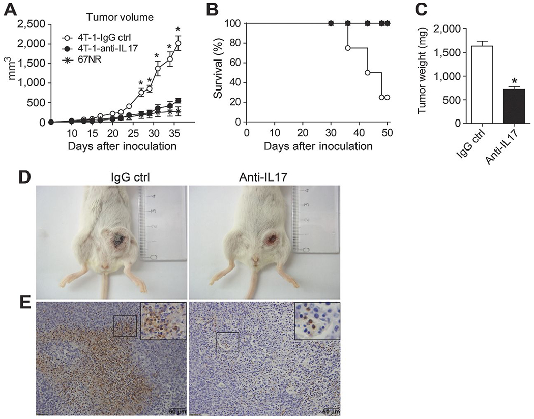
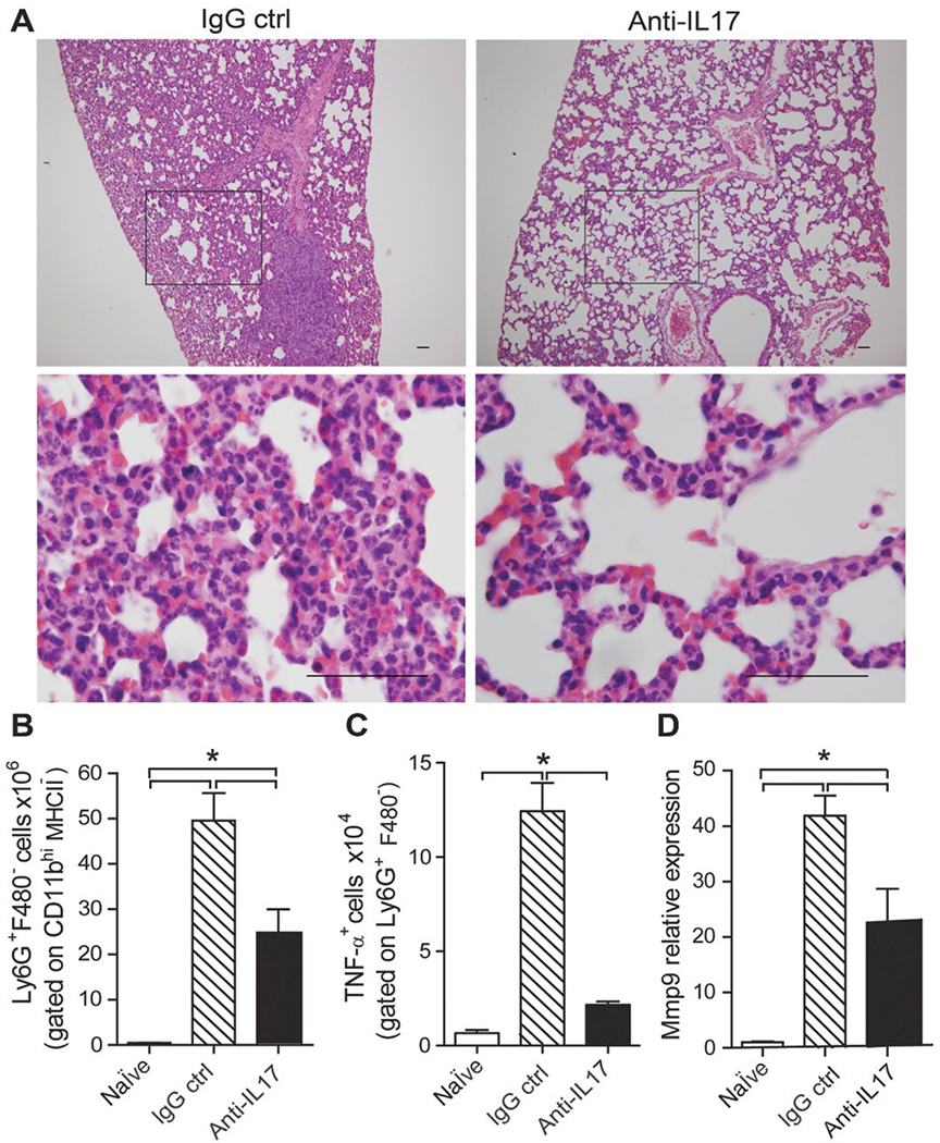
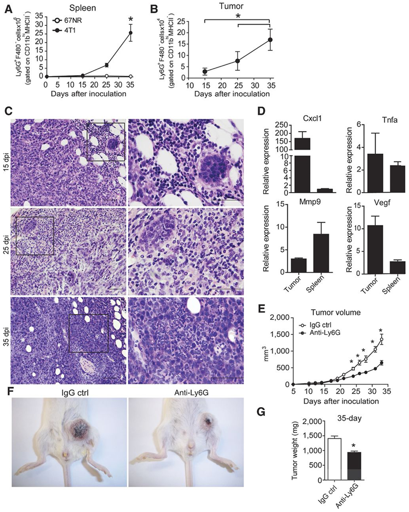
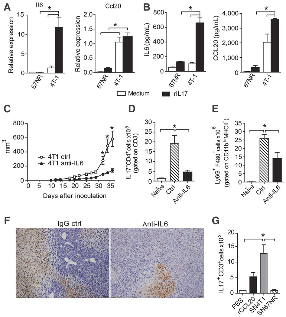
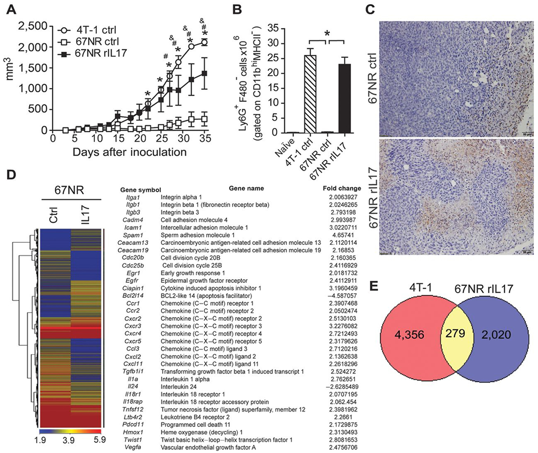
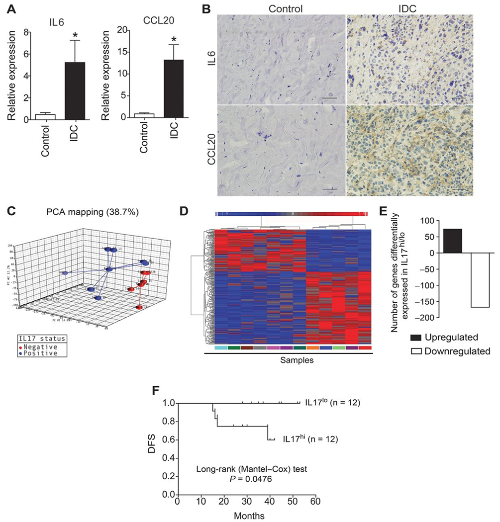
The aggressiveness of invasive ductal carcinoma (IDC) of the breast is associated with increased IL17 levels. Studying the role of IL17 in invasive breast tumor pathogenesis, we found that metastatic primary tumor-infiltrating T lymphocytes produced elevated levels of IL17, whereas IL17 neutralization inhibited tumor growth and prevented the migration of neutrophils and tumor cells to secondary disease sites. Tumorigenic neutrophils promote disease progression, producing CXCL1, MMP9, VEGF, and TNFα, and their depletion suppressed tumor growth. IL17A also induced IL6 and CCL20 production in metastatic tumor cells, favoring the recruitment and differentiation of Th17. In addition, IL17A changed the gene-expression profile and the behavior of nonmetastatic tumor cells, causing tumor growth in vivo, confirming the protumor role of IL17. Furthermore, high IL17 expression was associated with lower disease-free survival and worse prognosis in IDC patients. Thus, IL17 blockade represents an attractive approach for the control of invasive breast tumors.


Conflict of interest statement

Disclosure of Potential Conflicts of Interest

No potential conflicts of interest were disclosed.

Figures

Figure 1.
Figure 1.. IL17A is produced in murine metastatic mammary tumors.
A, cytokine production by tumor-infiltrating cells isolated from BALB/c mice subjected to an induction protocol with nonmetastatic (67NR) and metastatic (4T-1) mammary tumors on day 15 postinoculation (p.i.). Cells were stimulated with PMA + ionomycin, and the expression of IL17A and IFNγ was analyzed in CD3+ cells using flow cytometry. B, IL17A and IFNγ levels (measured by ELISA) in the supernatants of splenocytes isolated at day 0, 15, 25, and 35 postinoculation with 4T-1 and stimulated with anti-CD3 + anti-CD28 (closed bars) or unstimulated (open bars) for 72 hours. C and D, total numbers of PMA/ionomycin-stimulated, CD3-gated, CD4+, CD8+, and TCRγδ+ T cells producing IL17A (C) and IFNγ (D) in isolates from the tumor microenvironment (measured by flow cytometry). The data shown are representative of three independent experiments with similar results and are shown as the mean ± SEM of five mice (*, P < 0.05, comparing periods of 15–25 days; +, P < 0.05, comparing periods of 25–35 days; #, P < 0.05, comparing periods of 15–35 days).
Figure 2.
Figure 2.. Treatment with anti-IL17 inhibits the growth of murine mammary tumors.
BALB/c mice were subjected to an induction protocol with mammary tumor cells (4T1, squares) or treated with neutralizing anti-IL17A (filled squares) or rat immunoglobulin (IgG control; open squares) antibodies or 67NR (asterisks). A, the volume of the tumors was measured at 10 dpi. B, the percentage of survival was assessed daily. C, at 35 dpi, the weight of the tumors was measured. The data shown are representative of two independent experiments with similar results and are shown as the mean ± SEM of five mice per group (*, P < 0.05). D, photographs show representative tumors in mice treated with the IgG control (left) or anti-IL17 antibodies (right) at 35 dpi. E, immunohistochemical staining in tumor tissue of the mice treated with the IgG control (left) or anti-IL17 mAb (right) at 35 dpi was used to detect Ly6G protein; scale bars, 50 μm.
Figure 3.
Figure 3.. Blockade of IL17 inhibits neutrophil recruitment to the lung in a metastatic mammary tumor model.
BALB/c mice were subjected to an induction protocol with mammary tumors and treated with anti-IL17A or IgG control antibodies. A, representative H&E-stained sections of lung tissue collected at 35 dpi; original magnification, ×10 (top) and ×100 (bottom); scale bars, 50 μm. B and C, flow cytometric analysis of the numbers of neutrophils (Ly6G+F4/80) gated on CD11b+MHCII cells (B) and TNFα+ cells gated on Ly6G+MHCII cells (C) after isolation from the lung and stimulation with 100 μg/mL of LPS for 6 hours. D, mRNA was isolated from fragments of lung tissue from both groups at 35 dpi, and the relative expression of MMP-9 was evaluated by qPCR. The data shown are representative of two independent experiments with similar results and are shown as the mean ± SEM of five mice per group (*, P < 0.05).
Figure 4.
Figure 4.. Increased numbers of neutrophils and expression of genes related to the tumorigenic neutrophils phenotype in metastatic murine mammary tumors.
BALB/c mice subjected to an induction protocol with nonmetastatic (67NR) and metastatic (4T-1) mammary tumor cells at day 0, 15, 25, and 35 dpi. The numbers of neutrophils (Ly6G+F480) gated on CD11b+MHCII cells were analyzed in the population of granulocytes isolated from the spleens (A) and tumors (B) by flow cytometry. C, representative, H&E-stained sections of the tumor microenvironment collected at 15, 25, and 35 dpi; original magnification, ×10 (left) and ×100 (right); scale bars, 50 μm. D, Ly6G+F4/80 cells gated on CD11bhiMHCII cells were sorted from the spleens and tumors of 4T-1 tumor-bearing mice, and the expression of MMP-9, VEGF, TNFα, and CXCL1 was analyzed by qPCR. The data shown are normalized to β-actin expression. The data shown represent the means ± SEM compared with Ly6G+F4/80 cells isolated from the spleens of naïve mice. E, the volume of the tumors obtained from BALB/c mice subjected to an induction protocol with mammary tumor cells and treated with anti-Ly6G or rat immunoglobulin (IgG control) antibodies was measured at 10 dpi. F, photographs are representative of tumors in mice treated with IgG control (left) or anti-Ly6G (right) antibodies. G, at 35 dpi, the weight of the tumors was measured. Data are representative of two independent experiments with similar results and are shown as the mean ± SEM of four mice per group (*, P < 0.05).
Figure 5.
Figure 5.. IL17A induces the expression of IL6 and CCL20 in 4T-1 tumor cells lines.
67NR and 4T-1 cells were either treated or not treated with rIL17. After 48 hours, the mRNA was extracted and the culture supernatants were collected. A, the relative expression of IL6 and CCL20 was measured by qPCR. B, the IL6 and CCL20 levels were measured in the culture supernatants by ELISA. The data are representative of two independent experiments with similar results (*, P < 0.05). C, the volume of tumors obtained from BALB/c mice subjected to an induction protocol with metastatic (4T-1) mammary tumor cells and treated with anti-IL6 or rat immunoglobulin (IgG control) antibodies was measured at 10 dpi. At 35 dpi, the numbers of Th17 cells (IL17+CD4+) gated on CD3+ cells (D) and neutrophils (Ly6G+F4/80) gated on CD11b+MHCII cells isolated from the spleen (E) were assessed by flow cytometry. F, immunohistochemical staining in tumor tissues of the mice treated with the IgG control (left) or anti-IL6 mAb (right) at 35 dpi was used to detect Ly6G protein. The data are representative of two independent experiments with similar results and are shown as the mean ± SEM of four mice per group (*, P < 0.05). BALB/c mice received PBS, 400 ng of CCL20, and/or a 4T-1 or 67NR supernatant injected into the air pouch, and the cell infiltrates were harvested at 20 hours after injection. G, the total number of IL17A-producing CD3+ cells harvested per pouch was analyzed by flow cytometry. The data are shown as the mean ± SEM of three mice per group (*, P < 0.05).
Figure 6.
Figure 6.. IL17A directly affects breast cancer tumor cell behavior in vivo.
67NR cells were either treated or not treated with rIL17 in vitro, and after 48 hours, these cells were used to inoculate BALB/c mice to induce mammary tumors. A, the volumes of the tumors were measured at 5 dpi and monitored daily. B, at 35 dpi, the numbers of neutrophils (Ly6G+F4/80) gated on CD11b+MHCII cells were analyzed in the population of granulocytes isolated from the spleen by flow cytometry. C, immunohistochemical staining in tumor tissues of the mice receiving untreated 67NR cells (top) or treated with rIL17 (bottom) at 35 dpi was used to detect Ly6G protein. The data are representative of two independent experiments with similar results and are shown as the mean ± SEM of four mice per group (*, P < 0.05). 67NR cells were either treated or not treated with rIL17. After 48 hours, the mRNA was extracted, and the gene-expression profiles were analyzed by a microarray. D, heat map displaying the hierarchical clustering of the gene-expression profiles. The table presents selected differentially expressed genes of interest. The complete list of the differentially expressed immune response is presented in Supplementary Tables S2 and S3. E, the Venn diagram shows the number of upregulated genes in the 4T-1 cells and IL17-pretreated 67NR cells in relation to the untreated 67NR cells. The intersection of the diagram indicates the number of upregulated genes in common between the 4T-1 and IL17-pretreated 67NR cells.
Figure 7.
Figure 7.. Expression of Th17-promoting cytokines in the tumor microenvironment of patients with IDC.
RNA from breast tumor and healthy control tissue was extracted and analyzed by qPCR. A, the relative expression levels of IL6 and CCL20 are shown. Data are reported as the mean ± SEM of patients (N = 23) or healthy control subjects (N = 8) who were tested individually. The control values were significantly different compared with those of patients with IDC (*, P < 0.05; t test). B, immunohistochemical staining of healthy control (left) and breast tumor tissue (right) was used to detect IL6 and CCL20 protein; scale bars, 50 μm. RNA from breast tumor tissue was extracted, and the gene-expression profiles of samples with low and high IL17A expression were analyzed by microarray. C, principal component analysis of gene-expression microarray 3D scatter plot showing the first three principal components; each patient sample is connected to a centroid for each group (IL17-positive and IL17-negative). D, heat map showing the hierarchical clustering of the gene-expression profiles of samples with low and high IL17A expression as analyzed by microarray. Values are expressed as the relative units of induction (positive values in red), repression (negative values in blue), and no modulation (gray). E, the bar graph shows the number of genes that were down- and upregulated. F, Kaplan–Meier curves illustrate the DFS of patients according to the expression of IL17A. Log-rank P values for the DFS of patients with low and high gene expression were calculated. The data represent the mean ± SEM of 23 patients with IDC (*, P < 0.05).

References

    1. Ferlay J, Parkin DM, and Steliarova-Foucher E, Estimates of cancer incidence and mortality in Europe in 2008. Eur J Cancer, 2010. 46(4): p. 765–81. - PubMed
    1. Schmitt CA, Senescence, apoptosis and therapy--cutting the lifelines of cancer. Nat Rev Cancer, 2003. 3(4): p. 286–95. - PubMed
    1. Tlsty TD and Coussens LM, Tumor stroma and regulation of cancer development. Annu Rev Pathol, 2006. 1: p. 119–50. - PubMed
    1. Pierce BL, et al., Elevated biomarkers of inflammation are associated with reduced survival among breast cancer patients. J Clin Oncol, 2009. 27(21): p. 3437–44. - PMC - PubMed
    1. Chen Z, Laurence A, and O’Shea JJ, Signal transduction pathways and transcriptional regulation in the control of Th17 differentiation. Semin Immunol, 2007. 19(6): p. 400–8. - PMC - PubMed

Publication types

MeSH terms