Case Reports
. 2015 Aug 25;85(8):665-74.
doi: 10.1212/WNL.0000000000001864. Epub 2015 Jul 24.

SQSTM1 splice site mutation in distal myopathy with rimmed vacuoles

Affiliations
Case Reports

SQSTM1 splice site mutation in distal myopathy with rimmed vacuoles

Robert C Bucelli et al. Neurology. .

Abstract

Objective: To identify the genetic etiology and characterize the clinicopathologic features of a novel distal myopathy.

Methods: We performed whole-exome sequencing on a family with an autosomal dominant distal myopathy and targeted exome sequencing in 1 patient with sporadic distal myopathy, both with rimmed vacuolar pathology. We also evaluated the pathogenicity of identified mutations using immunohistochemistry, Western blot analysis, and expression studies.

Results: Sequencing identified a likely pathogenic c.1165+1 G>A splice donor variant in SQSTM1 in the affected members of 1 family and in an unrelated patient with sporadic distal myopathy. Affected patients had late-onset distal lower extremity weakness, myopathic features on EMG, and muscle pathology demonstrating rimmed vacuoles with both TAR DNA-binding protein 43 and SQSTM1 inclusions. The c.1165+1 G>A SQSTM1 variant results in the expression of 2 alternatively spliced SQSTM1 proteins: 1 lacking the C-terminal PEST2 domain and another lacking the C-terminal ubiquitin-associated (UBA) domain, both of which have distinct patterns of cellular and skeletal muscle localization.

Conclusions: SQSTM1 is an autophagic adaptor that shuttles aggregated and ubiquitinated proteins to the autophagosome for degradation via its C-terminal UBA domain. Similar to mutations in VCP, dominantly inherited mutations in SQSTM1 are now associated with rimmed vacuolar myopathy, Paget disease of bone, amyotrophic lateral sclerosis, and frontotemporal dementia. Our data further suggest a pathogenic connection between the disparate phenotypes.


Figures

Figure 1
Figure 1. SQSTM1 variant in 2 unrelated patients with distal myopathy
Pedigrees of family 1 (A) and a sporadic patient (B) are shown. Affected patients are in gray and an arrow denotes the proband. *Indicates patients clinically examined and variant carriers, **Indicates patients with muscle biopsies. (C) Representative chromatogram of the forward sequencing reaction in a control and in the proband of family 1. (D) T1-weighted MRI of the thighs (upper images) and calves (lower images) of the patient with sporadic distal myopathy. Note marked atrophy of the biceps femoris with relative sparing of adductors and vastus lateralis and marked atrophy of the soleus, medial gastrocnemius, and tibialis anterior with relative sparing of the extensor hallucis and digitalis longus, tibialis posterior, flexor hallucis, and digitorum longus.
Figure 2
Figure 2. Muscle histochemistry and immunohistochemistry of SQSTM1-associated distal myopathy with rimmed vacuoles
Muscle biopsy of patient III.1 demonstrates a rimmed vacuolar myopathy with lobulated fibers. (A) Hematoxylin and eosin, (B) modified Gomori trichrome staining of a vacuolated fiber, (C) nicotinamide adenine dinucleotide staining showing lobulated fibers. Immunohistochemical staining for (D) microtubule-associated protein 1A light chain-3 (LC3), (E) TAR DNA-binding protein 43 (TDP-43), and (F) SQSTM1. 4',6-Diamidino-2-phenylindole–stained nuclei are in blue. Scale bar is 15 μm. Electron micrograph of skeletal muscle from patient III.1 demonstrating (G) large focal areas of myofibrillar disorganization and (H) Z-band streaming. Scale bar is 500 nm.
Figure 3
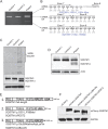
Figure 3. Cryptic splicing of SQSTM1 in a patient with distal myopathy
(A) Complementary DNA was isolated from primary fibroblasts of controls or patient III.1 from family 1 and was PCR amplified with primers spanning exon 6 to exon 9 of SQSTM1. In contrast to control fibroblasts, patient fibroblasts contain 3 SQSTM1 transcripts. (B) The sequence of SQSTM1 transcripts was identified by subcloning each PCR product into pGEM-T easy vector followed by Sanger sequencing. The sequence and resultant translated product are written below each transcript. (C) Lysates from fibroblasts of controls or patient III.1 were immunoblotted with antibodies to SQSTM1, ubiquitin, and actin. Note an increase in high-molecular-weight (HMW) ubiquitinated proteins in patient lysates and the presence of a truncated SQSTM1 protein (SQSTM1Δ). (D) Skeletal muscle lysates from controls or patient III.1 were immunoblotted with an anti-SQSTM1 antibody or actin as a loading control. Note that the patient has a reduction in full-length SQSTM1 and the appearance of a lower molecular weight product, SQSTM1Δ. (E) Diagrams of the 3 putative protein products generated in the patient's muscle: (1) is full-length SQSTM1, (2) lacks amino acids 351–388, which encode the second PEST domain (SQSTM1ΔPEST2), and (3) deletes the C-terminal ubiquitin-associated (UBA) domain with the addition of a C-terminal Asp and Lys (SQSTM1ΔUBA-DK). (F) Immunoblot for SQSTM1 or glyceraldehyde-3-phosphate dehydrogenase (GAPDH) from lysates of U2OS cells transiently transfected with plasmids expressing mCherry alone or mCherry fused to SQSTM1, SQSTM1ΔUBA-DK, or SQSTM1ΔPEST2.
Figure 4
Figure 4. Cryptically spliced mutant SQSTM1 proteins have distinct cellular and myofiber localization
(A, B) U2OS cells were transiently transfected with plasmids expressing mCherry alone or mCherry fused to SQSTM1, SQSTM1ΔUBA-DK, or SQSTM1ΔPEST2 and immunostained with an antibody to (A) microtubule-associated protein 1A light chain-3 (LC3) or (B) ubiquitin. SQSTM1ΔUBA-DK is present diffusely throughout the cell and does not colocalize with LC3 or ubiquitin, whereas SQSTM1ΔPEST2 is present as large perinuclear inclusions that colocalize with both LC3 and ubiquitin. (C) Mouse tibialis anterior muscle was electroporated with plasmids expressing mCherry alone or mCherry fused to SQSTM1, SQSTM1ΔUBA-DK, or SQSTM1ΔPEST2, sectioned, and subjected to fluorescence microscopy. SQSTM1 is present both throughout the sarcoplasm and as larger inclusions. SQSTM1ΔUBA-DK does not form inclusions and is present on myofibrillar structures. In contrast, SQSTM1ΔPEST2 is present as large subsarcolemmal and sarcoplasmic inclusions.

Comment in

References

    1. Udd B. Distal myopathies—new genetic entities expand diagnostic challenge. Neuromuscul Disord 2012;22:5–12. - PubMed
    1. Clemen CS, Herrmann H, Strelkov SV, Schroder R. Desminopathies: pathology and mechanisms. Acta Neuropathol 2013;125:47–75. - PMC - PubMed
    1. Palmio J, Sandell S, Suominen T, et al. Distinct distal myopathy phenotype caused by VCP gene mutation in a Finnish family. Neuromuscul Disord 2011;21:551–555. - PubMed
    1. Watts GD, Wymer J, Kovach MJ, et al. Inclusion body myopathy associated with Paget disease of bone and frontotemporal dementia is caused by mutant valosin-containing protein. Nat Genet 2004;36:377–381. - PubMed
    1. Ju JS, Fuentealba RA, Miller SE, et al. Valosin-containing protein (VCP) is required for autophagy and is disrupted in VCP disease. J Cell Biol 2009;187:875–888. - PMC - PubMed

Publication types

Substances