. 2015 Sep 18;43(16):7911-30.
doi: 10.1093/nar/gkv714. Epub 2015 Jul 23.

Processing by MRE11 is involved in the sensitivity of subtelomeric regions to DNA double-strand breaks

Affiliations

Processing by MRE11 is involved in the sensitivity of subtelomeric regions to DNA double-strand breaks

Keiko Muraki et al. Nucleic Acids Res. .

Abstract

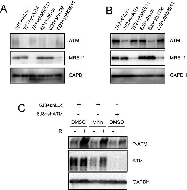
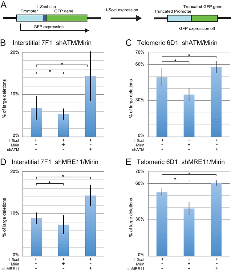
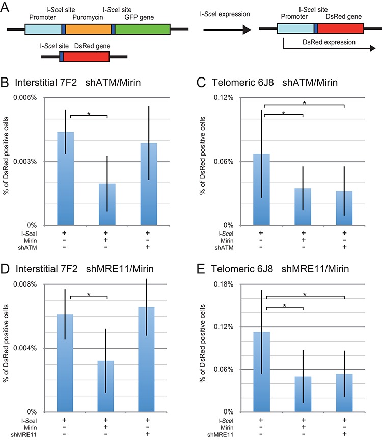
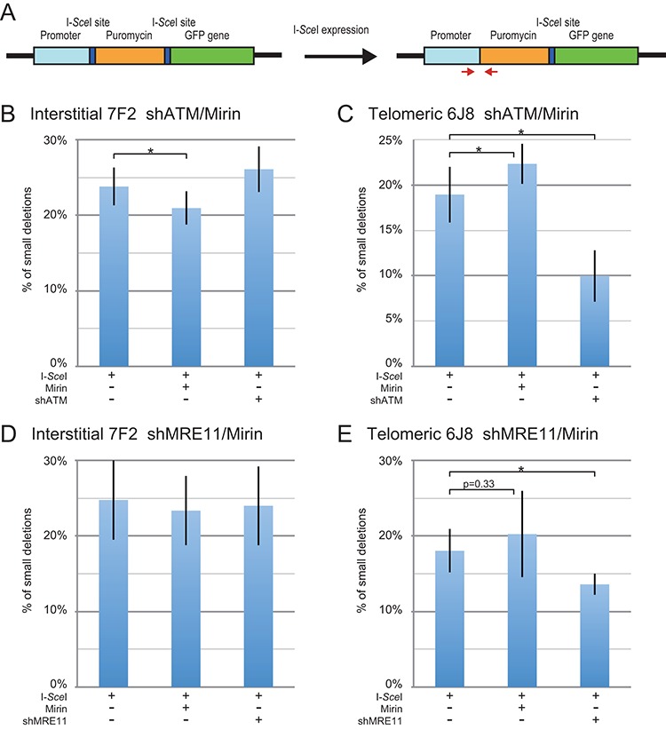
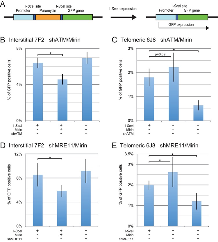
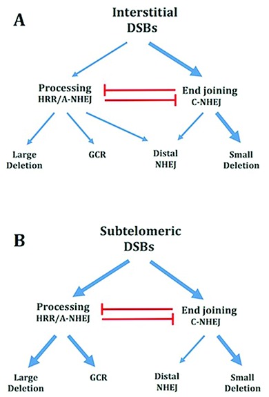
The caps on the ends of chromosomes, called telomeres, keep the ends of chromosomes from appearing as DNA double-strand breaks (DSBs) and prevent chromosome fusion. However, subtelomeric regions are sensitive to DSBs, which in normal cells is responsible for ionizing radiation-induced cell senescence and protection against oncogene-induced replication stress, but promotes chromosome instability in cancer cells that lack cell cycle checkpoints. We have previously reported that I-SceI endonuclease-induced DSBs near telomeres in a human cancer cell line are much more likely to generate large deletions and gross chromosome rearrangements (GCRs) than interstitial DSBs, but found no difference in the frequency of I-SceI-induced small deletions at interstitial and subtelomeric DSBs. We now show that inhibition of MRE11 3'-5' exonuclease activity with Mirin reduces the frequency of large deletions and GCRs at both interstitial and subtelomeric DSBs, but has little effect on the frequency of small deletions. We conclude that large deletions and GCRs are due to excessive processing of DSBs, while most small deletions occur during classical nonhomologous end joining (C-NHEJ). The sensitivity of subtelomeric regions to DSBs is therefore because they are prone to undergo excessive processing, and not because of a deficiency in C-NHEJ in subtelomeric regions.


Figures

Figure 1.
Figure 1.
Western blot analysis of the extent of knockdown MRE11 and ATM, and the lack of effect of Mirin activation of ATM. The extent of shRNA-mediated knockdown of MRE11 and ATM are shown for (A) clones GFP-7F1 and GFP-6D1 and (B) clones EDS-7F2 and EDS-6J8. The extent of knockdown was determined by analyzing the intensity of the ATM and MRE11 bands relative to the intensity of the loading control GAPDH bands (see Table 1). (C) The effect of Mirin on activation of ATM was determined by analysis of phosphorylation of ATM in response to ionizing radiation. Cultures treated with DMSO alone, 20 μM Mirin, or knockdown of ATM were analyzed by western blot 30 min after exposure to 10 Gy of ionizing radiation.
Figure 2.
Figure 2.
The effect of inhibition of MRE11 and ATM on large deletions at interstitial and subtelomeric DSBs. (A) Cell clones containing the pGFP-ISceI plasmid integrated at an interstitial (GFP-7F1) or telomeric (GFP-6D1) site were used for analysis of large deletions. The GFP gene in the integrated pGFP-ISceI plasmid is inactivated following large deletions of more than 28 bp at the I-SceI-induced DSB. The frequency of large deletions (GFP-negative cells) at the I-SceI-induced DSB was determined for clone GFP-7F1 (B, D) and clone GFP-6D1 (C, E) following infection with the pQCXIH-ISceI retrovirus vector and selection with hygromycin for 14 days. Large deletions were analyzed following (B, C) treatment with Mirin or knockdown of ATM (shATM), or (D, E) treatment with Mirin or knockdown of MRE11 (shMRE11). Control cultures for knockdown of ATM or MRE11 were treated with shRNA-mediated knockdown of luciferase, while control cultures for Mirin were treated with DMSO. The values shown in the graph represent the average of three independent experiments, each done in triplicate. Error bars represent the standard deviation of the three separate experiments. Statistical significance for comparisons between the indicated values (horizontal lines) was determined using the two-tailed Student's t-test, and an asterisk indicates statistically significant values of 0.05 or less.
Figure 3.
Figure 3.
The effect of inhibition of MRE11 and ATM on GCRs at interstitial and subtelomeric DSBs. (A) The analysis of GCRs was performed using cell clones that contain the pEJ5-GFP plasmid integrated at an interstitial (EDS-7F2) or telomeric (EDS-6J8) site, and a pDsRed-ISceI plasmid integrated at an interstitial site. The DsRed gene in the pDsRed-ISceI plasmid is initially inactive due to the lack of a promoter, but is activated following NHEJ between the I-SceI-induced DSBs in the pEJ5-GFP and pDsRed-ISceI plasmids. The frequency of GCRs (DsRed-positive cells) at the I-SceI-induced DSB was determined for clone EDS-7F2 (B, D) and clone EDS-6J8 (C, E) following infection with the pQCXIH-ISceI retrovirus vector and selection with hygromycin for 14 days for EDS-7F2 and 15 days for EDS-6J8. GCRs were analyzed following (B, C) treatment with Mirin or knockdown of ATM (shATM), or (D, E) treatment with Mirin or knockdown of MRE11 (shMRE11). Control cultures for knockdown of ATM or MRE11 were treated with shRNA-mediated knockdown of luciferase, while control cultures for Mirin were treated with DMSO. The values shown in the graph represent the average of more than three independent experiments, each done in triplicate (see Supplementary Figures S3 and S4, and Table S1 for raw data). Error bars represent the standard deviation of the more than three separate experiments. Statistical significance for comparisons between the indicated values (horizontal lines) was determined using the two-tailed Student's t-test, and an asterisk indicates statistically significant values of 0.05 or less.
Figure 4.
Figure 4.
The effect of inhibition of MRE11 and ATM on small deletions at interstitial and subtelomeric DSBs. (A) Cell clones containing the pEJ5-GFP plasmid integrated at an interstitial (EDS-7F2) or telomeric (EDS-6J8) site were used for analysis of small deletions. Small deletions were determined by first amplifying a PCR product that spans one of the I-SceI endonuclease recognition sites from genomic DNA isolated from the pooled population of cells expressing I-SceI endonuclease. The PCR product was then digested with I-SceI endonuclease to determine the frequency of cells in the population with small deletions at the I-SceI-induced DSB, as shown by the fraction of PCR product that is not cut with I-SceI endonuclease. The frequency of small deletions at the I-SceI-induced DSB was determined for clone EDS-7F2 (B, D) and clone EDS-6J8 (C, E) following infection with the pQCXIH-ISceI retrovirus vector and selection with hygromycin for 14 days for EDS-7F2 and 15 days for EDS-6J8. Small deletions were analyzed following (B, C) treatment with Mirin or knockdown of ATM (shATM), or (D, E) treatment with Mirin or knockdown of MRE11 (shMRE11). Control cultures for knockdown of ATM or MRE11 were treated with shRNA-mediated knockdown of luciferase, while control cultures for Mirin were treated with DMSO. The values shown in the graph represent the average of more than three independent experiments, each done in triplicate. Error bars represent the standard deviation of more than three separate experiments. Statistical significance for comparisons between the indicated values (horizontal lines) was determined using the two-tailed Student's t-test, and an asterisk indicates statistically significant values of 0.05 or less.
Figure 5.
Figure 5.
The effect of inhibition of MRE11 and ATM on distal NHEJ at interstitial and subtelomeric DSBs. (A) Cell clones containing the pEJ5-GFP plasmid integrated at an interstitial (EDS-7F2) or telomeric (EDS-6J8) site were used for analysis of distal NHEJ. The GFP gene in the pEJ5-GFP plasmid is initially inactive due to the presence of puromycin-resistance (puro) gene located between the GFP gene and its promoter, but is activated following NHEJ between the distal ends of the two I-SceI-induced DSBs, which results in the deletion of the puro gene. The frequency of distal NHEJ (GFP-positive cells) at the I-SceI-induced DSB was determined for clone EDS-7F2 (B, D) and clone EDS-6J8 (C, E) following infection with the pQCXIH-ISceI retrovirus vector and selection with hygromycin for 14 days for EDS-7F2 and 15 days for EDS-6J8. Distal NHEJ was analyzed following (B, C) treatment with Mirin or knockdown of ATM (shATM), or (D, E) treatment with Mirin or knockdown of MRE11 (shMRE11). Control cultures for knockdown of ATM or MRE11 were treated with shRNA-mediated knockdown of luciferase, while control cultures for Mirin were treated with DMSO. The values shown in the graph represent the average of the more than three independent experiments, each done in triplicate. Error bars represent the standard deviation of more than three separate experiments. Statistical significance for comparisons between the indicated values (horizontal lines) was determined using the two-tailed Student's t-test, and an asterisk indicates statistically significant values of 0.05 or less.
Figure 6.
Figure 6.
Model for the mechanisms of formation of mutations during repair of interstitial and subtelomeric I-SceI-induced DSBs. (A) Mechanisms of formation of mutations at interstitial DSBs. DSB repair occurs either directly through C-NHEJ, or following the processing of the ends of the DSB. As with HRR, large deletions and GCRs also involve the processing of DSBs, however repair occurs by A-NHEJ. Importantly, the GCR assay does not detect GCRs that also involve large deletions. However, large deletions near telomeres also commonly result in GCRs, so that the major difference between the large deletion and GCR assays is the extent of degradation involved in the GCR. Small deletions of a few base pairs occur during end joining involving C-NHEJ. Distal NHEJ (deletions resulting from joining two closely positioned DSBs) occurs both by end joining by C-NHEJ and following processing and A-NHEJ. (B) Mechanisms of formation of mutations at subtelomeric DSBs. End joining by C-NHEJ at subtelomeric DSBs occurs with the same efficiency as at interstitial DSBs, as shown by the fact that small deletions at subtelomeric DSBs occur at the same frequency as at interstitial DSBs. The repair of subtelomeric DSBs by end joining by C-NHEJ is ATM-dependent. As at interstitial DSBs, large deletions and GCRs occur through processing of DSBs and A-NHEJ, although with a much greater frequency than at interstitial DSBs. The decreased frequency of distal NHEJ at subtelomeric DSBs appears to be due to a reduced contribution of A-NHEJ, possibly because most DSBs repaired by A-NHEJ at subtelomeric DSBs become large deletions and/or GCRs. Combined together, our results suggest that the sensitivity of subtelomeric regions to DSBs is a result of excessive processing by MRE11 and other nucleases, and is not due to a deficiency in C-NHEJ.

Similar articles

Cited by

References

    1. Thompson L.H. Recognition, signaling, and repair of DNA double-strand breaks produced by ionizing radiation in mammalian cells: The molecular choreography. Mutat. Res. 2012;751:158–246. - PubMed
    1. Lieber M.R. NHEJ and its backup pathways in chromosomal translocations. Nat Struct. Mol. Biol. 2010;17:393–395. - PMC - PubMed
    1. Lavin M.F. ATM and the Mre11 complex combine to recognize and signal DNA double-strand breaks. Oncogene. 2007;26:7749–7758. - PubMed
    1. Bothmer A., Robbiani D.F., Di Virgilio M., Bunting S.F., Klein I.A., Feldhahn N., Barlow J., Chen H.T., Bosque D., Callen E., et al. Regulation of DNA end joining, resection, and immunoglobulin class switch recombination by 53BP1. Mol. Cell. 2011;42:319–329. - PMC - PubMed
    1. Boersma V., Moatti N., Segura-Bayona S., Peuscher M.H., van der Torre J., Wevers B.A., Orthwein A., Durocher D., Jacobs J.J. MAD2L2 controls DNA repair at telomeres and DNA breaks by inhibiting 5′ end resection. Nature. 2015;521:537–540. - PMC - PubMed

Publication types