Review
. 2015 Aug;38(8):496-505.
doi: 10.1016/j.tins.2015.06.004. Epub 2015 Jul 21.

The cellular and molecular landscape of neuroligins

Affiliations
Review

The cellular and molecular landscape of neuroligins

Michael A Bemben et al. Trends Neurosci. 2015 Aug.

Abstract

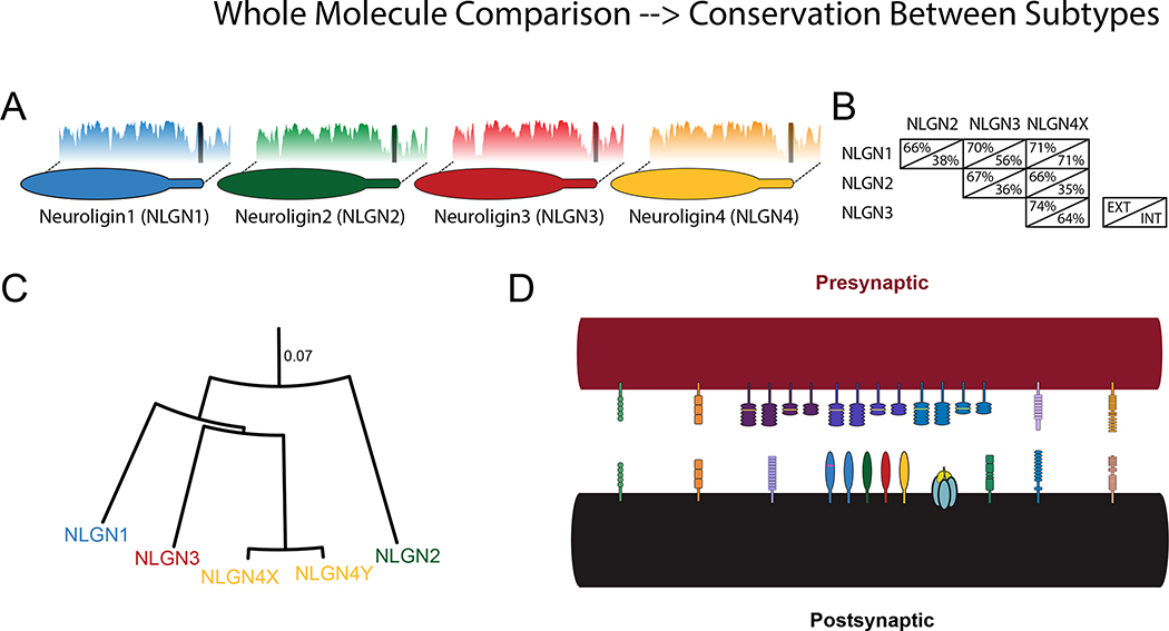
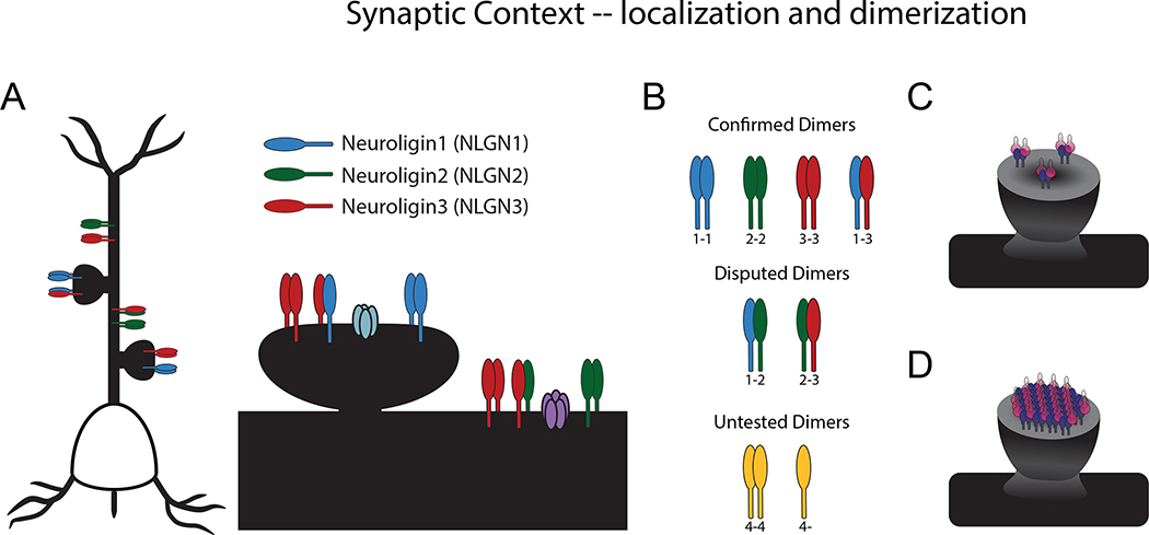
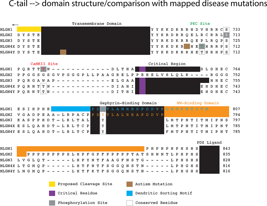
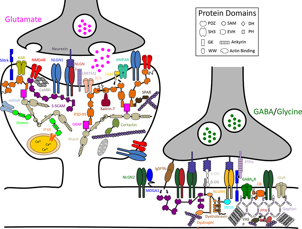
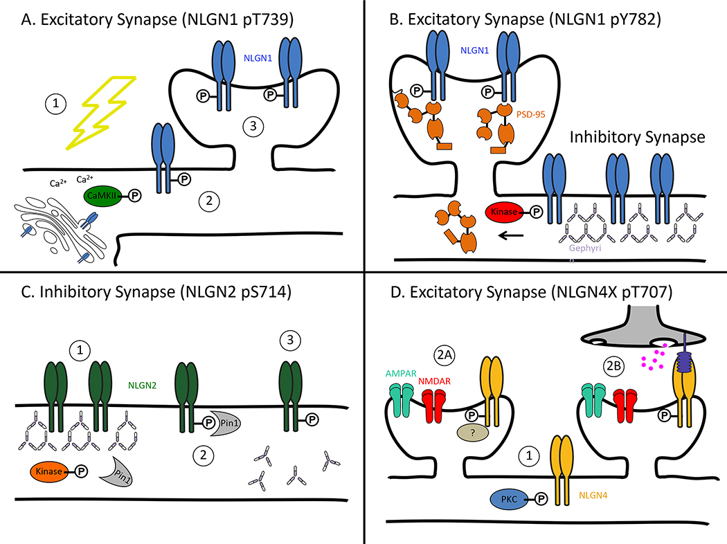
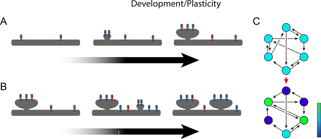
A fundamental physical interaction exists across the synapse. It is mediated by synaptic adhesion molecules, and is among the earliest and most indispensable of molecular events occurring during synaptogenesis. The regulation of adhesion molecules and their interactions with other synaptic proteins likely affect not only on synapse formation but also on ongoing synaptic function. We review research on one major family of postsynaptic adhesion molecules, neuroligins, which bind to their presynaptic partner neurexin across the synaptic cleft. We move from a structural overview to the broad cellular and synaptic context of neuroligins, intermolecular interactions, and molecular modifications that occur within a synapse. Finally, we examine evidence concerning the physiological functions of neuroligin in a cell and highlight areas requiring further investigation.


Figures

Figure 1.
Figure 1.. Neuroligin subtypes.
A. Subtypes of neuroligin with shared identity. Plots behind each molecule show percent identity to a group containing NLGNs 1–4 (blasp, sliding average 10 amino acids). Dark bar in the identity plot indicates the transmembrane domain. B. Matrix depicting the approximate percentage identity of the different human neuroligin protein sequences, excluding extracellular splice sites, separated by domain (EXT: extracellular; INT: intracellular). C. Distance model of neuroligin subtypes (Jukes-Cantor Model). Scale is given as residue substitutions per site. D. Schematic of synaptic adhesion superfamily (abbreviations: leukocyte common antigen-related receptor protein tyrosine phosphatase (LAR PTPR); leucine-rich repeat transmembrane protein (LRRTM); cerebellin (Clbn); CIRL1/latrophilin-1 (CL1); netrin-G ligand (NGL)).
Figure 2.
Figure 2.. Localization and dimerization.
A. Localization of neuroligins: NLGN1 and NLGN3 at excitatory synapses; NLGN2 and NLGN3 at inhibitory synapses. Shown also in the synapse are an AMPA receptor (in light blue) and GABA receptor (in purple). B. Confirmed, disputed, and untested dimers of neuroligin subtypes. C. Model of a synapse with only neuroligin dimers. D. Model of a synapse with higher-order neuroligin oligomers.
Figure 3.
Figure 3.. Neuroligin cytoplasmic domain comparison.
Alignment of the transmembrane and cytoplasmic domains of human neuroligins. Residues identified in rodent neuroligins are depicted on the analogous residues on the human isoforms for comparison purposes. Mapped residues and motifs are boxed. The autism mutation in NLGN4X is arginine (R) 704 mutated to a cysteine and in NLGN4Y isoleucine (I) 679 mutated to a valine. The critical region is necessary for neuroligin-mediated excitatory synaptic potentiation. (abbreviations: Ca2+/calmodulin-dependent protein kinase II (CaMKII); PSD-95/Discs large/ZO-1 (PDZ); protein kinase C (PKC))
Figure 4.
Figure 4.. Neuroligin binding partners at excitatory and inhibitory synapses.
Schematic diagram of a subset of protein constituents at excitatory and inhibitory synapses. Neuroligins bind a diverse set of proteins at glutamatergic and GABAergic/glycinergic synapses. (protein abbreviations are: alpha-dystroglycan (α-DG); α-amino-3-hydroxy-5-methyl-4-isoxazole propionic acid receptor (AMPAR); beta-dystroglycan (β-DG); Ca2+/calmodulin-dependent protein kinase II (CaMKII); collybistin (CB); cortical actin binding protein (Cortactin); gamma-aminobutyric acida receptor (GABAaR); guanylate kinase-associated protein (GKAP); glycine receptor (GlyR); immunoglobin superfamily member 9b (IgSF9b); inositol trisphosphate 3 receptor (IP3R); kainate receptor (KAR); potassium channel (KCh); leucine-rich transmembrane protein 2 (LRRTM2); MAM domain-containing glycosylphosphatidylinositol anchor 1 (MDGA1); metabotropic glutamate receptor (mGluR); N-methyl-D-aspartate receptor (NMDAR); profilin (PFN); postsynaptic density protein 95 (PSD-95); protein tyrosine phosphatase rho (PTPσ); synaptic scaffolding molecule (S-SCAM); SH3 and ankyrin repeat-containing protein (Shank); Slit- and NTRK-like family (Slitrk); syntrophin (SNTA); spine-associated RapGAP (SPAR); TCR gamma alternate reading from protein (TARP); vasodilator-stimulated phosphoprotein (VASP); protein domain abbreviations are: Dbl homology domain (DH); enabled/VASP homology domain (EVH); guanylate kinase-like domain (GK); PSD-95/Discs large/ZO-1 (PDZ); pleckstrin homology domain (PH); sterile alpha motif (SAM); Src homology domain (SH3))
Figure 5
Figure 5. Regulation of neuroligins by phosphorylation.
A. NLGN1 pT739. (1) Synaptic activity drives calcium influx thereby activating CaMKII. (2) CaMKII phosphorylates NLGN1, which leads to increased surface expression. (3) Increased NLGN1 surface expression promotes the creation of new synapses. B. NLGN1 pY782. Phosphorylation by an unknown kinase in the gephyrin-binding domain (Y782) drives NLGN1 to release from gephyrin and localize at excitatory synapses as opposed to inhibitory synapses. C. NLGN2 pS714. (1) An unknown kinase phosphorylates NLGN2 at S714. (2) Phosphorylation leads to recruitment and binding of Pin1. (3) Pin1 isomerizes NLGN2, which negatively affects the NLGN2-gephyrin interaction. D. NLGN4X pT707. (1) Activation of PKC phosphorylates NLGN4X at T707. Phosphorylation of NLGN4X promotes the genesis of excitatory synapses possibly through an unknown protein interaction (2A) or by increasing the recruitment of presynaptic terminals (2B). (abbreviations: α-amino-3-hydroxy-5-methyl-4-isoxazole propionic acid receptor (AMPAR); Ca2+/calmodulin-dependent protein kinase II (CaMKII); N-methyl-D-aspartate receptor (NMDAR); phosphorylation (P); protein kinase C (PKC); peptidyl-proly cis-trans isomerase (Pin1); postsynaptic density protein 95 (PSD-95))
Figure 6.
Figure 6.. Development and plasticity.
A. Model for ongoing synaptogenesis with increasing expression of neuroligin over development. B. Model for activity-dependent synaptogenesis following a Ca2+-mediated increase in the surface expression of neuroligin. C. Depiction of network plasticity. In this model, an initial network of neurons (above) has distributed connectivity, with each neuron receiving and sending an equal number of connections. Based on differential expression of neuroligin in each of the cells, this may evolve into a non-uniform network (below), in which certain neurons send and receive more connections than other neurons. Cells are color-coded from blue (sparsely connected) to green (densely connected) to depict the amount of connectivity, illustrating that certain cells may become over-connected “hubs” of the network.

References

    1. Antonelli R, Pizzarelli R, Pedroni A, Fritschy JM, Del Sal G, Cherubini E, and Zacchi P (2014) Pin1-dependent signalling negatively affects GABAergic transmission by modulating neuroligin2/gephyrin interaction. Nature communications 5, 5066. - PMC - PubMed
    1. Arac D, Boucard AA, Ozkan E, Strop P, Newell E, Sudhof TC, and Brunger AT (2007) Structures of neuroligin-1 and the neuroligin-1/neurexin-1 beta complex reveal specific protein-protein and protein-Ca2+ interactions. Neuron 56, 992–1003. - PubMed
    1. Arikkath J and Reichardt LF (2008) Cadherins and catenins at synapses: roles in synaptogenesis and synaptic plasticity. Trends Neurosci 31, 487–494. - PMC - PubMed
    1. Bemben MA, Nguyen QA, Wang T, Li Y, Nicoll RA, and Roche KW (2015) Autism-associated mutation inhibits protein kinase C-mediated neuroligin-4X enhancement of excitatory synapses. Proceedings of the National Academy of Sciences of the United States of America. - PMC - PubMed
    1. Bemben MA, Shipman SL, Hirai T, Herring BE, Li Y, Badger JD 2nd, . . . Roche KW (2014) CaMKII phosphorylation of neuroligin-1 regulates excitatory synapses. Nature neuroscience 17, 56–64. - PMC - PubMed