. 2015 Sep 3;126(10):1224-33.
doi: 10.1182/blood-2014-09-598722. Epub 2015 Jul 24.

Autophagy is induced upon platelet activation and is essential for hemostasis and thrombosis

Affiliations

Autophagy is induced upon platelet activation and is essential for hemostasis and thrombosis

Madhu M Ouseph et al. Blood. .

Erratum in

  • Blood. 2015 Oct 22;126(17):2072. Komatsu, Massaki [corrected to Komatsu, Masaaki]

Abstract

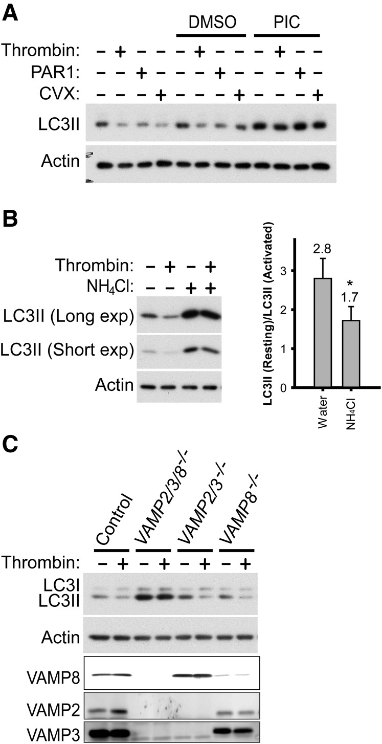
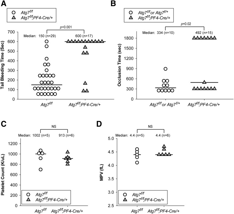
Autophagy is important for maintaining cellular homeostasis, and thus its deficiency is implicated in a broad spectrum of human diseases. Its role in platelet function has only recently been examined. Our biochemical and imaging studies demonstrate that the core autophagy machinery exists in platelets, and that autophagy is constitutively active in resting platelets. Moreover, autophagy is induced upon platelet activation, as indicated by agonist-induced loss of the autophagy marker LC3II. Additional experiments, using inhibitors of platelet activation, proteases, and lysosomal acidification, as well as platelets from knockout mouse strains, show that agonist-induced LC3II loss is a consequence of platelet signaling cascades and requires proteases, acidic compartments, and membrane fusion. To assess the physiological role of platelet autophagy, we generated a mouse strain with a megakaryocyte- and platelet-specific deletion of Atg7, an enzyme required for LC3II production. Ex vivo analysis of platelets from these mice shows modest defects in aggregation and granule cargo packaging. Although these mice have normal platelet numbers and size distributions, they exhibit a robust bleeding diathesis in the tail-bleeding assay and a prolonged occlusion time in the FeCl3-induced carotid injury model. Our results demonstrate that autophagy occurs in platelets and is important for hemostasis and thrombosis.


Figures

Figure 1
Figure 1
Autophagy machinery is present in murine and human platelets. (A) Western blot analyses of the autophagy machinery in freshly prepared mouse and human platelets and in banked human platelets. Extracts from mouse and human platelets were loaded in each lane and probed with antibodies to the indicated proteins. Atg5+/+ and Atg5−/− mouse embryonic fibroblasts (MEFs) were used as additional molecular weight markers for LC3I (unconjugated form) and LC3II (phosphatidylethanolamine-conjugated form), as Atg5−/− MEFs produced LC3I but not the faster-migrating LC3II. Atg5−/− MEFs also had higher p62 levels than Atg5+/+ MEFs because of impaired autophagy. Note that the loading amounts for the MEFs were much lower than those for platelets to have comparable LC3II levels. As a result, and as platelets have high actin content than the MEFs, both actin and p62 levels in the MEF samples are much lower than in platelets. The images shown are representative of at least 3 independent experiments. Full images of all blots are shown in supplemental Figure 1. (B) Platelets from the autophagy reporter mice (ie, GFP-LC3/+, Becn1-EGFP/+, EGFP-Atg5/+) but not wild-type mice show GFP-LC3-positive, EGFP-Atg5-positive, and Beclin 1-EGFP-positive puncta, respectively (arrows), which are autophagosome-related structures including isolation membranes, autophagosomes, and autolysosomes. Samples were visualized for GFP or EGFP fluorescence (fluorescein isothiocyanate [FITC] channel) and differential interference (DIC). The images shown are representative of at least 2 independent experiments.
Figure 2
Figure 2
Basal autophagy occurs in resting platelets. (A) Western blot analysis of LC3II in unstimulated human platelets treated with either NH4Cl (20 mM) or chloroquine (50 µM) to inhibit lysosomal activity. Two exposure times, short and long, are shown. Note that LC3I is visible only with longer exposures in this experiment. (B) GFP-LC3 puncta in unstimulated platelets without and with NH4Cl treatment (20 mM, 2 h). Samples were visualized for DIC and GFP fluorescence (GFP-LC3). Ten images (17∼74 platelets/field) were obtained at random and quantified for each condition. Platelets containing GFP-LC3-positive puncta are 21 ± 8% and 64 ± 10% without and with NH4Cl treatment, respectively. Statistical significance was evaluated with the Student t test. (C) Western blot analysis of LC3II in human platelets pretreated for 1 h with cycloheximide (100 µg/mL) or puromycin (10 µg/mL), in combination with pretreatment of NH4Cl (20 mM, 1 h) or protease inhibitor cocktail (1 h). The images shown are representative of at least 3 independent experiments.
Figure 3
Figure 3
Platelet activation leads to LC3II reduction. (A) Western blot analysis of LC3II in the murine and human platelets before and after stimulation (0.1 U/mL thrombin, 30 minutes), with various anti-LC3 antibodies. Cell extracts from Atg5+/+ and Atg5−/− MEFs were used as additional molecular weight markers to indicate the migration positions of LC3I and LC3II. Note that the loading volumes for the MEFs were optimized to have comparable LC3II levels, and platelets have high actin content. Thus, both actin and p62 levels in the MEF samples were below detection limits. The images shown are representative of at least 2 independent experiments. (B) Western blot analyses of LC3II in human platelets before and after stimulation with various agonists. Platelets were stimulated for 30 minutes with thrombin (0.1 U/mL), CVX (0.1 µg/mL), PAR1 or PAR4 peptide (100 µM), ADPβS (10 µM), collagen (1 µg/mL), A23187 (10 µM in DMSO), and U46619 (20 µM in DMSO). N = 2∼8 for various inhibitors. (C) Western blot analysis of LC3II, before and after stimulation, in human platelets pretreated with inhibitors of platelet activation. Platelets were pretreated for 60 minutes with U-73122 (20 µM), U-73343 (inactive analog of U-73122, 20 µM), BAPTA-AM (100 µM), Ro-31-8220 (10 µM), or PP2 (100 µM) and then stimulated for 30 minutes with either thrombin (0.1 U/ml) or CVX (0.1 µg/mL). Of note, U-73343, Ro-31-8220, and PP2 increased LC3II levels in resting platelets for an unknown reason. All inhibitor stock solutions were prepared in DMSO. N = 3 for all inhibitors. For B and C, *P < .05 by the Student t test.
Figure 4
Figure 4
Agonist-induced LC3II reduction involves autolysosomal proteolysis. (A) Western blot analysis of LC3II changes in response to agonists in banked human platelets pretreated with a PIC. Platelets were incubated with the PIC for 60 minutes before 30-minute agonist stimulation with thrombin (0.1 U/mL), PAR1 peptide (100 µM), and CVX (0.1 µg/mL), respectively. The images shown are representative of at least 3 independent experiments. (B) Western blot analysis of LC3II in thrombin-stimulated (0.1 U/mL, 30 minutes) mouse platelets after preincubation with NH4Cl (20 mM, 2 hours). The images shown are representative of 4 independent experiments. *P < .05. (C) Western blot analysis of LC3II in thrombin-stimulated (0.1 U/mL, 30 minutes) platelets isolated from mice lacking VAMP2, VAMP3, and/or VAMP8, and from wild-type control mice. The images shown are representative of 2 independent experiments.
Figure 5
Figure 5
Atg7-deficient platelets showed modest defects in ex vivo platelet function assays. (A) Representative western blot analysis showing reduced Atg7 and depleted LC3II levels in platelets from Atg7f/f;PF4-Cre/+ mice compared with platelets from Atg7f/f mice. For each genotype, results from 2 different mice are shown. (B) Lumi-aggregometry measurements of platelets from Atg7f/f (red traces) and Atg7f/f;PF4-Cre/+ (blue traces) mice when stimulated with thrombin (upper) or collagen (lower). Different agonist concentrations were titrated. The images shown are representative of at least 3 independent experiments. (C) Total levels of PF4 (left), total [3H]-serotonin uptake (middle), and total β-hexosaminidase activity (right) in platelets purified from Atg7f/f and Atg7f/f;PF4-Cre/+ mice. Data represent an average of 21 measurements. *P < .05, **P < .001, Student t test. (D) EM images of platelets from Atg7f/f (i-iii) and Atg7f/f;PF4-Cre/+ (iv-vi) mice. Samples were prepared by the freeze substitution method. α, α granules; arrows, dense granules; arrowheads and inset, isolation membranes. Scale bars, 500 nm. The images shown are representative micrographs from 2 independent experiments.
Figure 6
Figure 6
Megakaryocyte- and platelet-specific Atg7 deletion causes impaired hemostasis and thrombus formation without thrombocytopenia. (A) Tail-bleeding time measurements taken from Atg7f/f (○) and Atg7f/f;PF4-Cre/+ (filled gray triangles) mice. (B) Occlusion time measurements taken from Atg7f/f (○) and Atg7f/f;PF4-Cre/+ (filled gray triangles) mice in the FeCl3 carotid injury model of thrombosis. For A and B, each data point represents an individual mouse, with the horizontal line marking the median. Statistical significance was evaluated with the log-rank test using SigmaPlot12 software. (C) Platelet counts and (D) mean platelet volumes from Atg7f/f (○) and Atg7f/f;PF4-Cre/+ (filled gray triangles) mice. For C and D, statistical significance was evaluated with the Student t test. NS, not significant.

References

    1. Mizushima N, Levine B, Cuervo AM, Klionsky DJ. Autophagy fights disease through cellular self-digestion. Nature. 2008;451(7182):1069–1075. - PMC - PubMed
    1. Massey AC, Kaushik S, Cuervo AM. Lysosomal chat maintains the balance. Autophagy. 2006;2(4):325–327. - PubMed
    1. Kunz JB, Schwarz H, Mayer A. Determination of four sequential stages during microautophagy in vitro. J Biol Chem. 2004;279(11):9987–9996. - PubMed
    1. Uttenweiler A, Schwarz H, Neumann H, Mayer A. The vacuolar transporter chaperone (VTC) complex is required for microautophagy. Mol Biol Cell. 2007;18(1):166–175. - PMC - PubMed
    1. Suzuki K, Ohsumi Y. Molecular machinery of autophagosome formation in yeast, Saccharomyces cerevisiae. FEBS Lett. 2007;581(11):2156–2161. - PubMed

Publication types