. 2015 Nov;29(11):4654-69.
doi: 10.1096/fj.15-274340. Epub 2015 Jul 24.

Intracellular sphingosine kinase 2-derived sphingosine-1-phosphate mediates epidermal growth factor-induced ezrin-radixin-moesin phosphorylation and cancer cell invasion

Affiliations

Intracellular sphingosine kinase 2-derived sphingosine-1-phosphate mediates epidermal growth factor-induced ezrin-radixin-moesin phosphorylation and cancer cell invasion

Mohamad M Adada et al. FASEB J. 2015 Nov.

Abstract

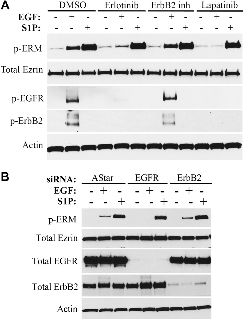
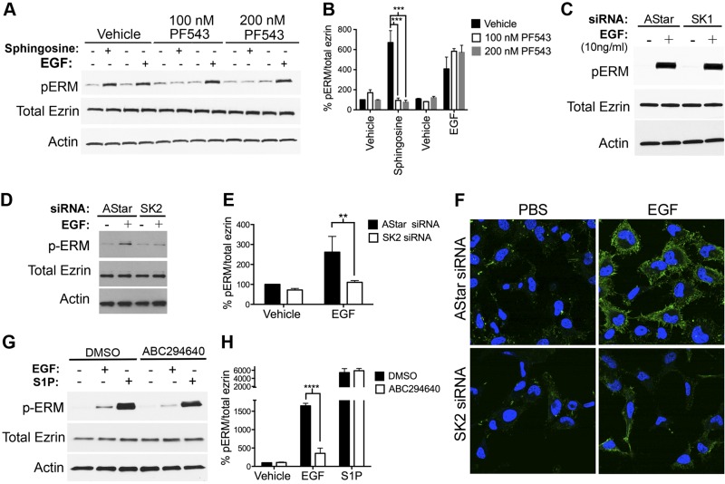
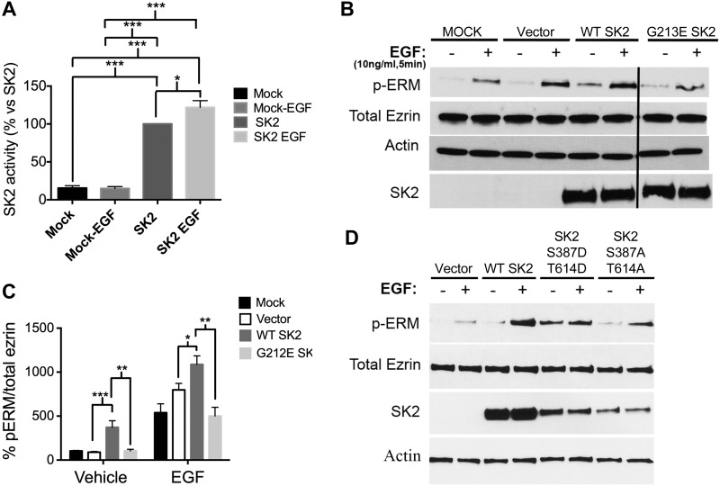
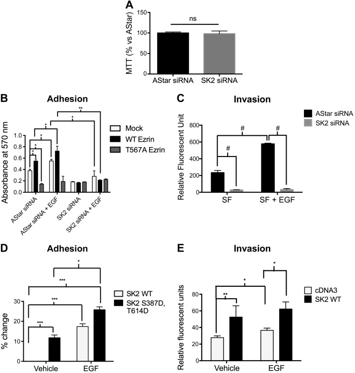
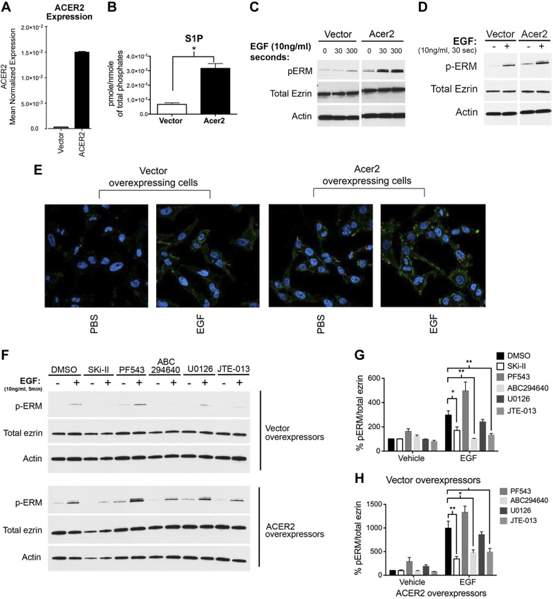
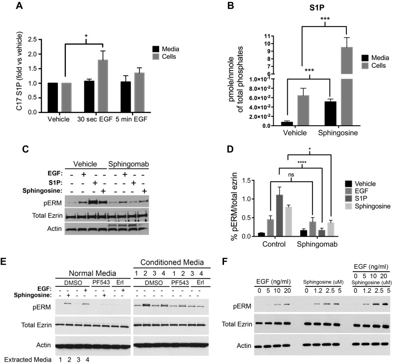
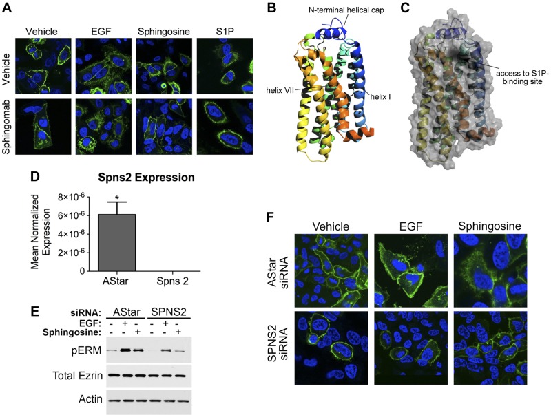
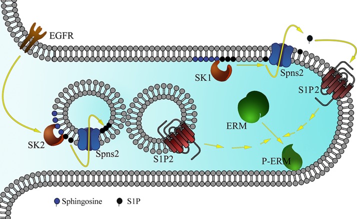
The bioactive sphingolipid sphingosine-1-phosphate (S1P) mediates cellular proliferation, mitogenesis, inflammation, and angiogenesis. These biologies are mediated through S1P binding to specific GPCRs [sphingosine-1-phosphate receptor (S1PR)1-5] and some other less well-characterized intracellular targets. Ezrin-radixin-moesin (ERM) proteins, a family of adaptor molecules linking the cortical actin cytoskeleton to the plasma membrane, are emerging as critical regulators of cancer invasion via regulation of cell morphology and motility. Recently, we identified S1P as an acute ERM activator (via phosphorylation) through its action on S1PR2. In this work, we dissect the mechanism of S1P generation downstream of epidermal growth factor (EGF) leading to ERM phosphorylation and cancer invasion. Using pharmacologic inhibitors, small interfering RNA technologies, and genetic approaches, we demonstrate that sphingosine kinase (SK)2, and not SK1, is essential and sufficient in EGF-mediated ERM phosphorylation in HeLa cells. In fact, knocking down SK2 decreased ERM activation 2.5-fold. Furthermore, we provide evidence that SK2 is necessary to mediate EGF-induced invasion. In addition, overexpressing SK2 causes a 2-fold increase in HeLa cell invasion. Surprisingly, and for the first time, we find that this event, although dependent on S1PR2 activation, does not generate and does not require extracellular S1P secretion, therefore introducing a potential novel model of autocrine/intracrine action of S1P that still involves its GPCRs. These results define new mechanistic insights for EGF-mediated invasion and novel actions of SK2, therefore setting the stage for novel targets in the treatment of growth factor-driven malignancies.

Keywords: Spns2; alkaline ceramidase 2; cell adhesion.


Figures

Figure 1.
Figure 1.
EGF-induced ERM phosphorylation is ErbB1 dependent. A) HeLa cells were pretreated with DMSO, erlotinib (100 nM), ErbB2 inhibitor (10 μM), or lapatinib (10 μM) for 1 h prior to treatment with EGF (10 ng/ml) or S1P (100 nM) for 5 min. pERM, phosphorylated EGFR (p-EGFR), and phosphorylated ErbB2 (p-ErbB2) levels were then assessed by immunoblotting. B) HeLa cells were treated with AStar, EGFR, or ErbB2 siRNA for 48 h. Cells were then starved for 4 h prior to treatment with EGF (10 ng/ml) for 5 min. pERM, total EGFR, and total ErbB2 levels were then assessed by immunoblotting.
Figure 2.
Figure 2.
SK2, and not SK1, is essential for EGF-mediated ERM phosphorylation. A) HeLa cells were pretreated with 100 or 200 nM PF543 for 1 h prior to stimulation with EGF (10 ng/ml) or sphingosine (5 μM) for 5 min. pERM levels were then assessed by immunoblotting. Total ezrin and actin were also included as loading controls. C) HeLa cells were treated with AStar or SK1 siRNA for 48 h. Cells were then starved for 4 h prior to treatment with EGF (10 ng/ml) for 5 min. pERM levels were then assessed by immunoblotting. D) HeLa cells were treated with AStar or SK2 siRNA for 48 h. Cells were then starved for 4 h prior to treatment with EGF (10 ng/ml) for 5 min. pERM levels were then assessed by immunoblotting (C) and confocal microscopy (F), where the green color corresponds to pERM levels labeled with Alexa 488 fluorophore antibody, and the blue color corresponds to DAPI staining the nuclei (F). Total ezrin and actin were also included as loading controls (C). G) HeLa cells were pretreated with DMSO or 10 μM ABC294640 for 1 h prior to stimulation with EGF (10 ng/ml) or S1P (100 nM) for 5 min. pERM levels were then assessed by immunoblotting. Total ezrin and actin were also included as loading controls. B, E, and H) Quantification of the ratio of pERM:total ezrin in (A), (D), and (G), respectively, was performed using ImageJ software. The data represent means ± se of 3 independent experiments. **P < 0.01; ***P < 0.001; ****P < 0.0001.
Figure 3.
Figure 3.
SK2 is sufficient to promote ERM activation. A) HeLa cells were transfected with mock or SK2 DNA for 24 h. Cells were then starved for 4 h prior to stimulation with EGF for 30 s. Cells were then lysed, and SK2 activity was measured using an SK2-specific activity assay as described in the Materials and Methods. B) HeLa cells were transfected with vector, WT SK2, and G213E SK2 DNA for 24 h. Cells were then starved for 4 h prior to treatment with EGF (10 ng/ml) for 5 min. pERM and total SK2 levels were then assessed by immunoblotting. Total ezrin and actin were also included as loading controls. C) Quantification of the ratio of pERM:total ezrin in (D) was performed using ImageJ software. The data represent means ± se of 3 independent experiments. *P < 0.05; **P < 0.01; ***P < 0.001. D) HeLa cells were transfected with vector, WT SK2, Ser387D;Tyr614D SK2, and Ser387A;Tyr614A SK2 DNA for 24 h. Cells were then starved for 4 h prior to treatment with EGF (10 ng/ml) for 5 min. pERM and total SK2 levels were then assessed by immunoblotting. Total ezrin and actin were also included as loading controls.
Figure 4.
Figure 4.
SK2 is required and sufficient for cell adhesion and invasion toward EGF. A) HeLa cells were treated with AStar or SK2 siRNA for 48 h. Cells were then starved for 4 h. An MTT assay was then performed as described in the Materials and Methods. B) HeLa cells were treated with AStar or SK2 siRNA for 24 h. Cells were then transfected with mock, WT ezrin, or Thr567A ezrin DNA for another 24 h. Cells were then starved for 4 h, trypsinized, and plated on fibronectin-coated plates. Cells were then treated with vehicle (PBS) or EGF (10 ng/ml) for 12 h. MTT assay was then performed, and absorbance was measured as a quantification of cell number. C) HeLa cells were treated with AStar or SK2 siRNA for 48 h. Cells were then starved for 4 h prior to their plating in the apical chamber of Matrigel-coated transwell inserts and allowed to invade for 36 h toward serum-free or EGF-supplemented medium. Invading cells were then stained with 4 μg/μl calcein AM, and absorbance was read in a plate reader as described in the Materials and Methods. D) HeLa cells were transfected with WT SK2 or Ser387D;Tyr614D SK2 DNA for 24 h. Cell adhesion was then assessed as described in (B). E) HeLa cells overexpressing cDNA3 or WT SK2 were plated in the apical chamber of Matrigel-coated transwell inserts and allowed to invade for 36 h toward serum-free or EGF-supplemented medium. Invaded cells were then quantified as previously described. The data represent means ± se of 3 independent experiments. ns, not significant; *P < 0.05; **P < 0.01; ***P < 0.001; #P < 0.0001.
Figure 5.
Figure 5.
Increased intracellular S1P production is sufficient to promote ERM activation. A) mRNA of HeLa cells overexpressing empty vector or ACER2 was assessed by quantitative RT-PCR. B) Cellular lipids were directly extracted in organic solvents from HeLa cells overexpressing empty vector or ACER2. S1P levels were analyzed by tandem liquid chromatography/MS. C) HeLa-ACER2-TET-ON and HeLa-vector control cells were treated with tetracycline (5 ng/ml) for 12 h. These cells were then starved for 4 h prior to treatment with EGF (10 ng/ml) for either 30 s or 5 min. pERM levels were then assessed by immunoblotting (C) and by confocal microscopy (E). The green color corresponds to pERM levels labeled with Alexa 488 fluorophore antibody, and the blue color corresponds to DAPI staining the nuclei (E). D) HeLa cells were transiently transfected with either vector control or ACER2 overexpressing DNA for 24 h. Cells were then starved for 4 h prior to treatment with EGF (10 ng/ml) for 5 min. pERM levels were then assessed by immunoblotting. Total ezrin and actin were also included as loading controls. F) HeLa cells stably overexpressing vector or ACER2 were starved overnight. These cells were then pretreated with Ski-II (1 μM), PF543 (100 nM), ABC294640 (10 μM), U0126 (10 μM), or JTE-013 (5 μM) prior to stimulation with EGF (10 ng/ml) for 5 min. G and H) Quantification of the ratio of pERM:total ezrin in (F) was performed using ImageJ software. The data represent means ± se of 3 independent experiments. *P < 0.05; **P < 0.01.
Figure 6.
Figure 6.
ERM phosphorylation does not require extracellular S1P production. A) HeLa cells were prelabeled for 30 min with 250 nM C17-Sph. EGF (10 ng/ml) was then added for 30 s or 5 min. C17-S1P levels from medium and cells were then analyzed via mass spectroscopy. B) HeLa cells were starved overnight, then treated with 5 μM sphingosine for 5 min. Endogenous S1P levels from medium and cells were then analyzed via mass spectroscopy. The data represent means ± se of 3 independent experiments performed in duplicates. C) HeLa cells were pretreated for 1 h with either PBS or 50 μg/well Sphingomab prior to treatment with EGF (10 ng/ml), or sphingosine (5 μM), or S1P (100 nM) for 5 min. pERM levels were then assessed by immunoblotting. Total ezrin and actin were also included as loading controls. D) Quantification of the ratio of pERM:total ezrin in (C) was performed using ImageJ software. The data represent means ± se of 2 independent experiments. E) HeLa cells were pretreated with DMSO, PF543 (100 nM), or erlotinib (Erl; 100 nM) for 1 h, then followed by treatment with either sphingosine (5 μM) or EGF (10 ng/ml) for 2 min. Media from DMSO-pretreated cells (with EGF, Sph, or vehicle treatment), named conditioned media and labeled 1–4, were then added on top of HeLa cells that are pretreated with DMSO, PF543 (100 nM), or erlotinib (100 nM). The reasoning lies in that PF543 treatment will inhibit sphingosine conversion to S1P and that erlotinib will inhibit EGF effect; thus, any effect seen after the addition of the conditioned media will be the result of S1P presence in these media. pERM levels were then assessed by immunoblotting as a marker of S1P presence. Total ezrin and actin were also included as loading controls. F) HeLa cells were plated for 24 h and then starved for 4 h prior to treatment with increasing doses of sphingosine, EGF, or both. pERM levels were then assessed by Western blotting. Total ezrin and actin were also included as loading controls. ns, not significant; *P < 0.05; ***P < 0.001; ****P < 0.0001.
Figure 7.
Figure 7.
Spns2 is partially required for EGF-mediated ERM phosphorylation and S1PR2 internalization. A) HeLa cells were transfected with S1PR2-GFP for 24 h. Following transfection, cells were starved for 4 h and then pretreated for 1 h with PBS or Sphingomab. Cells were treated with 100 nM S1P, 10 nM EGF, or 5 μM sphingosine. Cells were then fixed, and nuclei were stained with DAPI (blue). Images were taken using the Leica SP8 confocal microscope. B and C) Structure model for S1PR2 generated using the I-TASSER server (B). Cartoon with surface overlay, demonstrating the proposed point of access for S1P from the membrane into its receptor-binding site, is shown in (C). D) HeLa cells were treated with 20 nM AStar or Spns2 siRNA for 48 h, and Spns2 mRNA was then assessed by quantitative RT-PCR. *P < 0.05. E) HeLa cells were treated with AStar or Spns2 siRNA for 48 h. Cells were then starved for 4 h prior to treatment with EGF (10 ng/ml) or sphingosine (5 μM) for 5 min. pERM levels were then assessed by immunoblotting. Total ezrin and actin were also included as loading controls. F) HeLa cells were treated with AStar or Spns2 siRNA for 48 h. HeLa cells were then transfected with S1PR2-GFP, 24 h following siRNA transfection. Cells were starved for 4 h and then treated with 10 nM EGF, or 5 μM sphingosine. Cells were then fixed, and nuclei were stained with DAPI (blue). Images were taken using a Leica SP8 confocal microscope.
Figure 8.
Figure 8.
Schematic representation of the proposed model for EGF-induced ERM phosphorylation. EGF treatment activates SK2 localized on the cytosolic side of intracellular vesicles. This will lead to S1P being produced and transported by Spns2 (as well as other ABC transporters) to the inner side of these vesicles. These vesicles will then fuse with S1PR2-containing vesicles or with the plasma membrane. This is followed by S1PR2 activation and ERM phosphorylation, and filopodia formation as shown in this cellular extrusion. On the other hand, sphingosine treatment causes S1P production by SK1 localized on the plasma membrane. S1P will then be exported to the extracellular milieu by Spns2, leading to S1PR2 activation and ERM phosphorylation.

References

    1. Hannun Y. A., Obeid L. M. (2008) Principles of bioactive lipid signalling: lessons from sphingolipids. Nat. Rev. Mol. Cell Biol. 9, 139–150 - PubMed
    1. Hannun Y. A., Bell R. M. (1989) Regulation of protein kinase C by sphingosine and lysosphingolipids. Clin. Chim. Acta 185, 333–345 - PubMed
    1. Hannun Y. A., Obeid L. M. (2011) Many ceramides. J. Biol. Chem. 286, 27855–27862 - PMC - PubMed
    1. Gandy K. A., Obeid L. M. (2013) Regulation of the sphingosine kinase/sphingosine 1-phosphate pathway. Handb. Exp. Pharmacol. 216, 275–303 - PubMed
    1. Schnute M. E., McReynolds M. D., Kasten T., Yates M., Jerome G., Rains J. W., Hall T., Chrencik J., Kraus M., Cronin C. N., Saabye M., Highkin M. K., Broadus R., Ogawa S., Cukyne K., Zawadzke L. E., Peterkin V., Iyanar K., Scholten J. A., Wendling J., Fujiwara H., Nemirovskiy O., Wittwer A. J., Nagiec M. M. (2012) Modulation of cellular S1P levels with a novel, potent and specific inhibitor of sphingosine kinase-1. Biochem. J. 444, 79–88 - PubMed

Publication types

MeSH terms

LinkOut - more resources