

Papillary fibroelastoma of the anterior leaflet of the mitral valve mimicking vegetation
- PMID: 26209756
- PMCID: PMC4573212
- DOI: 10.1016/j.ijscr.2015.07.003
Papillary fibroelastoma of the anterior leaflet of the mitral valve mimicking vegetation
Abstract
Introduction: The papillary fibroelastoma (PFE) is a rare and benign primary cardiac tumor, and the mostly frequently found tumor occurring in cardiac valves.
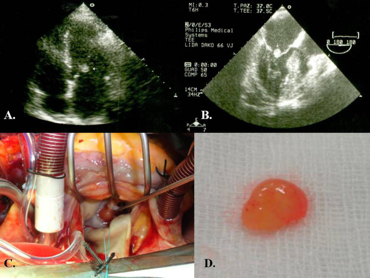
Case presentation: We describe a 52 year old female presenting a history of 2 weeks of fever due to wound infection after breast's surgery. A preoperative echocardiography demonstrated a mass >1cm(2) originating from the anterior leaflet of the mitral valve mimicking vegetation. The patient underwent successful surgical removal of the PFE. The histologic evaluation demonstrated a PFE.
Discussion: With the introduction of echocardiography, the diagnosis of these tumors in living patients has been reported sporadically. PFE have been found most often on valve leaflets, chordae tendineae, and both ventricles. The differential diagnosis of PFE includes other cardiac tumors, thrombus, vegetation, and Lambl's excrescences.
Conclusion: To summarize, we report a PFE of the anterior leaflet of the mitral valve. The diagnosis was confirmed by histopathological examination after surgical removal. Finally, careful echocardiographic analyses during evaluation of valvular masses are strongly recommended for differential diagnosis.
Keywords: Mitral valve; Papillary fibroelastoma; Transesophageal echocardiography.
Copyright © 2015 The Authors. Published by Elsevier Ltd.. All rights reserved.
Figures

References
-
- Yater W.M. Tumors of the heart and pericardium: pathology, symptomatology and report of nine cases. Arch. Intern. Med. 1931;48:627–666.
-
- Klarich K.W., Enriquez-Sarano M., Gura G.M., Edwards W.D., Tajik A.J., Seward J.B. Papillary fibroelastoma: echocardiographic characteristics for diagnosis and pathologic correlation. J. Am. Coll. Cardiol. 1997;30:784–790. - PubMed
-
- Kurup A.N., Tazelaar H.D., Edwards W.D., Burke A.P., Virmani R., Klarich K.W. Iatrogenic cardiac papillary fibroelastoma: a study of 12 cases (1990–2000) Hum. Pathol. 2002;33:1165–1169. - PubMed
-
- Liebeskind D.S., Buljubasic N., Saver J.L. Cardioembolic stroke due to papillary fibroelastoma. J. Stroke Cerebrovasc. Dis. 2001;10:94–95. - PubMed
-
- Gowda R.M., Khan I.A., Nair C.K., Mehta N.J., Vasavada B.C., Sacchi T.J. Cardiac papillary fibroelastoma: a comprehensive analysis of 725 cases. Am. Heart J. 2003;146:404–410. - PubMed
LinkOut - more resources
Full Text Sources
Other Literature Sources
