Review
. 2015 Nov;72(21):4111-26.
doi: 10.1007/s00018-015-1995-y. Epub 2015 Jul 26.

Macrophage polarization in pathology

Affiliations
Review

Macrophage polarization in pathology

Antonio Sica et al. Cell Mol Life Sci. 2015 Nov.

Abstract

Macrophages are cells of the innate immunity constituting the mononuclear phagocyte system and endowed with remarkable different roles essential for defense mechanisms, development of tissues, and homeostasis. They derive from hematopoietic precursors and since the early steps of fetal life populate peripheral tissues, a process continuing throughout adult life. Although present essentially in every organ/tissue, macrophages are more abundant in the gastro-intestinal tract, liver, spleen, upper airways, and brain. They have phagocytic and bactericidal activity and produce inflammatory cytokines that are important to drive adaptive immune responses. Macrophage functions are settled in response to microenvironmental signals, which drive the acquisition of polarized programs, whose extremes are simplified in the M1 and M2 dichotomy. Functional skewing of monocyte/macrophage polarization occurs in physiological conditions (e.g., ontogenesis and pregnancy), as well as in pathology (allergic and chronic inflammation, tissue repair, infection, and cancer) and is now considered a key determinant of disease development and/or regression. Here, we will review evidence supporting a dynamic skewing of macrophage functions in disease, which may provide a basis for macrophage-centered therapeutic strategies.

Keywords: Disease; Inflammation; Macrophage polarization; Tissue damage; Tissue homeostasis.


Figures

Fig. 1
Fig. 1
Role of macrophages in tissue repair. During the different phases of tissue repair, macrophages undergo dynamic changes, switching their phenotype from an M1- to an M2-phenotype. In the early inflammatory phase, M1 macrophages produce inflammatory cytokines and mediators, such as IL6, TNFα, IL1, and NO, stimulating innate immune cells (e.g., neutrophils) and defending host from pathogen colonization. During the resolution phase, macrophages initiate an M1 to M2 phenotype switch, gradually acquiring an anti-inflammatory phenotype, which includes down-regulation of inflammatory mediators, increased production of anti-inflammatory cytokines (e.g., TGFβ and IL10), phagocytosis of apoptotic neutrophils, and removal of damaged cells. In the proliferation phase, M2 macrophages produce a variety of growth factors, such as EGF, FGF, and VEGF, inducing the proliferation of various cell types involved in the healing process. Finally, in the remodeling phase macrophages contribute to the maturation of the regenerated tissue, reorganizing the extracellular matrix, the vasculature, and the scar tissue
Fig. 2
Fig. 2
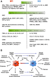
Mechanisms and markers of polarized macrophages in pathology. a Key signals driving M1 vs M2 polarization of macrophages. Inflammatory cytokines (IFNγ, TNFα), pathogen-associated molecular patterns (e.g., LPS), and damage-associated molecular patterns (e.g., HMGB1, HSP, Fetuin A, ATP) induce M1-polarized activation, whereas Th2 cytokines (IL-4, IL-13), anti-inflammatory molecules (IL-10, GC, AMP), and immunocomplexes (Ic) induce M2-polarized activation. IL-33 and thymic stromal lymphopoietin (TSLP) act as M2 amplifiers (asteriks). M1 and M2 signals engage signaling pathways involving different kinases (e.g., Akt1 and 2) and family of transcription factors (e.g., STATs, IRFs, NF-κB, KLFs, HIFs). Further, PPARγ and PPARδ control distinct aspects of M2 macrophage activation and the oxidative metabolism. c-Myc and c-Maf, respectively, regulate a subset of IL-4- and IL-10- inducible genes. Macrophage-polarized activation is also controlled at epigenetic levels by different miRNAs (miR-155, miR-124) and proteins involved in histone methylation and acetylation (bromodomain-containing BET proteins, JMJD3). Inflammatory cytokines (TNFα, IL-1β, IL-6, IL-12, IL-23, IL-27), Th1-recruiting chemokines (CXCL9, CXCL10, CXCL11), and the co-stimulatory receptor CD40, represent distinct markers of M1 polarization. M2 macrophages express higher levels of genes encoding anti-inflammatory cytokines (IL-10, TGFβ), Th2-recruiting chemokines (CCL17, CCL18, CCL22), c-type lectin (CD206, CD301, dectin-1), and scavenger receptors (CD163, Stabilin-1). Further, M1- and M2-polarized macrophages express distinct enzymes involved in iron and amino acid metabolism. Hence M1 macrophages exert anti-microbial activities by iron retention (ferritin, CP, DMT-1, Nramp-1) and ROS/NOS production (iNOS, gp91phox, p22phox), whereas M2 macrophages promote wound healing through iron recycling and release (Tfr, HO-1, Fpn) and polyamine production (Arg 1, Arg 2, ODC, SMO). b M1 and M2 macrophage polarization in disease. Association of M1 vs M2 macrophage polarization in distinct diseases. Dynamic reprogramming of macrophage polarization occurs during the progression of both infections (sepsis, protozoans, HIV) and cancer and mixed macrophage phenotypes can coexist (e.g., H. pylori infections). Systemic Inflammatory Response Syndrome (SIRS); Compensatory Anti-inflammatory Response Syndrome (CARS); Interferon γ (IFNγ) lipopolysaccharide (LPS) High Mobility Group Box 1 (HMGB1); Heat Shock Proteins (HSPs); glucocorticoids (GC) Adenosine monophosphate (AMP); Nuclear factor κB (NF-κB); Signal Transducer and Activator of Transcription (STAT); Interferon Regulatory Factor (IRF); Hypoxia Inducible Factor (HIF); Krüppel-like factor (KLF); peroxisome proliferator-activated receptors (PPAR); CCAAT/enhancer binding protein (C/EBP); Inducible nitric oxide synthase (iNOS); ceruloplasmin (CP); natural resistance-associated macrophage protein 1 (Nramp-1); divalent metal transporter-1 (DMT-1); Arginase (Arg); ornithine decarboxylase (ODC); spermidine oxidase (SMO); hemeoxygenase-1 (HO-1); ferroportin (Fpn); transferrin receptor (TfR)

References

    1. Gordon S, Taylor PR. Monocyte and macrophage heterogeneity. Nat Rev Immunol. 2005;5(12):953–964. doi: 10.1038/nri1733. - DOI - PubMed
    1. Mantovani A, Biswas SK, Galdiero MR, Sica A, Locati M. Macrophage plasticity and polarization in tissue repair and remodelling. J Pathol. 2013;229(2):176–185. doi: 10.1002/path.4133. - DOI - PubMed
    1. Biswas SK, Allavena P, Mantovani A. Tumor-associated macrophages: functional diversity, clinical significance, and open questions. Semin Immunopathol. 2013;35(5):585–600. doi: 10.1007/s00281-013-0367-7. - DOI - PubMed
    1. De Santa F, Totaro MG, Prosperini E, Notarbartolo S, Testa G, Natoli G. The histone H3 lysine-27 demethylase Jmjd3 links inflammation to inhibition of polycomb-mediated gene silencing. Cell. 2007;130(6):1083–1094. doi: 10.1016/j.cell.2007.08.019. - DOI - PubMed
    1. Ostuni R, Piccolo V, Barozzi I, et al. Latent enhancers activated by stimulation in differentiated cells. Cell. 2013;152(1–2):157–171. doi: 10.1016/j.cell.2012.12.018. - DOI - PubMed

Publication types