. 2015 Oct:67:84-92.
doi: 10.1016/j.biomaterials.2015.07.030. Epub 2015 Jul 17.

Effect of UV-photofunctionalization on oral bacterial attachment and biofilm formation to titanium implant material

Affiliations

Effect of UV-photofunctionalization on oral bacterial attachment and biofilm formation to titanium implant material

Erica Dorigatti de Avila et al. Biomaterials. 2015 Oct.

Abstract

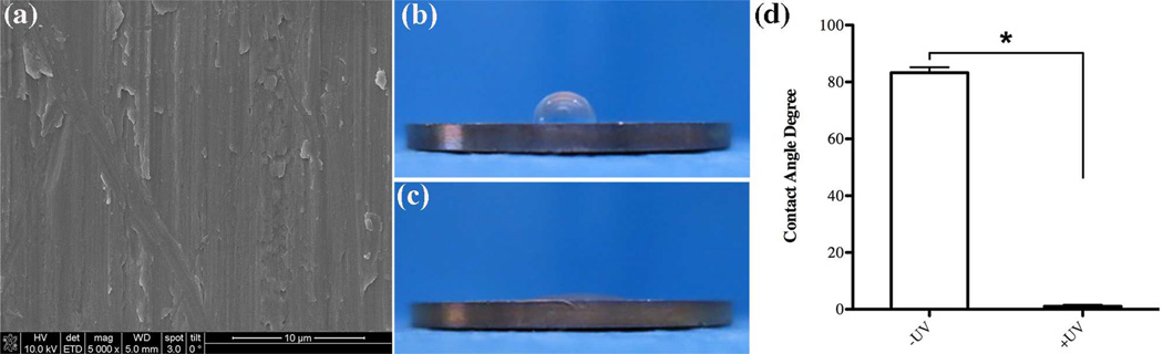
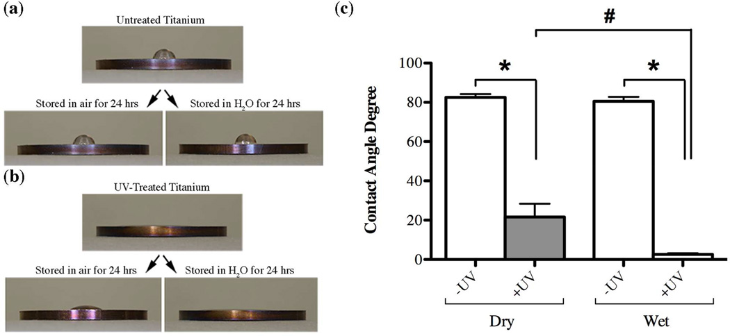
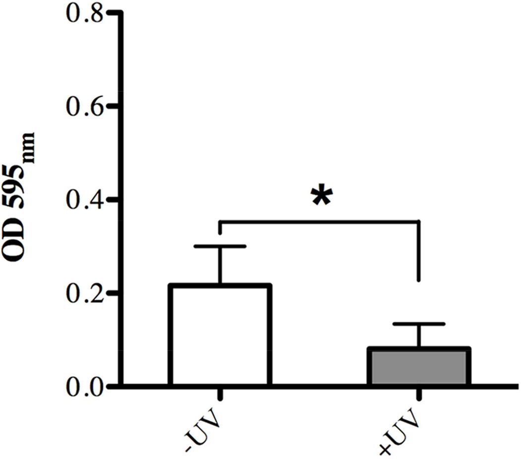
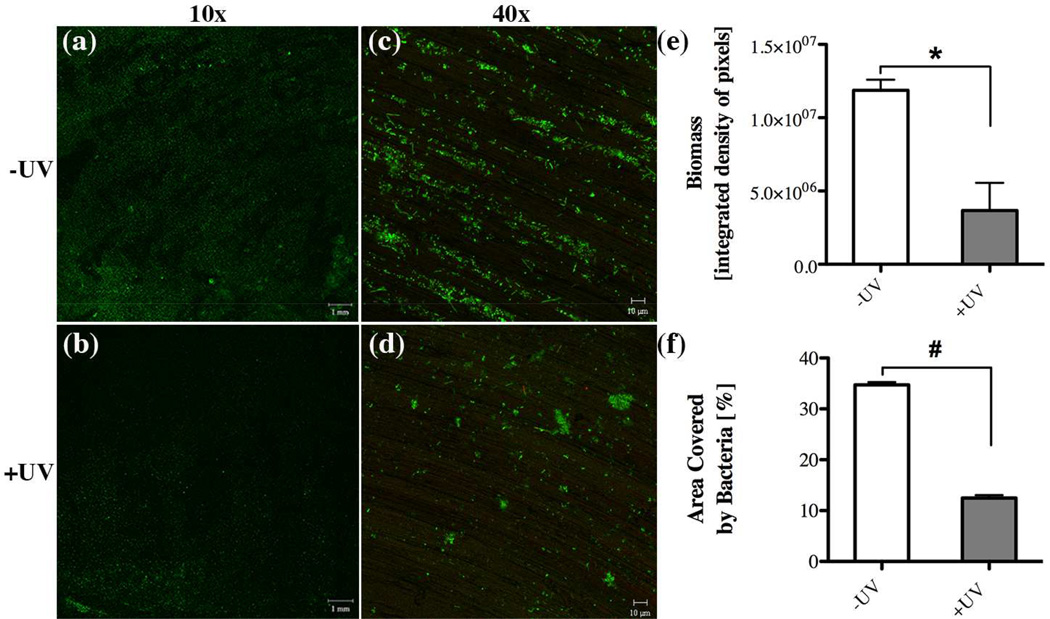
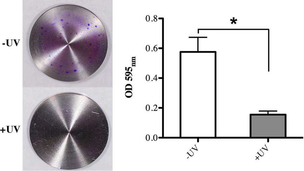
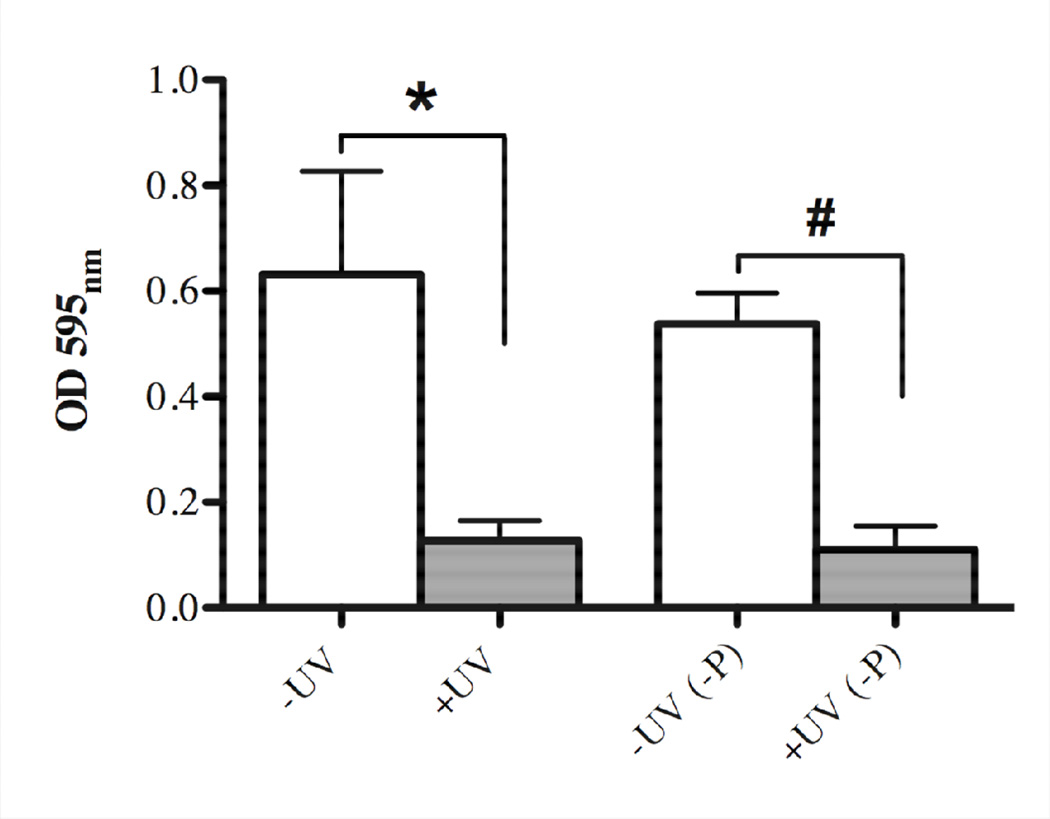
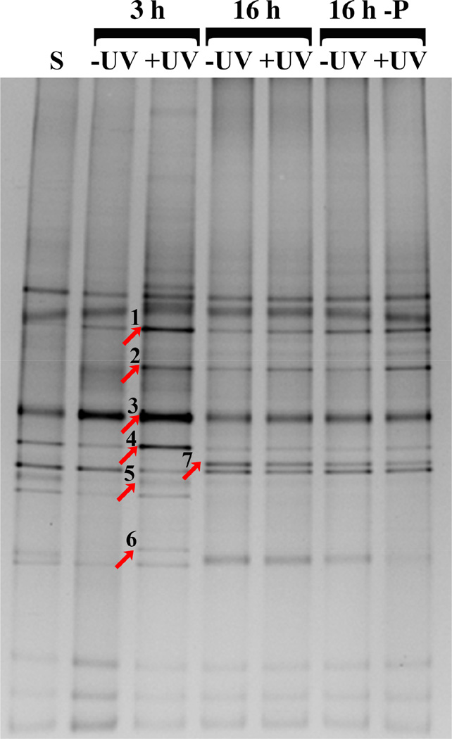
Bacterial biofilm infections remain prevalent reasons for implant failure. Dental implant placement occurs in the oral environment, which harbors a plethora of biofilm-forming bacteria. Due to its trans-mucosal placement, part of the implant structure is exposed to oral cavity and there is no effective measure to prevent bacterial attachment to implant materials. Here, we demonstrated that UV treatment of titanium immediately prior to use (photofunctionalization) affects the ability of human polymicrobial oral biofilm communities to colonize in the presence of salivary and blood components. UV-treatment of machined titanium transformed the surface from hydrophobic to superhydrophilic. UV-treated surfaces exhibited a significant reduction in bacterial attachment as well as subsequent biofilm formation compared to untreated ones, even though overall bacterial viability was not affected. The function of reducing bacterial colonization was maintained on UV-treated titanium that had been stored in a liquid environment before use. Denaturing gradient gel-electrophoresis (DGGE) and DNA sequencing analyses revealed that while bacterial community profiles appeared different between UV-treated and untreated titanium in the initial attachment phase, this difference vanished as biofilm formation progressed. Our findings confirm that UV-photofunctionalization of titanium has a strong potential to improve outcome of implant placement by creating and maintaining antimicrobial surfaces.

Keywords: Bacteria; Biofilm; Peri-implantitis; Photofunctionalization; Titanium.


Figures

Fig. 1
Fig. 1
Surface characterization of machined titanium. (a) scanning electron microscopy of UV-treated discs showing surface topography at a 5000x magnification, evaluation of hydrophilicity by contact angle measurement of 10µl ddH2O (b) before and (c) after UV-treatment on the Ti discs and (d) statistical significance testing of contact angle values for -UV and +UV titanium discs. Statistically significant differences are indicated as: * p<0.0001. Data represent the mean ± SD of triplicate data of one independent experiment.
Fig. 2
Fig. 2
Measurement of the contact angle of 10µl ddH2O to evaluate changes in hydrophilicity of titanium discs surfaces (a) without and (b) after UV-photofunctionalization treatment as well as after subsequent storage in air or a liquid environment for 24 hours. Comparison of contact angles values (c) between untreated (white bars, - UV) and UV-treated (gray bars, +UV) after storage in air (Dry) or a liquid environment (Wet). Statistically significant differences are indicated as: * p<0.0001, # p=0.0042. Data represent the mean ± SD of triplicate data from one independent experiment.
Fig. 3
Fig. 3
Effect of UV-treatment of titanium surfaces on bacterial attachment after 3 hours incubation evaluated via quantitative measurement of crystal violet staining as indicator of biomass accumulation on untreated (white bar, –UV) in comparison to UV-treated (gray bar, +UV) titanium discs. Each value represents the mean ± SD of nine samples comprised of three technical replicates of three independent biological experiments. * indicates a statistically significant difference (p<0.05).
Fig. 4
Fig. 4
Effect of UV-treatment of titanium surfaces on bacterial attachment after 3 hours incubation evaluated via confocal microscopy imaging through (a,b) 10x and (c,d) 40x objectives with representative images illustrating the live/dead distribution of bacterial cells (green for live cells, red for compromised cells) accumulated on (a,c) untreated and (b,d) UV-treated titanium discs. Quantitative comparison of (e) accumulated biomass and (f) the area covered by bacteria between untreated (white bar, –UV) in comparison to UV-treated (gray bar, +UV) titanium discs. Each value represents the mean ± SD of four samples comprised of two technical replicates of two independent biological experiments. Statistically significant differences are indicated as: * p=0.0145, # p=0.0003.
Fig. 5
Fig. 5
Effect of UV-treatment of titanium surfaces on bacterial biofilm formation after 16 hours incubation evaluated via quantitative measurement of crystal violet staining as indicator of biomass accumulation on untreated (white bar, –UV) in comparison to UV-treated (gray bar, +UV) titanium discs. Each value represents the mean ± SD of nine samples comprised of three technical replicates of three independent biological experiments. * indicates a statistically significant difference (p<0.001).
Fig. 6
Fig. 6
Effect of UV-treatment of titanium surfaces on bacterial biofilm formation after 16 hours incubation evaluated via confocal microscopy imaging through (a,b) 10x and (c,d) 40x objectives with representative images illustrating the live/dead distribution of bacterial cells (green for live cells, red for compromised cells) accumulated on (a,c) untreated (-UV) and (b,d) UV-treated (+UV) titanium discs. Scanning electron microscopy revealing the biofilm structure on (e) -UV and (f) +UV titanium discs. Quantitative comparison of (e) accumulated biomass and (f) the area covered by bacteria between untreated (white bar, –UV) in comparison to UV-treated (gray bar, +UV) titanium discs. Each value represents the mean ± SD of four samples comprised of two technical replicates of two independent biological experiments. Statistically significant differences are indicated as: * p<0.0116), # p<0.0461.
Fig. 7
Fig. 7
Sustainability of UV-treatment of titanium surfaces and the effect on 16 hours bacterial biofilm formation after immersion of titanium discs in liquid SHI-FSMS medium for 24 hours prior to incubation with bacteria. Quantitative measurement of crystal violet staining showing biomass accumulation on untreated (white bar, –UV) in comparison to UV-treated (gray bar, +UV) titanium discs for directly used samples and those pre-immersed in liquid SHI-FSMS (-P). Each value represents the mean ± SD of four samples comprised of two technical replicates of two independent biological experiments. Statistically significant differences are indicated as: * p=0.0012), # p<0.0001.
Fig. 8
Fig. 8
DGGE analysis of oral microbial communities formed on untreated (-UV) and UV-treated (+UV) titanium discs surfaces after 3 hours and with and without 24 hour pre-immersion in liquid SHI-FSMS (-P) medium after 16 hours. Bands that were excised for DNA sequencing are indicated by an arrow. Microbial identities are as follows: (1) Fusobacterium periodonticum, (2) Streptococcus (Mitis group), (3) Streptococcus (Mitis group), (4) Porphyromonas sp [HOT_279], (5) Gemella sp, (6) Peptostreptococcus stomatis, and (7) Streptococcus sp.

References

    1. Pjetursson BE, Asgeirsson AG, Zwahlen M, Sailer I. Improvements in implant dentistry over the last decade: comparison of survival and complication rates in older and newer publications. Int J Oral Maxillofac Implants. 2014;29(Suppl):308–324. - PubMed
    1. Simonis P, Dufour T, Tenenbaum H. Long-term implant survival and success: a 10–16-year follow-up of non-submerged dental implants. Clin Oral Implants Res. 2010;21:772–777. - PubMed
    1. Clementini M, Rossetti PH, Penarrocha D, Micarelli C, Bonachela WC, Canullo L. Systemic risk factors for peri-implant bone loss: a systematic review and meta-analysis. Int J Oral Maxillofac Surg. 2014;43:323–334. - PubMed
    1. Heitz-Mayfield LJ. Peri-implant diseases: diagnosis and risk indicators. J Clin Periodontol. 2008;35:292–304. - PubMed
    1. Aguirre-Zorzano LA, Estefania-Fresco R, Telletxea O, Bravo M. Prevalence of peri-implant inflammatory disease in patients with a history of periodontal disease who receive supportive periodontal therapy. Clin Oral Implants Res. 2014 - PubMed

Publication types

MeSH terms