Comparative Study
. 2015 Jul 26:15:551.
doi: 10.1186/s12885-015-1520-6.

Lymphocyte-specific protein tyrosine kinase (Lck) interacts with CR6-interacting factor 1 (CRIF1) in mitochondria to repress oxidative phosphorylation

Affiliations
Comparative Study

Lymphocyte-specific protein tyrosine kinase (Lck) interacts with CR6-interacting factor 1 (CRIF1) in mitochondria to repress oxidative phosphorylation

Shahrooz Vahedi et al. BMC Cancer. .

Abstract

Background: Many cancer cells exhibit reduced mitochondrial respiration as part of metabolic reprogramming to support tumor growth. Mitochondrial localization of several protein tyrosine kinases is linked to this characteristic metabolic shift in solid tumors, but remains largely unknown in blood cancer. Lymphocyte-specific protein tyrosine kinase (Lck) is a key T-cell kinase and widely implicated in blood malignancies. The purpose of our study is to determine whether and how Lck contributes to metabolic shift in T-cell leukemia through mitochondrial localization.

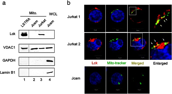
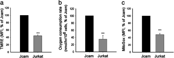
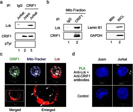
Methods: We compared the human leukemic T-cell line Jurkat with its Lck-deficient derivative Jcam cell line. Differences in mitochondrial respiration were measured by the levels of mitochondrial membrane potential, oxygen consumption, and mitochondrial superoxide. Detailed mitochondrial structure was visualized by transmission electron microscopy. Lck localization was evaluated by subcellular fractionation and confocal microscopy. Proteomic analysis was performed to identify proteins co-precipitated with Lck in leukemic T-cells. Protein interaction was validated by biochemical co-precipitation and confocal microscopy, followed by in situ proximity ligation assay microscopy to confirm close-range (<16 nm) interaction.

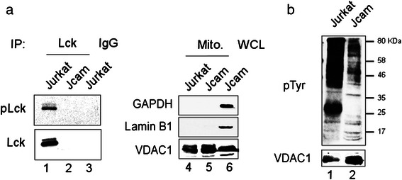
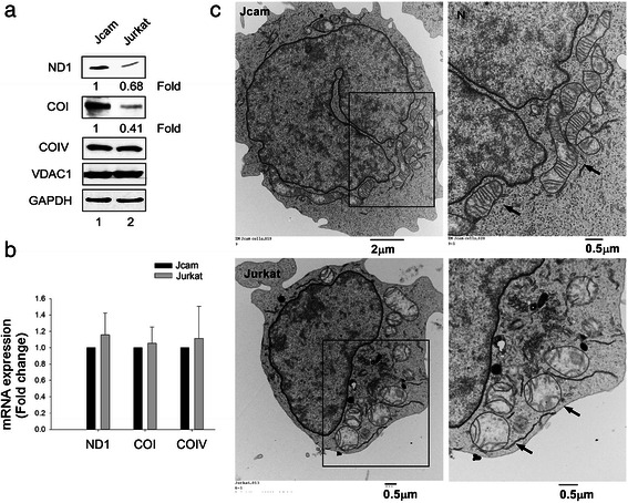
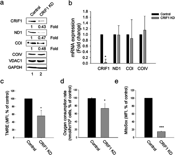
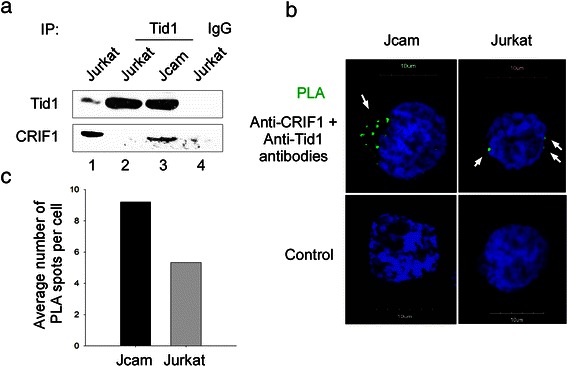
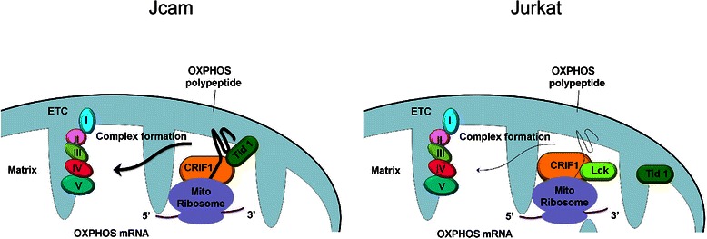
Results: Jurkat cells have abnormal mitochondrial structure and reduced levels of mitochondrial respiration, which is associated with the presence of mitochondrial Lck and lower levels of mitochondrion-encoded electron transport chain proteins. Proteomics identified CR6-interacting factor 1 (CRIF1) as the novel Lck-interacting protein. Lck association with CRIF1 in Jurkat mitochondria was confirmed biochemically and by microscopy, but did not lead to CRIF1 tyrosine phosphorylation. Consistent with the role of CRIF1 in functional mitoribosome, shRNA-mediated silencing of CRIF1 in Jcam resulted in mitochondrial dysfunction similar to that observed in Jurkat. Reduced interaction between CRIF1 and Tid1, another key component of intramitochondrial translational machinery, in Jurkat further supports the role of mitochondrial Lck as a negative regulator of CRIF1 through competitive binding.

Conclusions: This is the first report demonstrating the role of mitochondrial Lck in metabolic reprogramming of leukemic cells. Mechanistically, it is distinct from other reported mitochondrial protein tyrosine kinases. In a kinase-independent manner, mitochondrial Lck interferes with mitochondrial translational machinery through competitive binding to CRIF1. These findings may reveal novel approaches in cancer therapy by targeting cancer cell metabolism.


Figures

Fig. 1
Fig. 1
Mitochondrial localization of endogenous Lck protein in both mouse and human leukemia cell lines. (a) Mitochondrial (Mito) fractions isolated from three leukemia cell lines were analyzed by Lck immunoblotting. Immunoblotting for VDAC1 (mitochondrial marker), GAPDH (cytoplasmic marker), and lamin B1 (nuclear marker) was performed to verify purity of mitochondrial fractions. Jcam whole cell lysate (WCL) was used as the positive control for markers. LSTRA lysates were analyzed on a separate membrane as shown by the dotted lines. (b) Confocal microscopy of three-color fluorescence staining of Jurkat (top and middle panels) and Jcam (bottom panels) cells. An area of Jurkat microscopy (bordered with white lines) is enlarged and shown on the right. Lck (red) and mito-tracker (green) co-localization are shown as yellow dots and depicted by white arrows in the enlarged image. Nuclei are visualized with DAPI staining (blue). Scale bars are shown in the bottom
Fig. 2
Fig. 2
Association between Lck expression and decreased mitochondrial activity. Jurkat and Jcam cells were analyzed for mitochondrial membrane potentials (panel a), oxygen consumption rate (panel b), and mitochondrial superoxide level (panel c). Experimental details are described in Methods. Membrane potentials and mitochondrial superoxide levels are shown as mean fluorescence intensity (MFI) by flow cytometry. Data are presented as percentage of activity in Jurkat as compared to Jcam. Statistical analyses were perform on three independent experiments, ***p < 0.001
Fig. 3
Fig. 3
Lck interacts with mitochondrial CRIF1. (a) Jurkat and Jcam whole cell lysates were immunoprecipitated (IP) with anti-CRIF1 antibody, followed by Lck and CRIF1 immunoblotting. CRIF1 immunoblot was stripped and then reblotted with anti-phosphotyrosine (pTyr) antibody. Equal amounts of Jurkat whole cell lysate were also immunoprecipitated with normal IgG as a negative control (lane 1). (b) Mitochondrial proteins isolated from Jurkat cells were immunoprecipitated with either anti-CRIF1 antibody or control IgG, and then subjected to Lck and CRIF1 immunoblotting (left panels). A fraction of mitochondrial lysate was analyzed by lamin B1 and GAPDH immunoblotting to confirm the absence of nuclear and cytosolic contamination, respectively (right panels, lane 1). Jurkat whole cell lysate was used as a positive control (right panels, lane 2). (c) Jurkat cells were subjected to immunofluorescence microscopy with three-color staining for CRIF1 (green), mito-tracker (blue), and Lck (red). Cells were also stained with DAPI to visualize nuclei (grey on upper panels). An area of three-color merged image bordered with white lines is enlarged on the right to show better resolution (lower panels). White arrows indicate co-localization of Lck and CRIF1 in mitochondria (white dots). White arrowheads depict co-localization of Lck and CRIF1 in the nucleus (yellow dots). (d) Jurkat and Jcam cells were subjected to PLA microscopy using primary antibodies specific for Lck and CRIF1 (upper panels). Green fluorescence indicates Lck and CRIF1 interaction in situ (white arrows). Secondary antibodies alone were used as negative controls (lower panels). Scale bars of 10 μm are shown in the bottom of microscopy images
Fig. 4
Fig. 4
Active Lck kinase activity in Jurkat mitochondria. (a) Equal amount of proteins isolated from Jurkat and Jcam mitochondria were immunoprecipitated by anti-Lck (lanes 1 and 2) or control IgG (lane 3). Immunoprecipitates were blotted sequentially with antibodies specific for Tyr394-phosphorylated Lck (pLck) and total Lck (lanes 1-3). A small fraction of mitochondrial lysates were blotted for GAPDH, lamin B1 and VDAC1 to confirm fraction purity (lanes 4 and 5). Jcam whole cell lysate was included as a positive control for markers (lane 6). (b) Total proteins from mitochondrial fractions of Jurkat and Jcam cells were subjected to anti-phosphotyrosine immunoblotting (upper panel). Molecular weight markers are denoted on the right. VDAC1 immunoblot was used as a loading control (lower panel)
Fig. 5
Fig. 5
Lower levels of mitochondrion-encoded OXPHOS proteins and abnormal mitochondrial structure in Jurkat.( a) Normalized whole cell lysates from Jurkat and Jcam cells were analyzed by Western blot using antibodies specific for ND1, COI, COIV, VDAC1 and GAPDH. Signal intensity was quantitated for ND1 and COI and fold change is indicated below the images. (b) Total RNAs isolated from Jurkat and Jcam cells were subjected to real-time PCR using primers specific for human ND1, COI and COIV. Data from triplicates were normalized to actin and expressed as fold change of Jurkat in comparison to Jcam. Statistical analyses show no significant difference from three independent studies. (c) Transmission electron microscopy of Jurkat (lower panels) and Jcam (upper panels) cells. An area with enriched mitochondria is bordered with black lines and enlarged on the right to better visualize detailed intramitochondrial structure. Black arrows denote several representative mitochondria. The position of nucleus at the upper-left corner is also labeled as “N”. Scale bars are shown in the lower-right corners of microscopy images
Fig. 6
Fig. 6
CRIF1 is required for normal expression of mitochondrion-encoded proteins. Comparisons are made between Jcam cells expressing scrambled shRNA (Control) and CRIF1-specific shRNA (CRIF1 KD). (a) Normalized whole cell lysates were subjected to immunoblotting for CRIF1, ND1, COI, COIV, VDAC1 and GAPDH. Signal intensity was quantitated for CRIF1, ND1 and COI and fold change is indicated below the images. (b) Total RNAs were subjected to real-time PCR using primers specific for human CRIF1, ND1, COI and COIV. Data from triplicates were normalized to actin and expressed as fold change of CRIF1 knock-down (CRIF1 KD) in comparison to control Jcam. (c, d, e) Mitochondrial membrane potentials, oxygen consumption rate, and mitochondrial superoxide levels were analyzed as described for Fig. 2. Data are presented as percentage of change from CRIF1 knock-down in comparison to control Jcam. Statistical analyses show the results from three independent studies, *p < 0.05, ***p < 0.001
Fig. 7
Fig. 7
Decreased interaction of CRIF1 and Tid1 in Jurkat cells. (a) Whole cell lysates from Jurkat and Jcam cells were subjected to Tid1 immunoprecipitation and subsequent immunoblotting with anti-Tid1 and anti-CRIF1 antibodies. Jurkat lysate immunoprecipitated with normal IgG was used as a negative control (lane 4). A fraction of Jurkat lysate was loaded as a positive control (lane 1). (b) Jurkat and Jcam cells were analyzed by PLA microscopy using primary antibodies specific for CRIF1 and Tid1 (upper panels). Green fluorescence indicates CRIF1 and Tid1 interaction in situ (white arrows). Secondary antibodies alone were used as negative controls (lower panels). Scale bars of 10 μm are also shown. (c) The number of CRIF1 and Tid1 interacting complex were counted in multiple cells to obtain average number per cell
Fig. 8
Fig. 8
Schematic diagrams illustrating the potential mechanism of how Lck functions in mitochondria. As shown in Jcam, CRIF1 is a crucial component of the mitochondrial translational machinery and stabilizes the electron transport chain (ETC) complex. In Jurkat, Lck interaction with CRIF1 in mitochondria may disrupt CRIF1’s association with Tid1 and other translational components in the mitoribosome. Reduced levels of mitochondrion-encoded OXPHOS polypeptide and a defect in subsequent insertion into the inner membrane may lead to a decrease in mitochondrial respiration. The level of mitochondrion-encoded OXPHOS mRNA, however, remains unchanged in the mitochondrial matrix between Jurkat and Jcam cells

Similar articles

Cited by

References

    1. Scatena R. Mitochondria and cancer: a growing role in apoptosis, cancer cell metabolism and dedifferentiation. Adv Exp Med Biol. 2012;942:287–308. doi: 10.1007/978-94-007-2869-1_13. - DOI - PubMed
    1. Seyfried TN, Shelton LM. Cancer as a metabolic disease. Nutr Metab (Lond) 2010;7:7. doi: 10.1186/1743-7075-7-7. - DOI - PMC - PubMed
    1. Chatterjee A, Mambo E, Sidransky D. Mitochondrial DNA mutations in human cancer. Oncogene. 2006;25(34):4663–74. doi: 10.1038/sj.onc.1209604. - DOI - PubMed
    1. Carew JS, Huang P. Mitochondrial defects in cancer. Mol Cancer. 2002;1:9. doi: 10.1186/1476-4598-1-9. - DOI - PMC - PubMed
    1. Vander Heiden MG, Cantley LC, Thompson CB. Understanding the Warburg effect: The metabolic requirements of cell proliferation. Science. 2009;324:1029–33. doi: 10.1126/science.1160809. - DOI - PMC - PubMed

Publication types

MeSH terms

Substances

LinkOut - more resources