

Cardiovascular imaging in children and adults following Kawasaki disease
- PMID: 26210915
- PMCID: PMC4656233
- DOI: 10.1007/s13244-015-0422-0
Cardiovascular imaging in children and adults following Kawasaki disease
Abstract
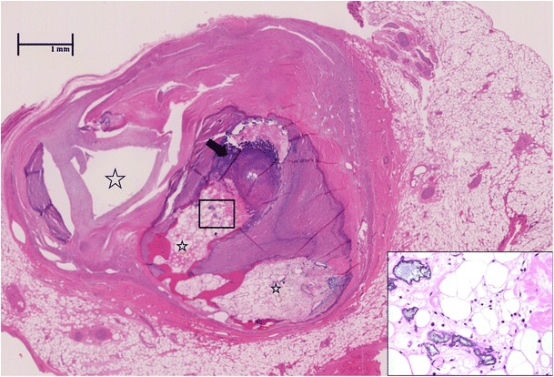
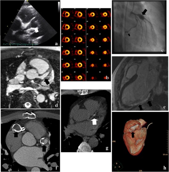
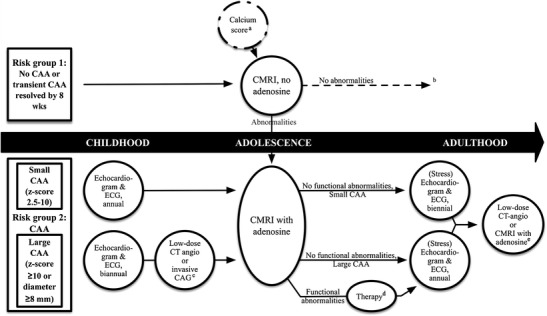
Kawasaki disease (KD) is a paediatric vasculitis with coronary artery aneurysms (CAA) as its main complication. Two guidelines exist regarding the follow-up of patients after KD, by the American Heart Association and the Japanese Circulation Society. After the acute phase, CAA-negative patients are checked for cardiovascular risk assessment or with ECG and echocardiography until 5 years after the disease. In CAA-positive patients, monitoring includes myocardial perfusion imaging, conventional angiography and CT-angiography. However, the invasive nature and high radiation exposure do not reflect technical advances in cardiovascular imaging. Newer techniques, such as cardiac MRI, are mentioned but not directly implemented in the follow-up. Cardiac MRI can be performed to identify CAA, but also evaluate functional abnormalities, ischemia and previous myocardial infarction including adenosine stress-testing. Low-dose CT angiography can be implemented at a young age when MRI without anaesthesia is not feasible. CT calcium scoring with a very low radiation dose can be useful in risk stratification years after the disease. By incorporating newer imaging techniques, detection of CAA will be improved while reducing radiation burden and potential complications of invasive imaging modalities. Based on the current knowledge, a possible pathway to follow-up patients after KD is introduced. Key Points • Kawasaki disease is a paediatric vasculitis with coronary aneurysms as major complication. • Current guidelines include invasive, high-radiation modalities not reflecting new technical advances. • Cardiac MRI can provide information on coronary anatomy as well as cardiac function. • (Low-dose) CT-angiography and CT calcium score can also provide important information. • Current guidelines for follow-up of patients with KD need to be revised.
Keywords: (Cardiac) MRI; Cardiac imaging; Coronary aneurysm; Kawasaki disease; MDCT.
Figures
Similar articles
-
Dissecting Kawasaki disease: a state-of-the-art review.Eur J Pediatr. 2017 Aug;176(8):995-1009. doi: 10.1007/s00431-017-2937-5. Epub 2017 Jun 27. Eur J Pediatr. 2017. PMID: 28656474 Free PMC article. Review.
-
Cardiac magnetic resonance imaging for noninvasive assessment of cardiovascular disease during the follow-up of patients with Kawasaki disease.Circ Cardiovasc Imaging. 2011 Nov;4(6):712-20. doi: 10.1161/CIRCIMAGING.111.965996. Epub 2011 Sep 15. Circ Cardiovasc Imaging. 2011. PMID: 21921132
-
CT Angiography or Cardiac MRI for Detection of Coronary Artery Aneurysms in Kawasaki Disease.Front Pediatr. 2021 Feb 4;9:630462. doi: 10.3389/fped.2021.630462. eCollection 2021. Front Pediatr. 2021. PMID: 33614558 Free PMC article.
-
European Association of Cardiovascular Imaging/Cardiovascular Imaging Department of the Brazilian Society of Cardiology recommendations for the use of cardiac imaging to assess and follow patients after heart transplantation.Eur Heart J Cardiovasc Imaging. 2015 Sep;16(9):919-48. doi: 10.1093/ehjci/jev139. Epub 2015 Jul 2. Eur Heart J Cardiovasc Imaging. 2015. PMID: 26139361 Review.
-
Computed tomography coronary angiography is the way forward for evaluation of children with Kawasaki disease.Glob Cardiol Sci Pract. 2017 Oct 31;2017(3):e201728. doi: 10.21542/gcsp.2017.28. Glob Cardiol Sci Pract. 2017. PMID: 29564349 Free PMC article. Review.
Cited by
-
Congenital anomalies of coronary artery misdiagnosed as coronary dilatations in Kawasaki disease: A clinical predicament.World J Clin Pediatr. 2025 Mar 9;14(1):99177. doi: 10.5409/wjcp.v14.i1.99177. eCollection 2025 Mar 9. World J Clin Pediatr. 2025. PMID: 40059891 Free PMC article.
-
Society for Cardiovascular Magnetic Resonance 2022 Cases of SCMR case series.J Cardiovasc Magn Reson. 2024 Summer;26(1):100007. doi: 10.1016/j.jocmr.2023.100007. Epub 2023 Dec 23. J Cardiovasc Magn Reson. 2024. PMID: 38211509 Free PMC article. Review.
-
Dissecting Kawasaki disease: a state-of-the-art review.Eur J Pediatr. 2017 Aug;176(8):995-1009. doi: 10.1007/s00431-017-2937-5. Epub 2017 Jun 27. Eur J Pediatr. 2017. PMID: 28656474 Free PMC article. Review.
-
Role of CT Coronary Angiography at Initial Presentation in Kawasaki Disease-Insights from a Tertiary Care Center in North India.Diagnostics (Basel). 2025 Jul 17;15(14):1806. doi: 10.3390/diagnostics15141806. Diagnostics (Basel). 2025. PMID: 40722554 Free PMC article.
-
Kawasaki disease: etiopathogenesis and novel treatment strategies.Expert Rev Clin Immunol. 2017 Mar;13(3):247-258. doi: 10.1080/1744666X.2017.1232165. Epub 2016 Sep 13. Expert Rev Clin Immunol. 2017. PMID: 27590181 Free PMC article. Review.
References
LinkOut - more resources
Full Text Sources
Other Literature Sources
