. 2015 Sep 7;54(37):10778-82.
doi: 10.1002/anie.201503417. Epub 2015 Jul 23.

Synthesis and Evaluation of Gd(III) -Based Magnetic Resonance Contrast Agents for Molecular Imaging of Prostate-Specific Membrane Antigen

Affiliations

Synthesis and Evaluation of Gd(III) -Based Magnetic Resonance Contrast Agents for Molecular Imaging of Prostate-Specific Membrane Antigen

Sangeeta Ray Banerjee et al. Angew Chem Int Ed Engl. .

Abstract

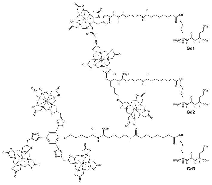
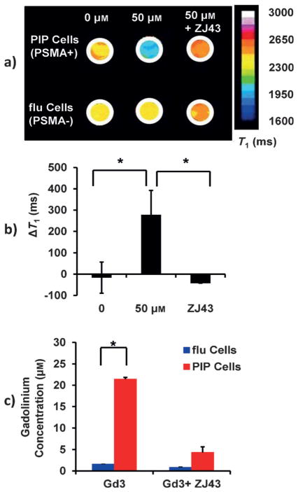
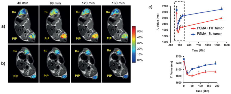
Magnetic resonance (MR) imaging is advantageous because it concurrently provides anatomic, functional, and molecular information. MR molecular imaging can combine the high spatial resolution of this established clinical modality with molecular profiling in vivo. However, as a result of the intrinsically low sensitivity of MR imaging, high local concentrations of biological targets are required to generate discernable MR contrast. We hypothesize that the prostate-specific membrane antigen (PSMA), an attractive target for imaging and therapy of prostate cancer, could serve as a suitable biomarker for MR-based molecular imaging. We have synthesized three new high-affinity, low-molecular-weight Gd(III) -based PSMA-targeted contrast agents containing one to three Gd(III) chelates per molecule. We evaluated the relaxometric properties of these agents in solution, in prostate cancer cells, and in an in vivo experimental model to demonstrate the feasibility of PSMA-based MR molecular imaging.

Keywords: cancer; gadolinium; imaging agents; magnetic resonance imaging.


Figures

Figure 1
Figure 1
Structures of PSMA-targeted MR contrast agents Gd1–Gd3.
Figure 2
Figure 2
T1 contrast enhancement generated by Gd3 in an isogenic human PC3 prostate cancer cell pair, PSMA + PIP and PSMA− flu cells. a) Color-coded T1 maps of PC3 PIP and PC3 flu cells. Relaxation rates were determined at 25 °C at 9.4 T. b) Quantification of ΔT1 in PIP and flu cells (n =4, P <0.05) following treatment with Gd3. c) Cellular uptake of Gd3 in PC3 PIP and PC3 flu cells. The amount of GdIII associated with PIP cell pellets was significantly higher than for the flu cell pellets. The accumulation of Gd3 in PIP cells was blocked by preincubating with ZJ43 (n =4, P <0.05).
Figure 3
Figure 3
Gd3 MR imaging of human PC3 prostate cancer PIP (PSMA +) and flu (PSMA−) tumor xenografts in male NOD/SCID mice. a) Enhancement (ΔR1%) maps in PC3 PIP (PSMA +) and PC3 flu (PSMA−) tumors are superimposed upon T2-weighted images during 40–160 min after a single bolus injection of Gd3 into the tail vein. b) ΔR1% maps in PSMA + and PSMA− tumors of a trimeric Gd contrast agent without a PSMA-targeting moiety. c) T1 time courses calculated for the entire volume of each tumor during 1–1600 min post injection (top). Enlarged region of the time course at 0–200 min (bottom).

References

    1. Caravan P. Acc Chem Res. 2009;42:851–862. - PubMed
    2. Huang CH, Tsourkas A. Curr Top Med Chem. 2013;13:411–421. - PMC - PubMed
    3. Lanza GM, Winter PM, Caruthers SD, Morawski AM, Schmieder AH, Crowder KC, Wickline SA. J Nucl Cardiol. 2004;11:733–743. - PubMed
    4. Pais A, Gunanathan C, Margalit R, Biton IE, Yosepovich A, Milstein D, Degani H. Cancer Res. 2011;71:7387– 7397. - PMC - PubMed
    1. Artemov D, Mori N, Ravi R, Bhujwalla ZM. Cancer Res. 2003;63:2723–2727. - PubMed
    2. Konda SD, Aref M, Wang S, Brechbiel M, Wiener EC. Magma. 2001;12:104–113. - PubMed
    3. Aime S, Cabella C, Colombatto S, Geninatti Crich S, Gianolio E, Maggioni F. J Magn Reson Imaging. 2002;16:394– 406. - PubMed
    1. Major JL, Meade TJ. Acc Chem Res. 2009;42:893–903. - PMC - PubMed
    2. Artemov D. J Cell Biochem. 2003;90:518– 524. - PubMed
    1. Hanaoka K, Lubag AJ, Castillo-Muzquiz A, Kodadek T, Sherry AD. Magn Reson Imaging. 2008;26:608–617. - PMC - PubMed
    2. De León-Rodríguez LM, Lubag A, Udugamasooriya DG, Proneth B, Brekken RA, Sun X, Kodadek T, Sherry AD. J Am Chem Soc. 2010;132:12829– 12831. - PMC - PubMed
    1. Wu X, Burden-Gulley SM, Yu GP, Tan M, Lindner D, Brady-Kalnay SM, Lu ZR. Bioconjugate Chem. 2012;23:1548– 1556. - PMC - PubMed