. 2015 Aug 4;12(5):864-78.
doi: 10.1016/j.celrep.2015.06.063. Epub 2015 Jul 23.

Quantitative Proteomics Identifies Serum Response Factor Binding Protein 1 as a Host Factor for Hepatitis C Virus Entry

Affiliations

Quantitative Proteomics Identifies Serum Response Factor Binding Protein 1 as a Host Factor for Hepatitis C Virus Entry

Gisa Gerold et al. Cell Rep. .

Abstract

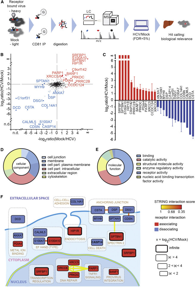
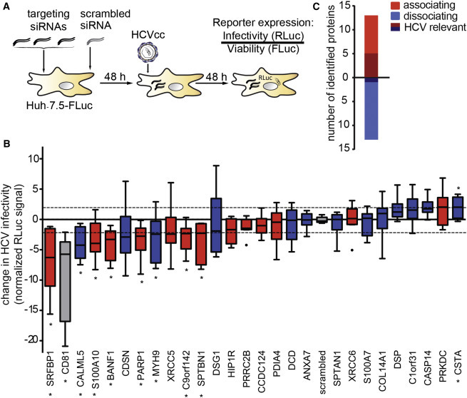
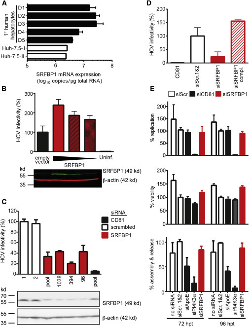
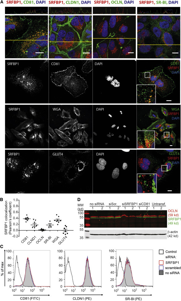
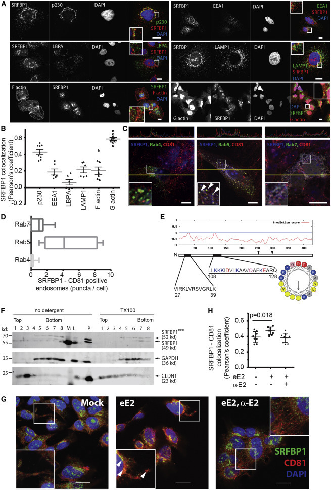
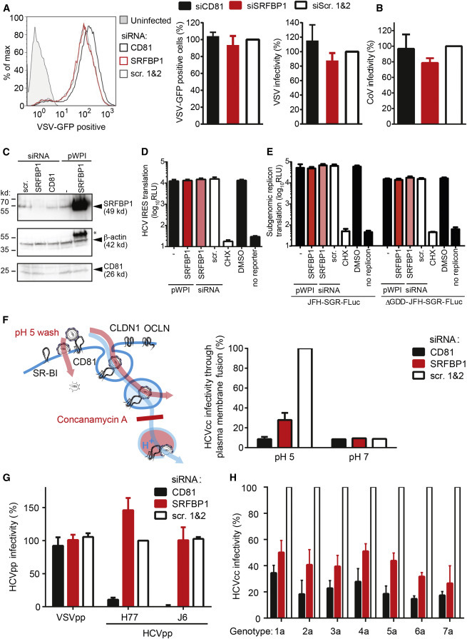
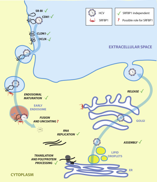
Hepatitis C virus (HCV) enters human hepatocytes through a multistep mechanism involving, among other host proteins, the virus receptor CD81. How CD81 governs HCV entry is poorly characterized, and CD81 protein interactions after virus binding remain elusive. We have developed a quantitative proteomics protocol to identify HCV-triggered CD81 interactions and found 26 dynamic binding partners. At least six of these proteins promote HCV infection, as indicated by RNAi. We further characterized serum response factor binding protein 1 (SRFBP1), which is recruited to CD81 during HCV uptake and supports HCV infection in hepatoma cells and primary human hepatocytes. SRFBP1 facilitates host cell penetration by all seven HCV genotypes, but not of vesicular stomatitis virus and human coronavirus. Thus, SRFBP1 is an HCV-specific, pan-genotypic host entry factor. These results demonstrate the use of quantitative proteomics to elucidate pathogen entry and underscore the importance of host protein-protein interactions during HCV invasion.


Figures

None
Graphical abstract
Figure 1
Figure 1
High-Resolution Quantitative MS Reveals Transient HCV Entry Factor Interactions (A) Outline of the virus entry interaction proteomics procedure. (B) CD81 interactome upon HCV exposure. Depicted are the mean log2 SILAC ratios of CD81-interacting proteins in HCV versus mock-treated samples from forward (y axis) and reverse experiments (x axis). Reverse label ratios are inverted, so that a positive correlation indicates reproducible interaction upon label swap. Significant (FDR < 5%) outliers are colored in red (CD81-associating proteins) and blue (CD81-dissociating proteins). Infinite ratio, interaction partners exclusively found in the presence of HCV. n.d., not quantified in either forward or reverse experiment. (C) SILAC log2 ratios for each of the 13 CD81-associating and 13 CD81-dissociating factors. Shown are means ± SEM of four biological replicates with inverted reverse label ratios. (D and E) Enrichment of Gene Ontology cellular component (GOCC) and molecular function (GOMF) annotations. (F) Functional map of host factors transiently interacting with the HCV receptor CD81 during virus entry. Functional clusters (white boxes) and previously reported interactions (bold lines) of the here identified transient CD81-binding partners are depicted. We assigned individual proteins to the highest scoring DAVID cluster. Yellow lines between genes of different clusters indicate high-confidence (>0.9) STRING interactions. Within a functional annotation cluster, also lower confidence (>0.35) STRING interactions are shown. Proteins are placed in their predominant cellular location; SRFBP1 is shown twice as it localizes to nucleus and cytoplasm. The box size indicates the degree of CD81 association or dissociation upon HCV binding. Associating factors (red) and dissociating factors (blue) are shown. See also Figure S1 and Tables S1 and S2–S4.
Figure 2
Figure 2
A Subset of CD81 Interaction Partners Is Required for HCV Infection (A) Outline of the RNAi screen on transient CD81 interaction partners relevant for HCV infection. (B) Functional RNAi follow-up screen on 26 selected transient CD81 interaction partners identifies nine putative host factors. We silenced the indicated transcript with a pool of three siRNAs in Huh-7.5 FLuc cells, infected 48 hr later with Renilla luciferase reporter HCV (JcR2A), and determined cell viability and HCV infectivity 48 hpi. Shown is the RLuc signal after normalization for cell viability and plate effects. Eight siRNA pools significantly decreased and one increased HCV infectivity (p ≤ 0.05; abs [z score] ≥ 2; ). Associating factors (red), dissociating factors (blue), CD81 and scrambled controls (gray) are shown. Box and whisker plot of nine biological replicates is shown. (C) The combined SILAC co-IP RNAi strategy reveals a bias for CD81-associating factors to act as HCV host factors. Out of 26 HCV-dependent CD81-binding partners, six decreased HCV infectivity upon RNAi with a minimum transcript reduction of 75% (shaded color). See also Figure S2 and Tables S5 and S6.
Figure 3
Figure 3
The CD81-Binding Partner SRFBP1 Is Expressed in Human Liver and Required for HCV Infection (A) SRFBP1 transcript levels in primary human hepatocytes are up to 6-fold higher than in Huh-7.5 cells. Absolute transcript numbers of SRFBP1 in hepatocytes from five donors (D1–D5) and in two independent passages of human hepatoma cells (Huh-7.5) were determined in technical triplicates and displayed as mean + SD. (B) HCV (JcR2A) infectivity increases in a dose-dependent manner in Huh-7.5 FLuc cells upon overexpression of full-length SRFBP1. Cells were transduced with lentiviruses encoding SRFBP1 or a blasticidin resistance gene (empty vector), 72 hr later infected with HCV, and infectivity measured 48 hpi by luciferase assay. Immunoblot analysis of lysates 72 post-transduction shows dose-dependent SRFBP1 overexpression (green). Actin served as loading control (red). The immunoblot is representative of three biological replicates. (C) HCV (JcR2A) infectivity is reduced in Huh-7.5 FLuc cells 48 hp silencing of SRFBP1 or CD81. We used a pool of three siRNAs or individual siRNAs targeting the indicated ORF position and measured infectivity at 48 hpi by luciferase assay. Two scrambled siRNAs (1 and 2) served as controls. Immunoblot analysis confirms reduced SRFBP1 protein levels 48 hp RNAi. Mean + SD of three technical replicates are shown. Infectivity data and immunoblot are representative of three biological replicates. (D) Lentiviral transduction with siRNA-resistant SRFBP1 rescues HCV infection in SRFBP1-silenced Huh-7.5 FLuc cells. Cells were transfected with siRNAs (SRFBP1: siRNA 394), 24 hr later transduced with blasticidin resistance gene encoding lentivirus (siSRFBP1) or siRNA-resistant SRFBP1 encoding lentivirus (siSRFBP1 compl.), and 24 hr later infected with HCV (JcR2A). Infectivity at 48 hpi measured by luciferase assay is shown. (E) SRFBP1 is dispensable for HCV replication, assembly, and release. Huh-7.5 FLuc cells were transfected with genomic HCV RNA (JcR2A) and the indicated gene silenced 5 hr later (SRFBP1: siRNA 394). At 72 and 96 hp transfection (hpt), supernatants were harvested, cells lysed, and replication efficiency in lysates measured by luciferase assay (upper panel). Viability of HCV-replicating cells upon RNAi was determined using the cellular FLuc reporter at 72 or 96 hpt (middle panel). Supernatants from HCV-transfected and SRFBP1-silenced cells were titrated on naive Huh-7.5 cells to determine virus particle assembly and release rates (bottom panel). Values were normalized to a scrambled siRNA control. Unless stated otherwise, all experiments are displayed as mean + SD of three independent biological replicates each performed in technical triplicates. See also Figure S3.
Figure 4
Figure 4
SRFBP1 Colocalizes with CD81 without Affecting Entry Factor Surface Expression (A) SRFBP1 partially colocalizes with CD81 and the membrane marker WGA but only weakly with CLDN1, OCLN, SR-BI, and GLUT4. Huh-7.5 cells were stained with Alexa-conjugated membrane marker WGA (panel 3) for 1 min or left unstained (panels 1, 2, and 4), fixed, permeabilized, and stained for SRFBP1 and the indicated protein. Nuclei were stained with DAPI. Colocalization across a section (yellow line in panel 1) is depicted above the respective image. Representative confocal images; insert 2.2-fold magnification; scale bars 10 μm. (B) Pearson’s correlation coefficient for SRFBP1 and the indicated cellular protein or the membrane marker WGA calculated by intensity correlation analysis. Each symbol represents an individual frame; horizontal lines indicate the mean ± SEM. (C) SRFBP1-silenced cells (siRNA 394) express CD81, CLDN1, and SR-BI at the plasma membrane. Surface expression of CD81, CLDN1, and SR-BI on Huh-7.5 cells was analyzed 48 hpt with the indicated siRNAs. Cells were stained with antibodies against HCV entry factors followed by flow cytometric analysis of 10,000 cells per sample. For quantification and additional controls, see Figure S4. Control is directly conjugated isotype antibody (histogram 1) or secondary antibody only (histograms 2 and 3). (D) OCLN expression levels are stable after SRFBP1 silencing (siRNA 394). Immunoblot analysis of OCLN (red) and SRFBP1 (green) after siRNA mediated silencing for 48 hr is shown. Actin served as loading control. Data are representative of at least three independent experiments. See also Figure S4.
Figure 5
Figure 5
A Pool of SRFBP1 Localizes to CD81 on Endosomes and Is Recruited to CD81 upon HCV Glycoprotein Exposure (A) SRFBP1 localizes to the trans-GOLGI, endosomes, and actin. Huh-7.5 cells were stained for SRFBP1; the trans-GOLGI marker p230; and the endosomal markers EEA1, LBPA, and LAMP1 as described in Figure 4A. F and G actin were stained with Alexa-conjugated phalloidin and DNase I, respectively. (B) Pearson’s correlation coefficient for SRFBP1 and indicated cellular proteins calculated by intensity correlation analysis. Each symbol represents an individual frame; horizontal lines indicate the mean ± SEM. (C) SRFBP1 localizes to CD81 on endosomes. Huh-7.5 cells were transfected with expression plasmids for EGFP-Rab4, -Rab5, and -Rab7 and stained for SRFBP1 and CD81. Colocalization of SRFBP1, CD81, and Rab proteins across a section (yellow line) is depicted in the upper panels. Arrowheads indicate colocalization. (D) SRFBP1 and CD81 colocalize at early endosomes. Quantification of SRFBP1, CD81, and Rab triple-positive puncta is shown. Box and whisker plot showing median, minimum, and maximum values from six independent frames. (E) Bioinformatics prediction of two weak amphipathic helices for SRFBP1 (black bars) with the second helix (aas 108–128) showing a small hydrophobic face of five amino acids (FLLVI). The hydrophobic face is highlighted in light gray in the primary sequence and in yellow in the helix model. Two cysteine residues (aa 254 and aa 300), which could serve as palmitoylation sites, are indicated by arrowheads. (F) Membrane flotation assay suggests membrane association of SRFBP1. Huh-7.5 cells were transduced with mycDDK-tagged SRFBP1, 48 hr later lysed in hypotonic buffer, and analyzed by Nycodenz gradient ultracentrifugation followed by immunoblot analysis against SRFBP1, GAPDH, and CLDN1. TX-100-treated lysates served as solubilization control. L, precleared lysate; M, marker; P, pellet after lysate preclearing. One out of three independent experiments is shown. (G) Exposure to soluble HCV glycoprotein (eE2) increases SRFBP1-CD81 colocalization in Huh-7.5 cells. Cells were incubated with eE2, with eE2 and an E2 blocking antibody (α-E2), or with PBS (mock) for 15 min; fixed; and stained for SRFBP1 and CD81 as described in Figure 4A. Arrowheads indicate colocalization. (H) Pearson’s correlation coefficient for SRFBP1 and CD81 calculated by intensity correlation analysis. Each symbol represents an individual frame; horizontal lines indicate the mean ± SEM; p value is indicated. Representative images; inserts show magnification; scale bars 10 μm (A and C) and 20 μm (G). See also Figure S5.
Figure 6
Figure 6
SRFBP1 Is a Pan-genotypic and HCV-Specific Host Entry Factor (A) SRFBP1 is dispensable for VSV infection. SRFBP1-silenced Huh-7.5 cells were infected with VSVMQ (MOI 0.1) and analyzed for GFP expression by flow cytometry 20 hpi. Histogram is representative of biological triplicates (left panel). Quantification of VSVMQ infectivity 20 hpi in SRFBP1-silenced cells is determined as percentage of GFP-positive cells (middle panel) or by normalization of the mean fluorescence intensity (MFI) of VSV-infected SRFBP1- or CD81-silenced cells to MFI of scrambled siRNA-transfected cells (right panel). (B) SRFBP1 is dispensable for coronavirus infection. SRFBP1-silenced cells were infected with HCoV229E-luc (MOI 0.1) and RLuc activity in cell lysates measured 24 hpi. Infectivity relative to a scrambled siRNA control is shown. (C) Immunoblot analysis of SRFBP1 and CD81 48 hp siRNA transfection. Huh-7.5 cells were transfected with siRNA or transduced with the indicated pWPI expression construct as in (A)–(H), 48 hr later lysed, and analyzed by immunoblot. Actin served as loading control. , residual SRFBP1 signal. (D) Bicistronic translational reporter assay with HCV IRES-driven RLuc and cap-dependent FLuc (see also Figures S6A and S6B). SRFBP1 silencing and overexpression was performed as in (C), and 48 hr later, cells were transfected with translational reporter RNA. Eight hours after reporter transfection, luciferase activity in lysates was monitored. (E) Early replication reporter assay using a subgenomic HCV genome expressing FLuc. SRFBP1 silencing and overexpression was performed as in (C), and 48 hr later, JFH-SGR-FLuc RNA was transfected into cells; cells lysed after 8 hr; and luciferase activity monitored. A polymerase mutant JFH-SGR-FLuc replicon (ΔGDD) was used to assess translation of HCV genomes independent of de novo replication. See also Figures S6C and S6D for additional controls. (F) SRFBP1 is required in a plasma membrane fusion assay of HCV infection. Huh-7.5 cells silenced for SRFBP1 were pretreated with concanamycin A (5 nM; 1 hr) to block vacuolar type H+-ATPases, incubated with HCV (JcR2A) for 2 hr at 4°C in the presence of concanamycin A, shifted to 37°C, and washed with a pH 5 or pH 7 buffer for 5 min. After incubation with concanamycin A for 4 hr, medium was changed and endosomal acidification independent infectivity measured at 48 hpi. See also Figures S6E and S6F for additional controls. (G) Lentiviral pseudotypes infect Huh-7.5 cells independently of SRFBP1. Cells in which SRFBP1 had been silenced (48 hr) were infected with HIV-1 pseudotypes encoding FLuc and displaying glycoproteins from HCV genotype 1 (H77), HCV genotype 2 (J6), VSV, or no glycoprotein. At 72 hpi, cells were lysed and FLuc activity measured. Infectivity was calculated by subtraction of background read for glycoprotein-free particles and relative to VSVG particles. (H) Silencing of SRFBP1 reduces infectivity of chimeric HCV viruses with glycoproteins from all seven genotypes. Huh-7.5 FLuc cells were subjected to siRNA-mediated silencing followed by infection with intergenotypic HCV chimeras (MOI 0.1) expressing RLuc. Forty-eight hours post-infection, infectivity was determined by RLuc activity measurement. Cells treated with CD81 targeting or scrambled siRNAs served as controls. SRFBP1-targeting siRNA 394 was used in all experiments. Data from three to five biological replicates are displayed as mean + SD. See also Figure S6.
Figure 7
Figure 7
HCV Life Cycle and Possible Role for SRFBP1 during HCV Entry Our data point toward a role for SRFBP1 in the last step of entry; i.e., nucleocapsid uncoating.

Comment in

  • SRFBP1, an Additional Player in HCV Entry.
    Fénéant L, Cocquerel L. Fénéant L, et al. Trends Microbiol. 2015 Oct;23(10):590-593. doi: 10.1016/j.tim.2015.08.003. Epub 2015 Aug 26. Trends Microbiol. 2015. PMID: 26319372

Similar articles

Cited by

References

    1. Benali-Furet N.L., Chami M., Houel L., De Giorgi F., Vernejoul F., Lagorce D., Buscail L., Bartenschlager R., Ichas F., Rizzuto R., Paterlini-Bréchot P. Hepatitis C virus core triggers apoptosis in liver cells by inducing ER stress and ER calcium depletion. Oncogene. 2005;24:4921–4933. - PubMed
    1. Berger K.L., Cooper J.D., Heaton N.S., Yoon R., Oakland T.E., Jordan T.X., Mateu G., Grakoui A., Randall G. Roles for endocytic trafficking and phosphatidylinositol 4-kinase III alpha in hepatitis C virus replication. Proc. Natl. Acad. Sci. USA. 2009;106:7577–7582. - PMC - PubMed
    1. Blanchard E., Belouzard S., Goueslain L., Wakita T., Dubuisson J., Wychowski C., Rouillé Y. Hepatitis C virus entry depends on clathrin-mediated endocytosis. J. Virol. 2006;80:6964–6972. - PMC - PubMed
    1. Brazzoli M., Bianchi A., Filippini S., Weiner A., Zhu Q., Pizza M., Crotta S. CD81 is a central regulator of cellular events required for hepatitis C virus infection of human hepatocytes. J. Virol. 2008;82:8316–8329. - PMC - PubMed
    1. Brimacombe C.L., Wilson G.K., Hübscher S.G., McKeating J.A., Farquhar M.J. A role for CD81 and hepatitis C virus in hepatoma mobility. Viruses. 2014;6:1454–1472. - PMC - PubMed

Publication types

Substances