. 2015 Aug 6;59(3):399-412.
doi: 10.1016/j.molcel.2015.06.033. Epub 2015 Jul 23.

Conformational Differences between Open and Closed States of the Eukaryotic Translation Initiation Complex

Affiliations

Conformational Differences between Open and Closed States of the Eukaryotic Translation Initiation Complex

Jose L Llácer et al. Mol Cell. .

Abstract

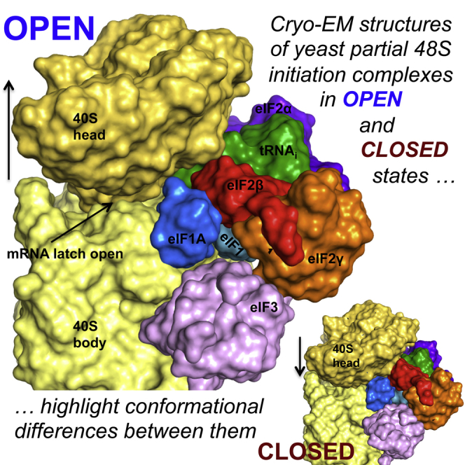
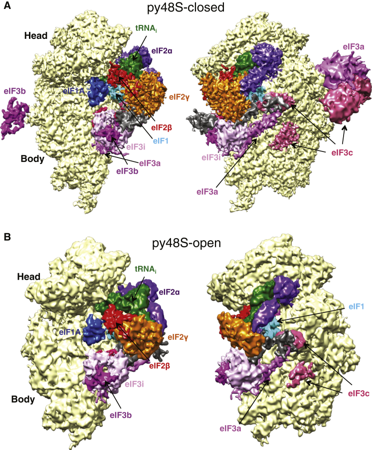
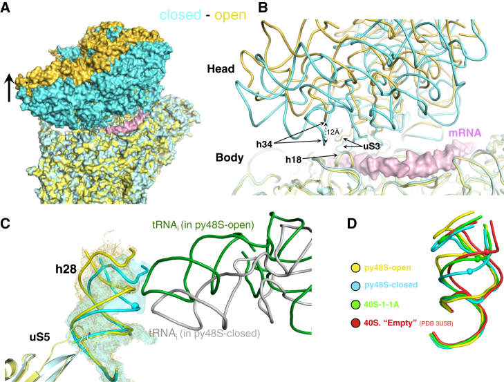
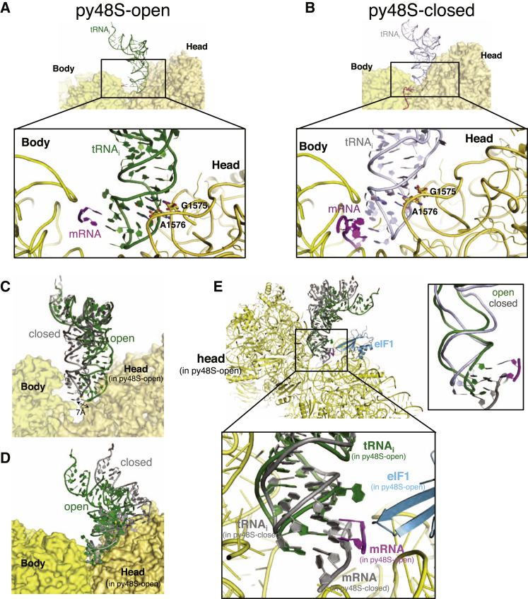
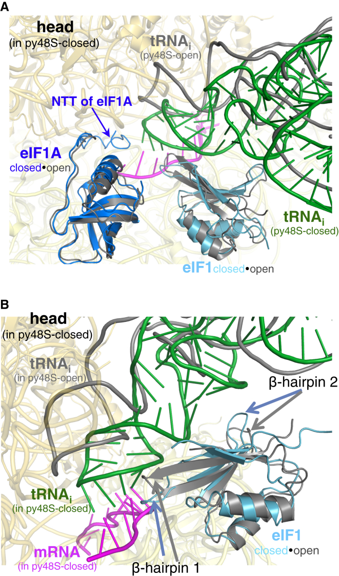
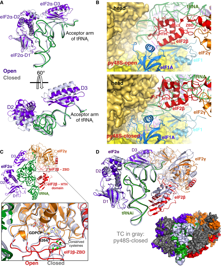
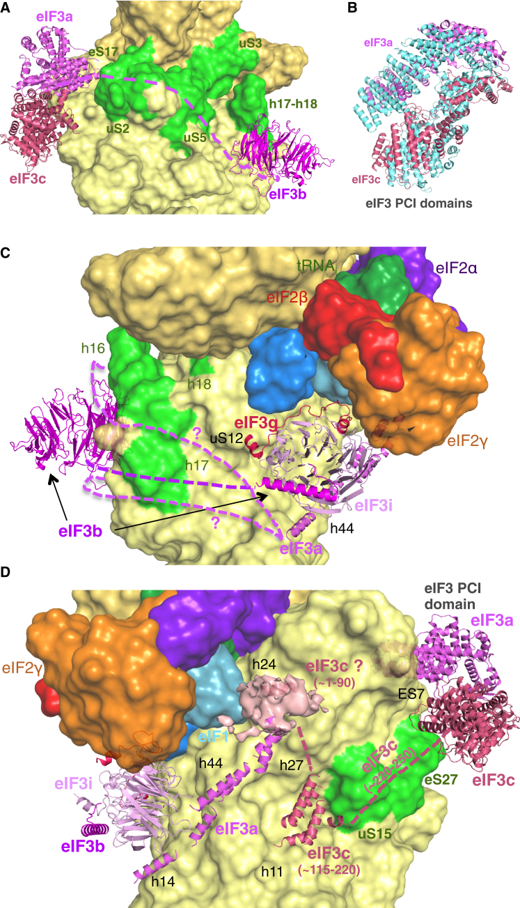
Translation initiation in eukaryotes begins with the formation of a pre-initiation complex (PIC) containing the 40S ribosomal subunit, eIF1, eIF1A, eIF3, ternary complex (eIF2-GTP-Met-tRNAi), and eIF5. The PIC, in an open conformation, attaches to the 5' end of the mRNA and scans to locate the start codon, whereupon it closes to arrest scanning. We present single particle cryo-electron microscopy (cryo-EM) reconstructions of 48S PICs from yeast in these open and closed states, at 6.0 Å and 4.9 Å, respectively. These reconstructions show eIF2β as well as a configuration of eIF3 that appears to encircle the 40S, occupying part of the subunit interface. Comparison of the complexes reveals a large conformational change in the 40S head from an open mRNA latch conformation to a closed one that constricts the mRNA entry channel and narrows the P site to enclose tRNAi, thus elucidating key events in start codon recognition.


Figures

None
Graphical abstract
Figure 1
Figure 1
Cryo-EM Maps of Eukaryotic 48S PICs (A) Two views of py48S-closed. (B) Two views of py48S-open. Density for eIF3 is Gaussian-filtered. Unassigned density is in dark gray. See also Figures S1–S4; Table S1; and Movies S1 and S2.
Figure 2
Figure 2
Distinct Position of the 40S Head Widens the mRNA Entry Channel and Opens the Latch in the Open Complex (A) Front view of superposition of py48S-open (yellow) and py48S-closed (cyan), showing mRNA (pink) from py48S (Hussain et al., 2014) to highlight the complete mRNA channel. (B) Superposition of refined models of py48S-open (yellow) and py48S-closed (cyan), indicating elements forming the latch. (C) h28 in py48S-open (yellow) and py48S-closed (cyan) based on superposition of the two complexes, viewed from the A site. tRNAi and uS5 for the two complexes are also shown. (D) h28 in empty 40S (red, PDB: 3U5B), 40S•eIF1•eIF1A (green), py48-open (yellow), and py48S-closed (cyan) complexes. Equivalent atoms in h28 are shown as spheres. See also Figures S4 and S2 and Movies S3 and S4.
Figure 3
Figure 3
tRNAi Is Not Engaged with P-Site Elements of the 40S Body in the Open Complex (A) tRNAi in py48S-open, viewed from E site. The body and head of 40S are shown in lighter and darker shades of yellow. The zoomed view shows mRNA at the P site and recognition of conserved GC base pairs in ASL by rRNA bases. For clarity, 40S proteins and other factors are not shown. (B) The tRNAi in py48S-closed viewed as in (A). (C) Superposition of the 40S body reveals distinct locations of tRNAi in the P site of py48S-open (green) and py48S-closed (gray). The body and head of py48S-open complex are shown. The two ASLs are separated by about 7 Å in the P site. (D) Superposition of the 40S head of py48S-open (green) and py48S-closed (gray). (E) Superposition of two complexes as in (D), viewed from the A site. The mRNA of py48S-closed is in gray. Inset shows the superposition of the two positions of tRNAi and interacting mRNA codon. See also Figure S5.
Figure 4
Figure 4
Contacts of eIF1A-NTT and eIF1 with tRNAi Restricted to the Closed Complex (A) Superposition of the open and closed complexes with the ligands of py48S-closed shown in color while those of py48S-open are in gray. Only the 40S of py48S-closed is shown (yellow). The zoomed view shows the NTT of eIF1A in the two complexes. (B) Superposition of the open and closed complexes as in (A).
Figure 5
Figure 5
Distinctive Interactions of eIF2β with eIF1, eIF1A, and tRNAi Occlude the mRNA Channel in py48S-Open (A) Conformational changes in eIF2α based on superposition of the TC coordinates using tRNAi as the reference. The eIF2α and tRNAi of py48S-open are shown in color and those of py48S-closed are in gray. (B) Position of eIF2β with respect to tRNAi, eIF1, eIF1A, and 40S head in py48S-open and py48S-closed. (C) Similar position for the ZBD of eIF2β in both complexes with respect to eIF2γ. The eIF2β, eIF2γ and tRNAi of py48S-open are shown in color while those of py48S-closed are in gray. Ser264 is shown as sticks near conserved cysteines. (D) Cartoon and surface representations of the superimposition of TC coordinates in py48S-open (color) and closed (gray) complexes based on tRNAi as reference. It shows the internal conformational change within TC during transition from the open to the closed conformation. While D2 and the helix connecting the D1 and the D2 domains of eIF2α experience an internal rearrangement, eIF2α-D3, eIF2γ and eIF2β rotate together around the acceptor arm of the tRNA. See also Figures S6 and S7 and Movie S5
Figure 6
Figure 6
Genetic Evidence that eIF2β Interactions with the tRNAi ASL and eIF1 Preferentially Stabilize py48S-Open to Impede Initiation at Near-Cognate UUG Codons In Vivo (A) Positions of eIF1, eIF2β, and tRNAi in the py48S-open and py48S-closed, with residues substituted in genetic studies shown as spheres. (B) Expression of HIS4-lacZ reporters with AUG or UUG start codons in strains of the indicated SUI3 or SUI1 genotypes, expressed as mean (± SEM) ratios of UUG- to AUG-reporter expression with fold-changes relative to WT in parentheses. (C) Expression of the GCN4-lacZ reporter expressed as mean (± SEM) units of β-galactosidase. (D and E) Western analysis of eIF1 (D) or eIF2β (E) proteins in whole-cell extracts (WCEs), with eIF3j or eIF2Bε analyzed as loading controls, reported as mean (± SEM) eIF1:eIF3j ratios or eIF2β/eIF2Bε ratios, normalized to the WT ratios, determined from biological replicates. Lanes have been cropped from the same gels. (F) WCEs were immunoprecipitated with FLAG affinity resin and immune complexes subjected to western analysis to detect Flag-eIF2β and co-immunoprecipitated eIF2γ, eIF2α, and eIF2Bε, resolving 1×, 2×, or 3× amounts in successive lanes. In, 20% input WCEs. Western signals were quantified to yield mean (± SEM) recoveries of eIF2γ, eIF2α, or eIF2Bε normalized to Flag-eIF2β. Lanes have been cropped from the same gels. Error bars represent the SEM from three biological replicates.
Figure 7
Figure 7
Structural Arrangement of eIF3 Components in 48S PICs (A) Locations of the eIF3a/eIF3c PCI domains and β-propeller of eIF3b at different positions on the solvent-exposed surface of the 40S, highlighting rRNA helices and ribosomal proteins (green) predicted to bind to eIF3a. The proposed path of the unassigned central portion of the eIF3a-CTD connecting the PCI domain to the subunit interface is shown as a dashed purple line. (B) Lateral displacement of eIF3a/eIF3c PCI domains in py48S-closed versus their positions in yeast 40S•eIF1•eIF1A•eIF3 (PDB: 4UER). (C) Trimeric eIF3b-CTD/eIF3i/eIF3g-NTD subcomplex is shown near h44 and interacting with eIF2γ and the 40S interface surface. The β-propeller of eIF3b is also shown. Two alternative proposed paths of the eIF3a-CTD connecting the PCI domain to the bundle of helices below the eIF3i β-propeller are shown as dashed purple lines. (D) A cluster of helices tentatively assigned to eIF3c is located near h11 and uS15 (green). A globular density with a single modeled helix is tentatively assigned to the eIF3c-NTD in proximity to eIF1 and h24. The proposed path of a linker connecting the cluster of helices to the eIF3c PCI domain is shown as a dashed magenta line. Long helices tentatively assigned to eIF3a bridge the eIF3i β-propeller and h44 with the putative eIF3c-NTD and h24.

References

    1. Algire M.A., Maag D., Lorsch J.R. Pi release from eIF2, not GTP hydrolysis, is the step controlled by start-site selection during eukaryotic translation initiation. Mol. Cell. 2005;20:251–262. - PubMed
    1. Asano K., Clayton J., Shalev A., Hinnebusch A.G. A multifactor complex of eukaryotic initiation factors, eIF1, eIF2, eIF3, eIF5, and initiator tRNA(Met) is an important translation initiation intermediate in vivo. Genes Dev. 2000;14:2534–2546. - PMC - PubMed
    1. Aylett C.H., Boehringer D., Erzberger J.P., Schaefer T., Ban N. Structure of a yeast 40S-eIF1-eIF1A-eIF3-eIF3j initiation complex. Nat. Struct. Mol. Biol. 2015;22:269–271. - PubMed
    1. Bai X.C., Fernandez I.S., McMullan G., Scheres S.H. Ribosome structures to near-atomic resolution from thirty thousand cryo-EM particles. Elife. 2013;2:e00461. - PMC - PubMed
    1. Ben-Shem A., Garreau de Loubresse N., Melnikov S., Jenner L., Yusupova G., Yusupov M. The structure of the eukaryotic ribosome at 3.0 Å resolution. Science. 2011;334:1524–1529. - PubMed

Publication types

MeSH terms