. 2015 Jul 27:6:7718.
doi: 10.1038/ncomms8718.

miR-218 is essential to establish motor neuron fate as a downstream effector of Isl1-Lhx3

Affiliations

miR-218 is essential to establish motor neuron fate as a downstream effector of Isl1-Lhx3

Karen P Thiebes et al. Nat Commun. .

Erratum in

Abstract

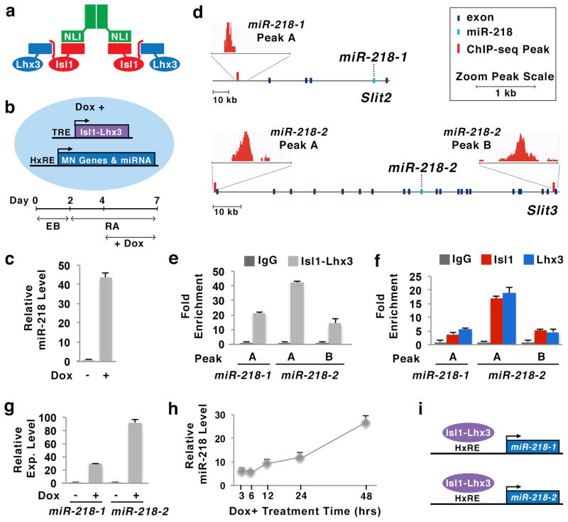
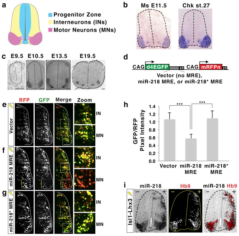
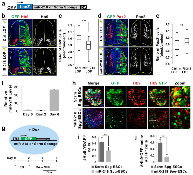
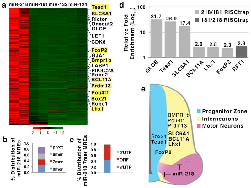
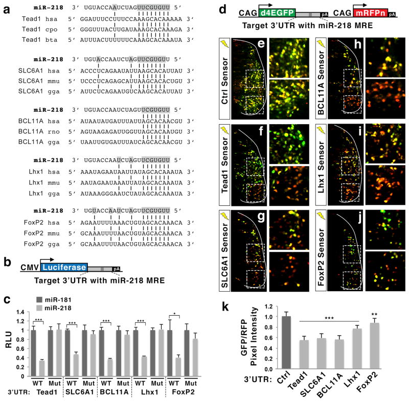
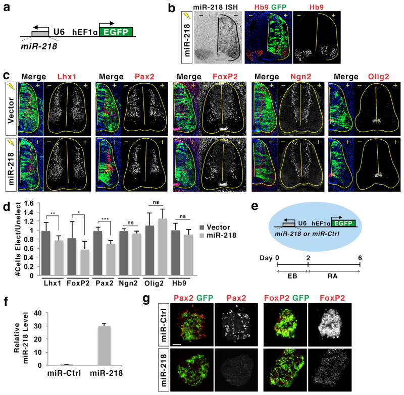
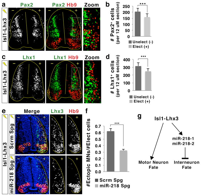
While microRNAs have emerged as an important component of gene regulatory networks, it remains unclear how microRNAs collaborate with transcription factors in the gene networks that determines neuronal cell fate. Here we show that in the developing spinal cord, the expression of miR-218 is directly upregulated by the Isl1-Lhx3 complex, which drives motor neuron fate. Inhibition of miR-218 suppresses the generation of motor neurons in both chick neural tube and mouse embryonic stem cells, suggesting that miR-218 plays a crucial role in motor neuron differentiation. Results from unbiased RISC-trap screens, in vivo reporter assays and overexpression studies indicated that miR-218 directly represses transcripts that promote developmental programs for interneurons. In addition, we found that miR-218 activity is required for Isl1-Lhx3 to effectively induce motor neurons and suppress interneuron fates. Together our results reveal an essential role of miR-218 as a downstream effector of the Isl1-Lhx3 complex in establishing motor neuron identity.


Conflict of interest statement

COMPETING FINANCIAL INTERESTS

The authors declare no competing financial interests.

Figures

Figure 1
Figure 1. Identification of miR-218 as a direct target miRNA of Isl1-Lhx3 in motor neurons
(a) Illustration of the Isl1-Lhx3 hexamer complex, consisting of two NLI, two Isl1 and two Lhx3 proteins. (b) Schematic model of Isl1-Lhx3 embryonic stem cell (Isl1-Lhx3-ESC) line and the experimental design to differentiate Isl1-Lhx3-ESCs to neurons. Treatment with doxycycline (Dox) induces the expression of Isl1-Lhx3, which is controlled by tetracycline response element (TRE), in Isl1-Lhx3-ESCs. Then, Isl1-Lhx3 upregulates its direct target genes that have hexamer response element (HxRE), such as motor neuron (MN) genes and miRNAs. EB, embryoid bodies; RA, retinoic acid. (c) Isl1-Lhx3 triggered the expression of mature miR-218 in Isl1-Lhx3-ESCs, as determined by qPCR using TaqMan probes. Error bars represent the standard deviation. n = 2, biological duplicates. (d) Isl1-Lhx3-bound ChIP-seq peaks were identified near miR-218-1 and miR-218-2 genes, within the introns of Slit2 and Slit3, respectively. (e) Isl1-Lhx3 bound to three Isl1-Lhx3-bound ChIP-seq peak regions near miR-218-1 and miR-218-2 genes in Isl1-Lhx3-ESCs. Error bars represent the standard deviation. n = 3. (f) Both Isl1 and Lhx3 were recruited to three Isl1-Lhx3-bound ChIP-seq peaks near miR-218-1 and miR-218-2 genes in E12.5 mouse spinal cord. Error bars represent the standard deviation. n = 3. (g) Isl1-Lhx3 induced the expression of pri-miR-218-1 and pri-miR-218-2 in Isl1-Lhx3-ESC-derived motor neurons, as determined by qPCR using TaqMan probes. Error bars represent the standard deviation. n = 2. (h) Isl1-Lhx3 strongly upregulated miR-218 expression within 48 hours in Isl1-Lhx3-ESCs cultured in monolayer without retinoic acid, as determined by qPCR using TaqMan probes. Error bars represent the standard deviation. n = 3. (i) Isl1-Lhx3 complex binds to hexamer response element (HxRE) near miR-218-1 and miR-218-2 genes and triggers the expression of miR-218 genes in differentiating MNs.
Figure 2
Figure 2. miR-218 is expressed and functional in embryonic motor neurons
(a) Illustration of the developing spinal cord. Stereotypical locations of progenitor cells, interneurons (INs), and motor neurons (MNs) in mouse E11.5 and chicken HH St.27 embryos are shown. (b) miR-218 is specifically expressed in motor neurons of mouse and chick embryos, as shown by in situ hybridization with a probe detecting mature miR-218. (c) miR-218 is induced at the onset of motor neuron differentiation and continues to be expressed in motor neurons throughout mouse embryonic development, as shown by in situ hybridization with a probe detecting mature miR-218. Scale bars represent 100 μm. (d) Illustrations of miRNA sensor plasmids. The multimerized microRNA response element (MRE) for miR-218 or miR-218* was inserted between the destabilized EGFP (d4EGFP) gene and polyA (pA) sequences. The expression of both d4EGFP and mononuclear RFP (mRFPn) is driven by two separate, ubiquitously active CAG promoters. (e–g) The expression pattern of GFP and RFP in the chick spinal cord electroporated with miRNA sensor vector (e), miR-218 MRE sensor (f) or miR-218* MRE sensor (g). Only electroporated side of the spinal cord is shown. GFP expression is regulated by endogenous miRNA that binds to the MREs present in 3′UTR of the GFP gene, while RFP expression depicts the electroporated cells. The areas of interneuron (IN) and motor neuron (MN) are magnified. The miR-218 MRE sensor shows drastically down-regulated GFP expression in motor neuron area (bracket in f), indicating that endogenous miR-218 in motor neurons suppresses the expression of GFP. (h) Quantification of relative pixel intensity of GFP/RFP in motor neurons, as quantified using ImageJ program. Error bars represent the standard deviation. ***p < 0.0001 in two-tailed Student’s t-test. n = 5 embryos. (i) Analyses using serial sections from the same chicken embryo electroporated with Isl1-Lhx3. In situ hybridization for miR-218 and immunohistochemical analyses with Hb9 antibody reveals that the expression of miR-218 was highly induced by Isl1-Lhx3 in the dorsal spinal cord, where Hb9+ ectopic motor neurons are formed. +, electroporated side; −, unelectroporated control side.
Figure 3
Figure 3. miR-218 is important for motor neuron differentiation
(a) Illustration of bulge sponge inhibitor constructs, which have either bulged miR-218 MRE or control scrambled (Scrm) sequences in the 3′UTR of a CMV-promoter driven LacZ gene. (b,d) Loss of function (LOF) analyses in chicks electroporated with control (Ctrl) LOF conditions (scrambled sponge inhibitor, 2′Ome-inhibitor control, and CMV-GFP) or miR-218 LOF conditions (miR-218 sponge inhibitor, miR-218 2′Ome-inhibitor, and CMV-GFP). Hb9 labels motor neurons, while Pax2 labels a broad population of interneurons in immunohistochemical analyses. +, electroporated side; −, unelectroporated control side. (c,e) The effect of LOF conditions was quantified by the ratio of Hb9+ cells or Pax2+ cells on the electroporated (elect) side over the unelectroporated (unelect) control side. ***p < 0.0001 in two-tailed Student’s t-test. ns, not significant; n = 19–21 embryos for (b) Hb9 counts and n = 15–18 embryos for (d) Pax2 counts. (f) The qPCR analysis using miR-218 TaqMan probe revealed that miR-218 expression is low in ESCs cultured in monolayer (day 0) or in embryoid bodies (day 2), but is highly induced in ESC-derived motor neurons (day 6). Error bars represent the standard deviation. (g) Illustration of doxycycline (Dox)-inducible sponge mouse ESC lines, in which Dox induces the expression of either miR-218 sponge inhibitor or scrambled (Scrm) sponge inhibitor, and the experimental design to differentiate ESCs to motor neurons. TRE, tetracycline response element; EB, embryoid body; RA, retinoic acid; Shh, a sonic hedgehog agonist Purmorphamine. (h) Immunohistochemical analyses in Dox-inducible sponge ESC-derived motor neurons cultured with Dox at differentiation day 6. Hb9 labels motor neurons, and GFP labels the cells, in which the expression of Scramble (Scrm) or miR-218 sponge inhibitor is induced by Dox. Zoom shows the magnified view of the areas marked by dotted lines in Hb9/GFP merged images. Scale bar represents 50 μm. (i,j) The effect of Dox-inducible sponge inhibitors was quantified by the ratio of Hb9+ cells over all cells (DAPI+) (i), or by the ratio of Hb9 and GFP double-positive motor neurons over the total number of GFP+ cells (j). Error bars represent the standard deviation. **p < 0.001 and ***p < 0.0001 in two-tailed Student’s t-test. n = 15 embryoid bodies.
Figure 4
Figure 4. Identification of direct miR-218 target mRNAs using RISC-trap screen
(a) RISC-trap screens identified direct target genes for miR-218, when analyzed against RISC-trap screens for miR-181, miR-132, miR-124. All identified miR-218 targets are sorted in a heatmap by fold-change and biological replicates, compared to miR-181 RISC-trap (FDR <0.05, fold enrichment ≥ 4). Previously published miR-218 targets identified in the RISC-trap screen are labeled, and selected miR-218 targets, which were newly identified by RISC-trap, are highlighted in yellow. Scale bar represents Z-score of row. (b,c) Analyses for miR-218 MREs in 1178 genes targeted by miR-218. (b) Percent distribution of miR-218 MREs was classified by the inclusion of at least 1 MRE motif in the order of 8mer > 7mer > 6mer > pivot. Each transcript was counted only once. (c) Percent distribution of miR-218 7mer MRE motifs per target in the 5′UTR, ORF, and 3′UTR. (d) Independent RISC-trap experiments followed by qRT-PCR analyses validated selected miR-218 target genes enriched against miR-181 RISC-trap. RFT1 is a miR-181 target mRNA identified in the RISC-trap screen. The qPCR results were shown as relative fold change in Log10 scale between miR-218 and miR-181 RISC-trap experiments. (e) Schematic model of the expression patterns of selected RISC-trap miR-218 target mRNAs. These target mRNAs include genes that are important for the differentiation and function of spinal interneurons, such as Lhx1, BCL11A, SLC6A1, FoxP2, Pou4f1, Prdm13, Sox21, and Bmpr1b, as well as genes that play roles in spinal neural progenitors, including Tead1, FoxP2 and Sox21.
Figure 5
Figure 5. 3′UTR region of Tead1, SLC6A1, BCL11A, Lhx1 and FoxP2 mediates miR-218-directed gene repression via miR-218 MRE
(a) Evolutionarily conserved miR-218 MREs in the 3′UTRs of selected RISC-trap target as identified by TargetScan. (b) Illustration of miRNA luciferase reporter plasmids, in which the partial 3′UTR of miR-218 targets is cloned downstream of the luciferase gene. (c) Luciferase assays using luciferase reporters linked with the 3′UTR of miR-218 targets in HEK293T cells. miR-218 inhibits the 3′UTRs in a miR-218 MRE-dependent manner. Each reporter was transfected with either miR-218 or miR-181. WT, luciferase reporters linked to the wild-type 3′UTR sequences of each gene; Mut, luciferase reporters linked to the mutated 3′UTR, in which miR-218 MRE is mutated to eliminate the binding of miR-218. RLU, relative luciferase unit. Error bars represent the standard deviation. *p < 0.05 and ***p < 0.0001 in two-tailed Student’s t-test. n = 3. (d) Illustration of miRNA sensor plasmids, in which the partial 3′UTR of miR-218 targets is cloned downstream of d4EGFP. The expression of both d4EGFP and mononuclear RFP (mRFPn) is driven by two separate, ubiquitously active CAG promoters. (e–j) The in vivo miRNA sensor analyses in the developing chick spinal cord electroporated with each miRNA sensor as indicated. Only the electroporated side of the spinal cord is shown. GFP expression is regulated by the 3′UTR of miR-218 target genes containing a miR-218 MRE, while RFP is ubiquitously expressed in all electroporated cells. Interneuron and motor neuron regions are magnified. The miRNA sensors show significant downregulation of GFP in motor neuron area compared to interneuron area. (k) Quantification of relative pixel intensity of GFP/RFP in motor neurons, as quantified using ImageJ program. Error bars represent the standard deviation. ***p < 0.0001 and **p < 0.005 in two-tailed Student’s t-test. n = 6–8 embryos.
Figure 6
Figure 6. miR-218 represses differentiation of spinal interneurons
(a) Illustration of miR-218 expression construct, in which the miR-218 sequence is cloned into the hairpin structure of the EFU6 shRNA plasmid. The expression of miR-218 is driven by the ubiquitously active U6 promoter, and the EGFP gene is regulated by a separate, ubiquitously active hEF1α promoter. (b) Electroporation of the miR-218 construct results in robust and overlapping expression of miR-218 and GFP, as determined by in situ hybridization (ISH) with miR-218. miR-218 expression did not trigger ectopic formation of Hb9+ motor neurons. +, electroporated side; −, unelectroporated control side. (c, d) Immunohistochemical analyses in chicks electroporated with miR-218 expression construct or vector. miR-218 expression led to the reduction of Lhx1+, Pax2+ and FoxP2+ interneurons, while it did not make a significant change in the number of Ngn2+ differentiating neurons, Olig2+ motor neuron progenitors, or Hb9+ motor neurons. +, electroporated (Elect) side; −, unelectroporated (Unelect) control side. ***p < 0.0001, **p < 0.001, and *p < 0.05 in two-tailed Student’s t-test. ns, not significant; n = 4–8 embryos. (e) Illustration of miR-218 and miR-control (Ctrl) ESC lines, and the protocol to differentiate miRNA ESCs into Pax2+ and FoxP2+ spinal interneurons. In these ESCs, the expression of miR-218 or miR-Ctrl and EGFP are constitutively driven by U6 promoter and hEF1α promoter, respectively. EB, embryoid body; RA, retinoic acid. (f) The miR-218 expression construct used to generate miR-218 ESCs triggers robust expression of miR-218 compared to the miR-Ctrl construct in HEK293T cells, as determined by qPCR analysis using miR-218 TaqMan probe. (g) Immunohistochemical analyses of miRNA ESCs at interneuron differentiation day 6. miR-218 effectively inhibited the generation of Pax2+ and FoxP2+ interneurons. Scale bar represents 50 μm.
Figure 7
Figure 7. miR-218 is required for efficient generation of motor neurons by Isl1-Lhx3
(a, c) Immunohistochemical analyses in the chick neural tube electroporated with Isl1-Lhx3. Isl1-Lhx3 suppresses the generation of Pax2+ or Lhx1+ interneurons, while promoting the formation of ectopic Hb9+ motor neurons in the dorsal spinal cord. The magnified images show that the Hb9+ motor neurons are largely exclusive with Pax2+ or Lhx1+ interneurons, suggesting that Isl1-Lhx3 drives motor neuron formation at the expense of interneurons. +, electroporated side; −, unelectroporated control side. (b, d) Quantification of the number of Pax2+ or Lhx1+ interneurons on the electroporated (+) and unelectroporated (−) sides of the spinal cord. Isl1-Lhx3 expression resulted in a decrease in Pax2+ or Lhx1+ interneurons. Error bars represent the standard deviation. ***p < 0.0001 in two-tailed Student’s t-test. n = 3 embryos. (e, f) The analyses of ectopic motor neuron formation by Isl1-Lhx3 in the presence of either miR-218 sponge inhibitor (miR-218 Spg) or scrambled sponge inhibitor (Scrm Spg) in the chick neural tube. +, electroporated side; −, unelectroporated control side. miR-218 inhibition reduces the efficiency of Isl1-Lhx3 in triggering ectopic motor neurons in dorsal neural tube. (f) The effect of miR-218 inhibition on Isl1-Lhx3-induced motor neuron differentiation was quantified by the ratio of ectopic Hb9+ motor neurons (MNs) over Lhx3-expressing transfected cells (Elect cells). Error bars represent the standard error of the mean. ***p < 0.0001 in two-tailed Student’s t-test. n = 5 embryos. (g) Model of the Isl1-Lhx3 and miR-218 gene regulatory network in motor neuron development. While triggering the expression of many motor neuron-specific genes required for motor neuron differentiation and maturation, Isl1-Lhx3 also directly induces the expression of miR-218-1 and miR-218-2, which are crucial to suppress unwanted interneuron genes in developing motor neurons.

References

    1. Jessell TM. Neuronal specification in the spinal cord: inductive signals and transcriptional codes. Nat Rev Genet. 2000;1:20–29. - PubMed
    1. Lee SK, Pfaff SL. Transcriptional networks regulating neuronal identity in the developing spinal cord. Nature Neuroscience. 2001;4 (Suppl):1183–1191. - PubMed
    1. Helms AW, Johnson JE. Specification of dorsal spinal cord interneurons. Current Opinion in Neurobiology. 2003;13:42–49. - PubMed
    1. Briscoe J, Pierani A, Jessell TM, Ericson J. A homeodomain protein code specifies progenitor cell identity and neuronal fate in the ventral neural tube. Cell. 2000;101:435–445. - PubMed
    1. Muhr J, Andersson E, Persson M, Jessell TM, Ericson J. Groucho-mediated transcriptional repression establishes progenitor cell pattern and neuronal fate in the ventral neural tube. Cell. 2001;104:861–873. - PubMed

Publication types

MeSH terms