. 2015 Jul 27:5:12562.
doi: 10.1038/srep12562.

A novel laminin β gene BmLanB1-w regulates wing-specific cell adhesion in silkworm, Bombyx mori

Affiliations

A novel laminin β gene BmLanB1-w regulates wing-specific cell adhesion in silkworm, Bombyx mori

Xiaoling Tong et al. Sci Rep. .

Abstract

Laminins are important basement membrane (BM) components with crucial roles in development. The numbers of laminin isoforms in various organisms are determined by the composition of the different α, β, and γ chains, and their coding genes, which are variable across spieces. In insects, only two α, one β, and one γ chains have been identified thus far. Here, we isolated a novel laminin β gene, BmLanB1-w, by positional cloning of the mutant (crayfish, cf) with blistered wings in silkworm. Gene structure analysis showed that a 2 bp deletion of the BmLanB1-w gene in the cf mutant caused a frame-shift in the open reading frame (ORF) and generated a premature stop codon. Knockdown of the BmLanB1-w gene produced individuals exhibiting blistered wings, indicating that this laminin gene was required for cell adhesion during wing development. We also identified laminin homologs in different species and showed that two copies of β laminin likely originated in Lepidoptera during evolution. Furthermore, phylogenetic and gene expression analyses of silkworm laminin genes revealed that the BmLanB1-w gene is newly evolved, and is required for wing-specific cell adhesion. This is the first report showing the tissue specific distribution and functional differentiation of β laminin in insects.


Figures

Figure 1
Figure 1. Phenotype of wild-type (Dazao) and cf mutant strain.
Pupal and moth stages of wild-type (A,B) and cf (C,D) are shown. Hematoxylin–eosin stained paraffin sections of silkworm wing discs in late larval stage (day 6 of 5th instar) of wild-type (E) and cf (F) are also shown. LC: larval cuticle, EP: epidermis, wc: wing cavity, ws: wing sac, wd: wing disc, TB: trachea tube.
Figure 2
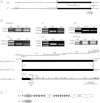
Figure 2. Mapping, bioinformatics and expression profile analysis of the cf locus.
(A) Preliminary mapping of the cf locus. The cf locus was mapped between markers S2213 and S1311. The marker S1309 was tightly linked with the cf locus. Marker search in the genome was carried out within a 1500 kb sequence length both upstream and downstream of S1309. (B) Fine mapping of the cf locus. The cf locus was mapped between M1 and M2 using 493 BC1M individuals. S1309 was still tightly linked with the cf locus. (C) Screening the candidate gene and genotyping using polymorphism markers. Microarray analysis showed that there were only two genes (BGIBMGA001218 and BGIBMGA000915) separated by a distance of 465 kb in the mapped region with different expression levels in the wild-type and the cf mutant. (a) qRT-PCR analysis of BGIBMGA001218 revealed no significant difference in gene expression between the cf mutant and wild-type (The P value equals 0.3300, Student’s t-test. N = 3). Gene BGIBMGA000915 within the mapped region was expressed much lower in cf than in wild-type based on both microarray and qRT-PCR analysis. Sequencing analysis of the two genes revealed that BGIBMGA001218 was not different between the cf mutant and wild-type, but BGIBMGA000915 differed. (b) Linkage analysis of the polymorphism marker M000915, which is 450 bp downstream of BGIBMGA000915. F1: first filial generation. BC1F: first backcross generation for linkage analysis. (c) Genotyping of the M000915 marker and cf locus using 493 BC1M (first backcross generation for recombination analysis) individuals. There is no recombination between M000915 and the cf locus. The results indicate that M000915 was also tightly linked with the cf locus.
Figure 3
Figure 3. Schematic diagram of BmLanB1-w in wild-type (Dazao) and cf mutant.
(A) Differences in the upstream and downstream sequences of BmLanB1-w between Dazao and cf. Gray rectangle indicates a non-LTR retrotransposon located 5.9 kb upstream of BmLanB1-w in the cf mutant. Gray box indicates the region upstream of BmLanB1-w in cf with sequences differing from Dazao. M000915 is a polymorphism marker 450 bp downstream of the BGIBMGA000915 gene. Bold arrows indicate the location and transcriptional orientation of BmLanB1-w. (B) Cloning and characterization of the BmLanB1-w transcripts in Dazao and cf strains. We cloned the full-length cDNA sequences of the BmLanB1-w in Dazao and cf by RT-PCR and RACE techniques, using six primers (5′RACE, P1, P2, P3, P4, 3′RACE). The black horizontal lines indicate the location of the cloned fragments in BmLanB1-w, respectively. There are two types of transcripts (BmLanB1-w-L and BmLanB1-w-S) in Dazao, but only one type in cf. Black rectangles and gray bars indicate coding and untranslated regions of the BmLanB1-w exon, respectively. A 2 bp (“TA”) deletion was found in BmLanB1-w in the cf strain, and this deletion introduced a premature stop codon (TGA). Downward-pointing triangles represent nucleotide substitution in the BmLanB1-w in the cf strain. The scale bar corresponds to 1000 bp. (C) Predicted protein structure of BmLanB1-w. Gray oval represents LanB1 N-terminal domain, diamond represents laminin-type EGF-like domains, black rectangle represents signal peptide, and white oval represents other conserved domains.
Figure 4
Figure 4. Temporal expression patterns of BmLanB1-w in Dazao and cf mutant.
Quantitative RT-PCR analysis of BmLanB1-w was performed in wing discs and wings of wild-type (Dazao) and cf at different developmental stages. The BmLanB1-w gene expression had similar fluctuating patterns in both strains but was expressed at a significantly lower level in the cf mutant compared to the wild-type. All data are mean ± S.D (n = 3). V1-V7 represent 1st day to 7th of 5th instar, respectively; W1-W3 represent day 1-day 3 of the wandering and spinning stages, respectively; P1, P4, P10 represent day 1, day 4, and day 10 of the pupae, respectively; M represents 1st day after eclosion. In wild-type (Dazao) the 5th instar lasts 7 days, while in cf it is only 5 days.
Figure 5
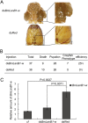
Figure 5. RNAi of BmLanB1-w.
(A) Phenotypes of BmLanB1-w RNAi: wings with blisters were similar to wings in the cf mutant. DsRed control did not show cf phenotype. (B) RNAi statistics. (C) Analysis of BmLanB1-w transcripts. All data are mean ± S.D (n = 3). Statistical analyses were performed using Student’s t-test (n = 3).
Figure 6
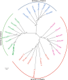
Figure 6. Phylogenetic analysis of laminins in various species.
MEGA 6.0 was used to construct the phylogenetic tree using the Maximum likelihood method. Numbers in the cladogram indicate bootstrap values. Abbreviations, Bm: Bombyx mori, Dm: Drosophila melanogaster, Am: Apis mellifera, Aa: Aedes aegypti, Tc: Tribolium castaneum, Hs: Homo sapiens, Ce: Caenorhabditis elegans. Accession numbers are listed in Table S2.
Figure 7
Figure 7. Spatial expression patterns of laminin genes in wild-type (Dazao).
Quantitative RT-PCR analysis of laminin genes in 12 tissues in the 3rd day of 5th instar. The BmLanB1-w gene had robust expression in the wing discs and weak expression in other tissues. In contrast, the BmLanB1 gene was expressed strongly in the hemocyte and weakly in wing discs and other organs, which was consistent with the expression patterns of laminin α (BmLanA) and laminin γ (BmLanB2) genes. Abbreviations, ASG: anterior silk glands, MSG: middle silk glands, Malpighian: malpighian tubule. The data indicate the mean ± S.D (n = 3).
Figure 8
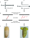
Figure 8. Schematic representation of the mechanism leading to cf phenotype.
(A) Laminin structure in wild-type and cf mutant. In the cf mutant, laminin trimer cannot form due to the defect in BmLanB1-w. (B) Model diagram depicting the adhesion of dorsal and ventral sides of the wings in wild-type and cf. In the cf mutant, dorsal and ventral sides of the wings are not linked because normal basement membrane cannot form due to dysfunctional laminin trimer, which does not allow wings to withstand the hemolymph pressure thus resulting in wing blisters. Light blue oval represents integrins, gray pattern represents laminin trimer, red line indicates proteins such as collagen IV that are linked to the laminin trimer to enable basement membrane assembly. (C) Phenotypes of wild-type and cf.

Similar articles

Cited by

References

    1. Burgeson R. E. et al. A new nomenclature for the laminins. Matrix Biol 14, 209–211 (1994). - PubMed
    1. Timpl R. et al. Laminin—a glycoprotein from basement membranes. J Biol Chem 254, 9933–9937 (1979). - PubMed
    1. Patarroyo M., Tryggvason K. & Virtanen I. Laminin isoforms in tumor invasion, angiogenesis and metastasis. Semin Cancer Biol 12, 197–207 (2002). - PubMed
    1. Adams J. C. & Watt F. M. Regulation of development and differentiation by the extracellular matrix. Development 117, 1183–1198 (1993). - PubMed
    1. Colognato H. & Yurchenco P. D. Form and function: the laminin family of heterotrimers. Dev Dyn 218, 213–234 (2000). - PubMed

Publication types

LinkOut - more resources