. 2015 Nov;135(4):666-73.
doi: 10.1111/jnc.13263. Epub 2015 Aug 25.

Live imaging of endogenous Ca²⁺/calmodulin-dependent protein kinase II in neurons reveals that ischemia-related aggregation does not require kinase activity

Affiliations

Live imaging of endogenous Ca²⁺/calmodulin-dependent protein kinase II in neurons reveals that ischemia-related aggregation does not require kinase activity

Kelsey Barcomb et al. J Neurochem. 2015 Nov.

Abstract

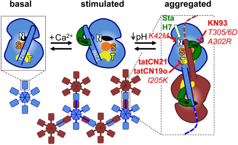
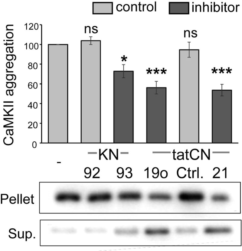
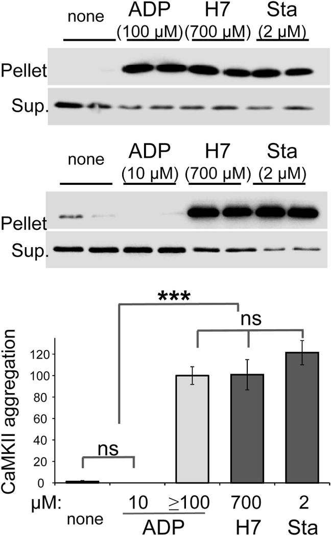
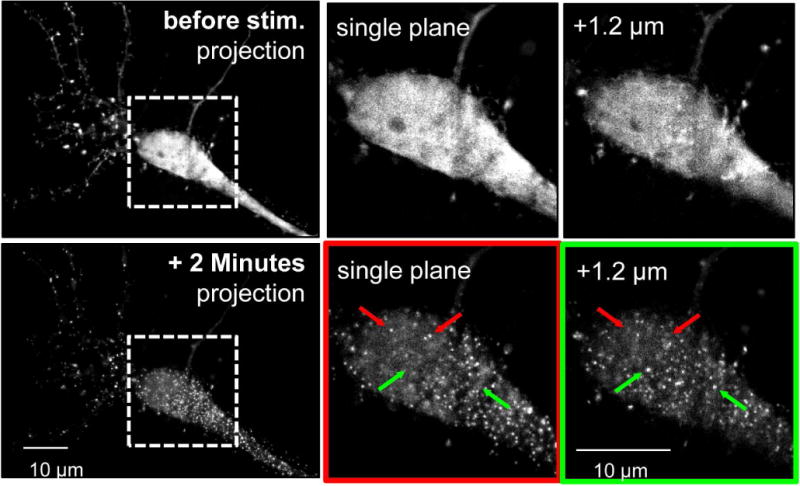
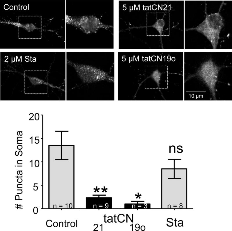
The Ca(2+) /calmodulin-dependent protein kinase II (CaMKII) forms 12meric holoenzymes. These holoenzymes cluster into larger aggregates within neurons under ischemic conditions and in vitro when ischemic conditions are mimicked. This aggregation is thought to be mediated by interaction between the regulatory domain of one kinase subunit with the T-site of another kinase subunit in a different holoenzyme, an interaction that requires stimulation by Ca(2+) /CaM and nucleotide for its induction. This model makes several predictions that were verified here: Aggregation in vitro was reduced by the CaMKII inhibitors tatCN21 and tatCN19o (which block the T-site) as well as by KN93 (which is CaM-competitive). Notably, these and previously tested manipulations that block CaMKII activation all reduced aggregation, suggesting an alternative mechanism that instead requires kinase activity. However, experiments with the nucleotide-competitive broad-spectrum kinase inhibitors staurosporin and H7 showed that this is not the case. In vitro, staurosporine and H7 enabled CaMKII aggregation even in the absence of nucleotide. Within rat hippocampal neurons, an intra-body enabled live monitoring of endogenous CaMKII aggregation. This aggregation was blocked by tatCN21, but not by staurosporine, even though both effectively inhibit CaMKII activity. These results support the mechanistic model for CaMKII aggregation and show that kinase activity is not required. CaMKII aggregation is prevented by inhibiting kinase activity with mutations (red italics; shown previously) or inhibitors (red bold; shown here), indicating requirement of kinase activity. However, we show here that nucleotide-competitive inhibitors (green) allow CaMKII aggregation (including endogenous CaMKII within neurons), demonstrating that kinase activity is not required and supporting the current mechanistic model for CaMKII aggregation.

Keywords: CaMKII; FingR; aggregation; glutamate; hippocampus; ischemia.


Conflict of interest statement

Conflict of Interest Statement: The University of Colorado holds patent rights for inhibitors used in this study (US patent US 8,816,046 B2). K.U.B is the owner of Neurexus Therapeutics, LLC.

Figures

Fig. 1
Fig. 1
The current mechanistic model for aggregation of CaMKII holoenzymes into larger clusters. In the basal state (shown for an individual kinase subunit without depiction of the C-terminal association domain), the regulatory domain blocks the substrate-binding S-site (S, orange) and the neighboring T-site (named for its interaction with the T286 region of the regulatory domain; T, yellow); the nucleotide-binding pocket (N, white) is also indicated. Ca2+/CaM binding replaces the regulatory domain to allow access to the S- and T-sites. Then, the T-site can interact with binding partners such as GluN2B. T-site interaction with the regulatory domain of another kinase subunit additionally requires a drop in pH to ~6.8 or lower and then causes aggregation of multiple 12meric holoenzymes into large aggregates (shown here as hexamers and in two different colors for better visualization of the aggregates). Both aggregation and GluN2B binding additionally require occupation of the nucleotide binding pocket. Both aggregation and GluN2B binding is prevented by inhibitory mutations that prevent nucleotide binding (K42M) or Ca2+/CaM binding (T305/306D) as well as by inhibitors that block the T-site (tatCN21, tatCN19o) or are competitive with Ca2+/CaM (KN93). By contrast, nucleotide competitive inhibitors (staurosporine, H7) block enzymatic kinase activity but can replace the nucleotide function for aggregation and GluN2B binding. The differential inhibitor effects on aggregation were elucidated by the results of this study.
Fig. 2
Fig. 2
CaMKII aggregation in vitro is reduced by the CaMKII inhibitors KN93, tatCN19o and tatCN21. CaMKII aggregation was induced at pH 6.5–6.8 by the addition of 1 mM ADP in the presence of Ca2+/CaM and 10 μM KN93, KN92, 5 μM tatCN19o, tatCtrl, or tatCN21, as indicated (with 10 μM KN compounds or 5 μM tat peptides). Aggregates were separated by centrifugation from soluble kinase, and both pellet and supernatant (Sup.) were analyzed for CaMKII content by Western blot. Aggregation was normalized to ADP-only control. CaMKII inhibitor (dark grey) versus control substance (light grey) conditions are indicated. One-Way ANOVA with post-hoc Tukey’s test indicated that the inhibitors KN93, tatCN19o and tatCN21 significantly reduced aggregation (* p<0.05, *** p<0.001, ns = non-significant as compared to ADP-only control; n=5–6).
Fig. 3
Fig. 3
The nucleotide-competitive inhibitors staurosporine (Sta) and H7 can substitute for nucleotide in inducing CaMKII aggregation in vitro. CaMKII aggregation was induced at pH 6.5–6.8 in the presence of Ca2+/CaM by addition of ADP, H7, or staurosporine at the concentrations indicated. Without addition of inhibitor or nucleotide (none), no aggregation was seen. Aggregates were separated by centrifugation from soluble kinase, and both pellet and supernatant (Sup.) was analyzed for CaMKII content by Western blot. Aggregation was normalized to ADP positive control. One-Way ANOVA with post-hoc Tukey’s test showed that ≥100 μM ADP (n=3), 700 μM H7 (n=4), 2 μM Sta (n=5) equally and significantly induced CaMKII aggregation, compared to either no nucleotide (n=4) or 10 μM ADP (n=2) (*** p<0.001, ns = non-significant).
Fig. 4
Fig. 4
A FingR that enables live imaging of aggregation of endogenous CaMKII within neurons. Primary hippocampal neuron cultures were transfected with a CaMKII FingR at 12 DIV, allowing for visualization of endogenous CaMKII (at 13 DIV). (Top) Basally CaMKII is not aggregated in the soma, as seen in a representative neuron. Images of neurons were collected in z-stacks over 1.8 μm with 0.3 μm between planes. Somal puncta are not seen in a flattened image of all of the planes or in individual planes; top and bottom planes of the boxed in region are shown on the right. (Bottom) Aggregation is clearly seen 2 minutes after glutamate/glycine stimulation (stim.) in the same neuron depicted in the top panel. Arrows in the right panels identify puncta that are present in either the top plane (red) or the bottom plane (green) but not both. The presence of unique puncta in individual planes throughout the z-stacks shows that they are cytoplasmic rather than membrane associated.
Fig. 5
Fig. 5
CaMKII aggregation within neurons does not require enzymatic kinase activity. CaMKII FingR transfected neurons were imaged in the presence of tatCN21 (5 μM; n=9), tatCN19o (5 μM; n=3), staurosporine (Sta; 2 μM; n=8), or no inhibitor (control; n=10). The top panels show representative images of cells in all four conditions 2 minutes after glutamate/glycine stimulation. Puncta – indicating aggregation – can clearly be seen in the control and staurosporine conditions, but not in either of the tat-peptide conditions. The images were quantified by counting the number of somal puncta, as defined by objects greater than 2 pixels in size whose intensity was greater than 3 standard deviations from the mean somal intensity at 2 minutes. This value was significantly less than control for tatCN19o and tatCN21, as determined by a One-Way ANOVA with post-hoc Tukey’s test (* p<0.05, ** p<0.01, ns = non-significant as compared to control).

References

    1. Aarts MM, Tymianski M. Molecular mechanisms underlying specificity of excitotoxic signaling in neurons. Curr Mol Med. 2004;4:137–147. - PubMed
    1. Ashpole NM, Hudmon A. Excitotoxic neuroprotection and vulnerability with CaMKII inhibition. Mol Cell Neurosci. 2011;46:720–730. - PubMed
    1. Barcomb K, Buard I, Coultrap SJ, Kulbe JR, O’Leary H, Benke TA, Bayer KU. Autonomous CaMKII requires further stimulation by Ca2+/calmodulin for enhancing synaptic strength. FASEB J. 2014;28:3810–3819. - PMC - PubMed
    1. Barcomb K, Coultrap SJ, Bayer KU. Enzymatic Activity of CaMKII Is Not Required for Its Interaction with the Glutamate Receptor Subunit GluN2B. Mol Pharmacol. 2013;84:834–843. - PMC - PubMed
    1. Bayer KU, De Koninck P, Leonard AS, Hell JW, Schulman H. Interaction with the NMDA receptor locks CaMKII in an active conformation. Nature. 2001;411:801–805. - PubMed

Publication types

MeSH terms