

Hepatocyte-Specific Expression of Human Lysosome Acid Lipase Corrects Liver Inflammation and Tumor Metastasis in lal(-/-) Mice
- PMID: 26212911
- PMCID: PMC4597280
- DOI: 10.1016/j.ajpath.2015.05.021
Hepatocyte-Specific Expression of Human Lysosome Acid Lipase Corrects Liver Inflammation and Tumor Metastasis in lal(-/-) Mice
Abstract
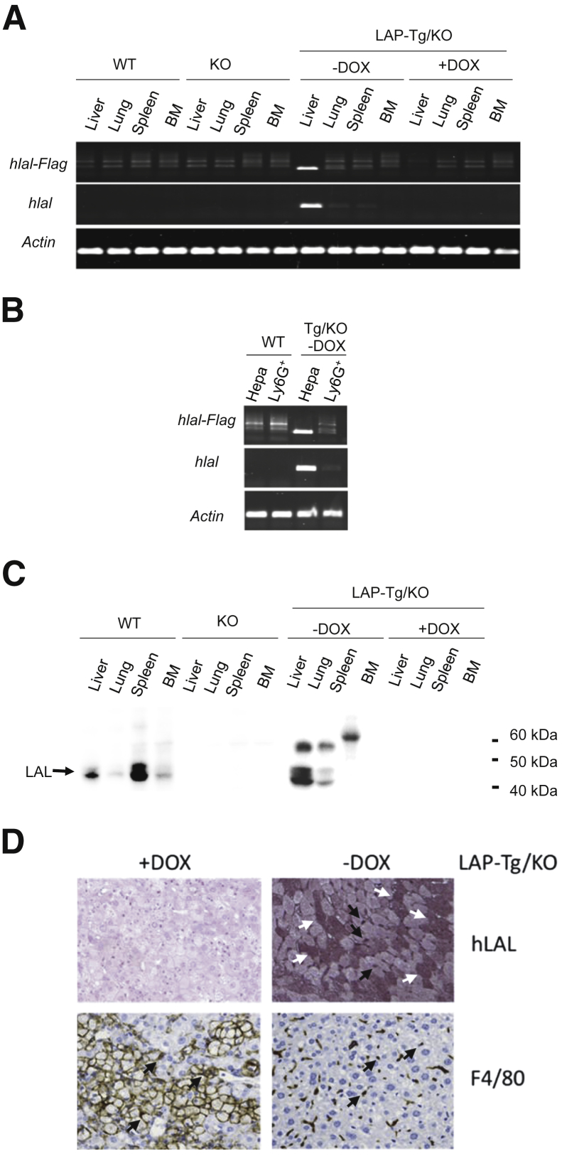
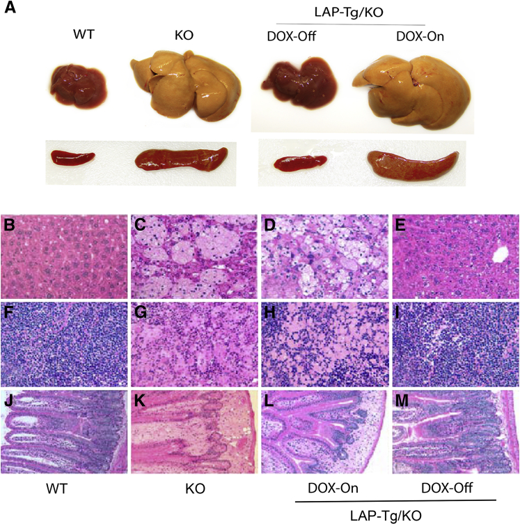
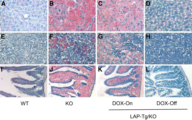
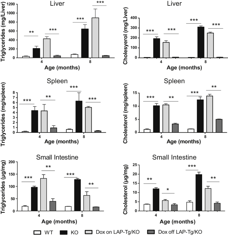
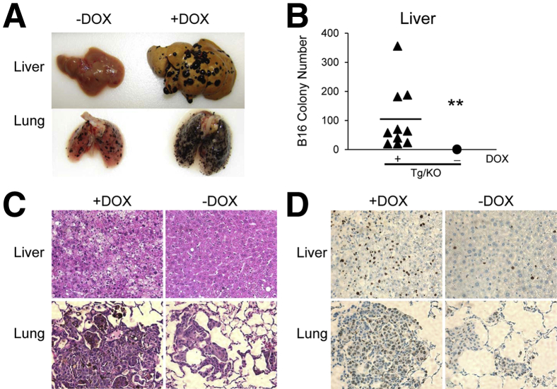
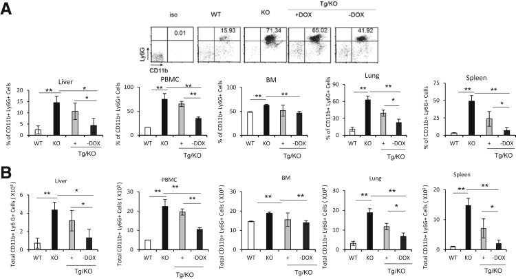
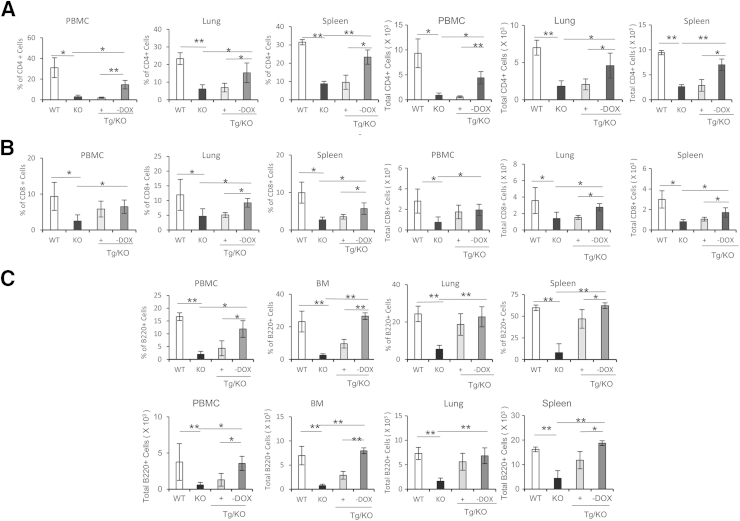
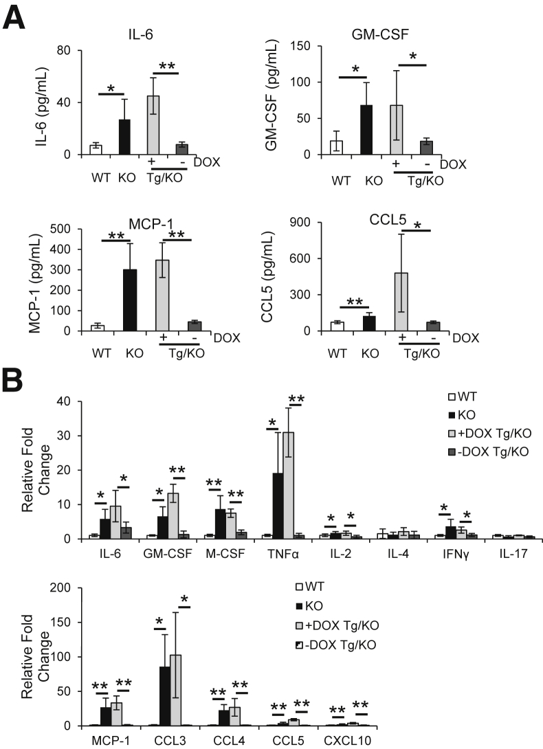
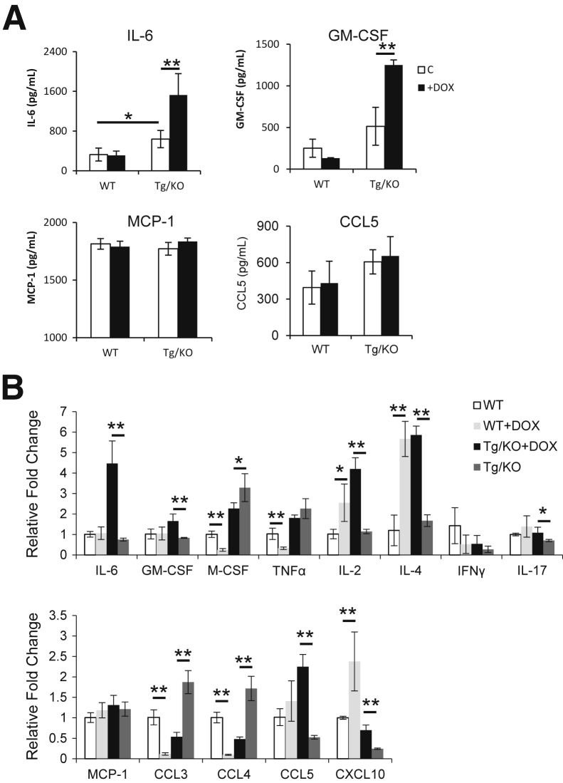
The liver is a major organ for lipid synthesis and metabolism. Deficiency of lysosomal acid lipase (LAL; official name Lipa, encoded by Lipa) in mice (lal(-/-)) results in enlarged liver size due to neutral lipid storage in hepatocytes and Kupffer cells. To test the functional role of LAL in hepatocyte, hepatocyte-specific expression of human LAL (hLAL) in lal(-/-) mice was established by cross-breeding of liver-activated promoter (LAP)-driven tTA transgene and (tetO)7-CMV-hLAL transgene with lal(-/-) knockout (KO) (LAP-Tg/KO) triple mice. Hepatocyte-specific expression of hLAL in LAP-Tg/KO triple mice reduced the liver size to the normal level by decreasing lipid storage in both hepatocytes and Kupffer cells. hLAL expression reduced tumor-promoting myeloid-derived suppressive cells in the liver of lal(-/-) mice. As a result, B16 melanoma metastasis to the liver was almost completely blocked. Expression and secretion of multiple tumor-promoting cytokines or chemokines in the liver were also significantly reduced. Because hLAL is a secretory protein, lal(-/-) phenotypes in other compartments (eg, blood, spleen, and lung) also ameliorated, including systemic reduction of myeloid-derived suppressive cells, an increase in CD4(+) and CD8(+) T and B lymphocytes, and reduced B16 melanoma metastasis in the lung. These results support a concept that LAL in hepatocytes is a critical metabolic enzyme in controlling neutral lipid metabolism, liver homeostasis, immune response, and tumor metastasis.
Copyright © 2015 American Society for Investigative Pathology. Published by Elsevier Inc. All rights reserved.
Figures
Similar articles
-
Hepatocyte-specific deletion of lysosomal acid lipase leads to cholesteryl ester but not triglyceride or retinyl ester accumulation.J Biol Chem. 2019 Jun 7;294(23):9118-9133. doi: 10.1074/jbc.RA118.007201. Epub 2019 Apr 25. J Biol Chem. 2019. PMID: 31023823 Free PMC article.
-
Lung Epithelial Cell-Specific Expression of Human Lysosomal Acid Lipase Ameliorates Lung Inflammation and Tumor Metastasis in Lipa(-/-) Mice.Am J Pathol. 2016 Aug;186(8):2183-2192. doi: 10.1016/j.ajpath.2016.04.014. Epub 2016 Jul 20. Am J Pathol. 2016. PMID: 27461363 Free PMC article.
-
Hepatocyte-specific lysosomal acid lipase deficiency protects mice from diet-induced obesity but promotes hepatic inflammation.Biochim Biophys Acta Mol Cell Biol Lipids. 2019 Apr;1864(4):500-511. doi: 10.1016/j.bbalip.2019.01.007. Epub 2019 Jan 9. Biochim Biophys Acta Mol Cell Biol Lipids. 2019. PMID: 30639734 Free PMC article.
-
Lysosomal Acid Lipase in Lipid Metabolism and Beyond.Arterioscler Thromb Vasc Biol. 2019 May;39(5):850-856. doi: 10.1161/ATVBAHA.119.312136. Arterioscler Thromb Vasc Biol. 2019. PMID: 30866656 Free PMC article. Review.
-
Lysosomal acid lipase and lipid metabolism: new mechanisms, new questions, and new therapies.Curr Opin Lipidol. 2018 Jun;29(3):218-223. doi: 10.1097/MOL.0000000000000507. Curr Opin Lipidol. 2018. PMID: 29547398 Free PMC article. Review.
Cited by
-
Role of the RAB7 Protein in Tumor Progression and Cisplatin Chemoresistance.Cancers (Basel). 2019 Aug 1;11(8):1096. doi: 10.3390/cancers11081096. Cancers (Basel). 2019. PMID: 31374919 Free PMC article. Review.
-
Hepatocyte-specific deletion of lysosomal acid lipase leads to cholesteryl ester but not triglyceride or retinyl ester accumulation.J Biol Chem. 2019 Jun 7;294(23):9118-9133. doi: 10.1074/jbc.RA118.007201. Epub 2019 Apr 25. J Biol Chem. 2019. PMID: 31023823 Free PMC article.
-
The Double-Edge Sword of Autophagy in Cancer: From Tumor Suppression to Pro-tumor Activity.Front Oncol. 2020 Oct 7;10:578418. doi: 10.3389/fonc.2020.578418. eCollection 2020. Front Oncol. 2020. PMID: 33117715 Free PMC article. Review.
-
Emerging role of lipophagy in liver disorders.Mol Cell Biochem. 2024 Jan;479(1):1-11. doi: 10.1007/s11010-023-04707-1. Epub 2023 Mar 21. Mol Cell Biochem. 2024. PMID: 36943663 Review.
-
Targeting the lysosome: Mechanisms and treatments for nonalcoholic fatty liver disease.J Cell Biochem. 2022 Oct;123(10):1624-1633. doi: 10.1002/jcb.30274. Epub 2022 May 23. J Cell Biochem. 2022. PMID: 35605052 Free PMC article. Review.
References
-
- Grabowski G.A., Du H., Charnas L. Lysosomal acid lipase deficiencies: the wolman disease/cholesteryl ester storage disease spectrum. In: Valle D., Voglstein B., Kinzler K.W., Antonarakis S.E., Ballabio A., editors. The Online Metabolic and Molecular Bases of Inherited Disease (OMMBID) ed 9. McGraw-Hill; New York: 2012.
-
- Assmann G., Seedorf U. 8th Edition. McGraw-Hill; New York: 2001. Metabolic and Molecular Bases of Inherited Diseases.
-
- Yan C., Du H. Lysosomal acid lipase is critical for myeloid-derived suppressive cell differentiation, development, and homeostasis. World J Immunol. 2014;4:42–51.
-
- Beaudet A.L., Ferry G.D., Nichols B.L., Jr., Rosenberg H.S. Cholesterol ester storage disease: clinical, biochemical, and pathological studies. J Pediatr. 1977;90:910–914. - PubMed
-
- Bernstein D.L., Hulkova H., Bialer M.G., Desnick R.J. Cholesteryl ester storage disease: review of the findings in 135 reported patients with an underdiagnosed disease. J Hepatol. 2013;58:1230–1243. - PubMed
Publication types
MeSH terms
Substances
Grants and funding
LinkOut - more resources
Full Text Sources
Other Literature Sources
Medical
Molecular Biology Databases
Research Materials
Miscellaneous
