. 2015 Jul 30;162(3):593-606.
doi: 10.1016/j.cell.2015.06.056. Epub 2015 Jul 23.

β-Neurexins Control Neural Circuits by Regulating Synaptic Endocannabinoid Signaling

Affiliations

β-Neurexins Control Neural Circuits by Regulating Synaptic Endocannabinoid Signaling

Garret R Anderson et al. Cell. .

Abstract

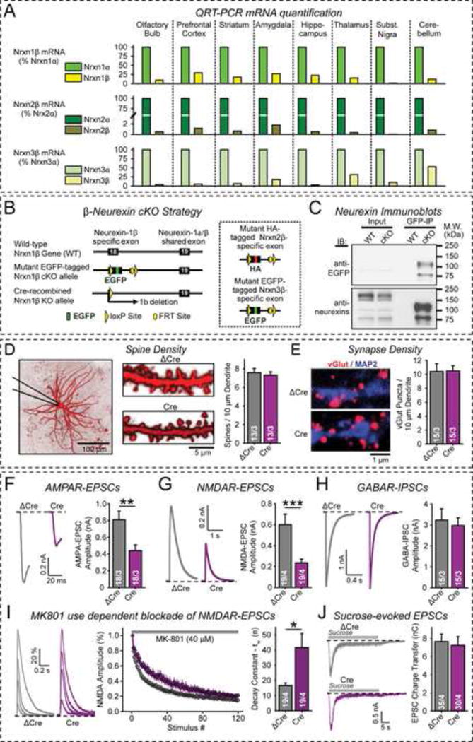
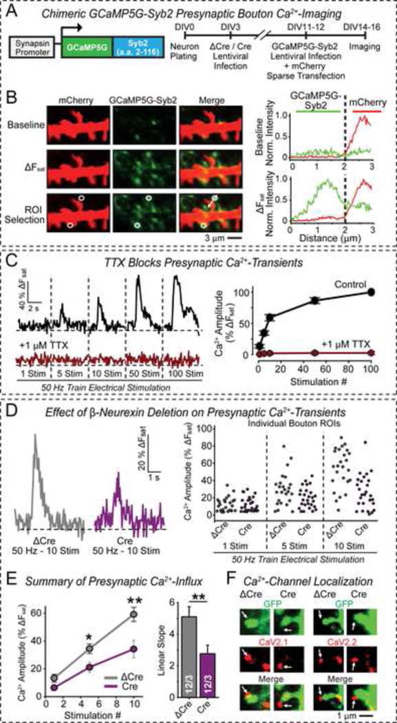
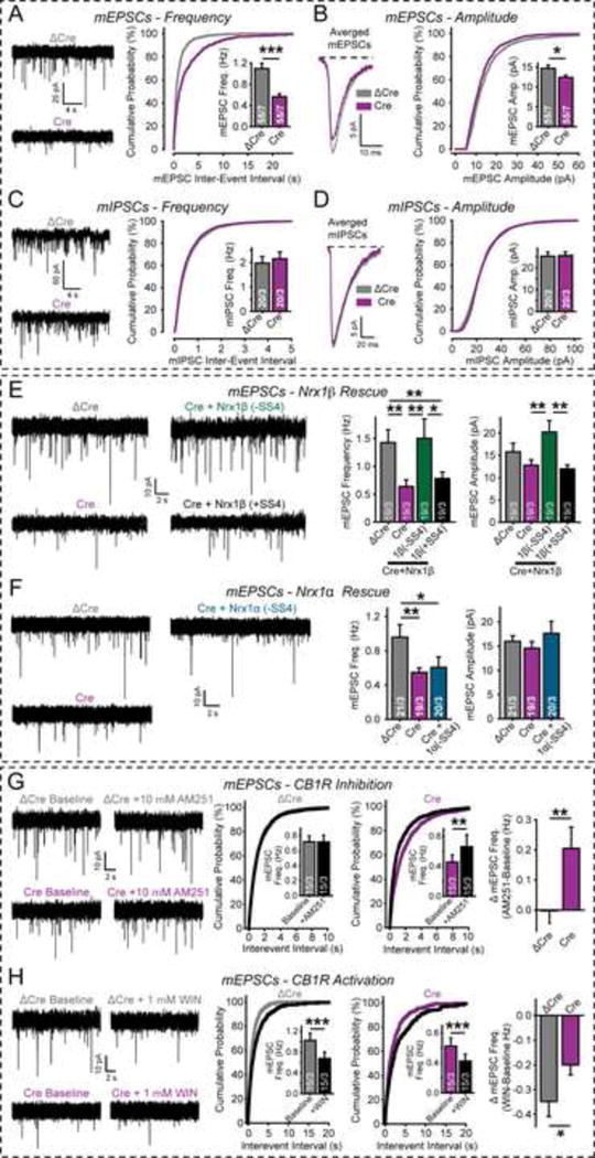
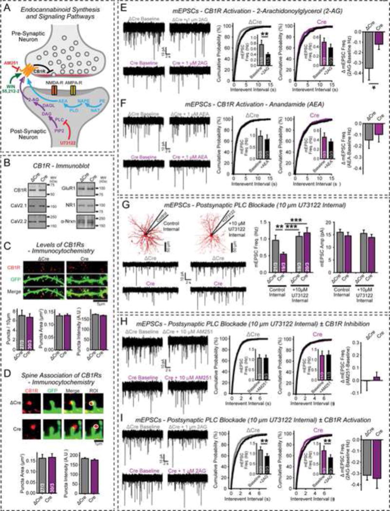
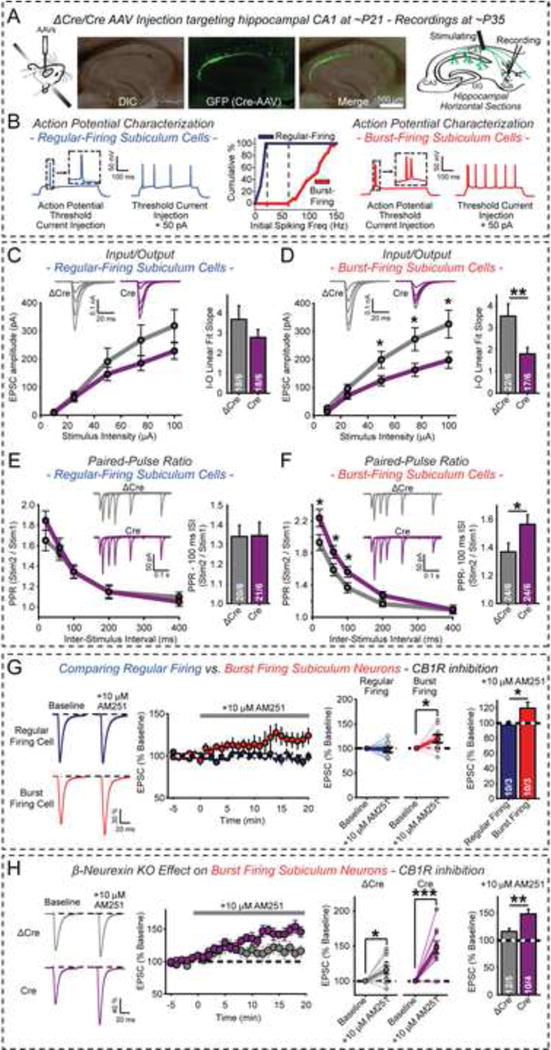
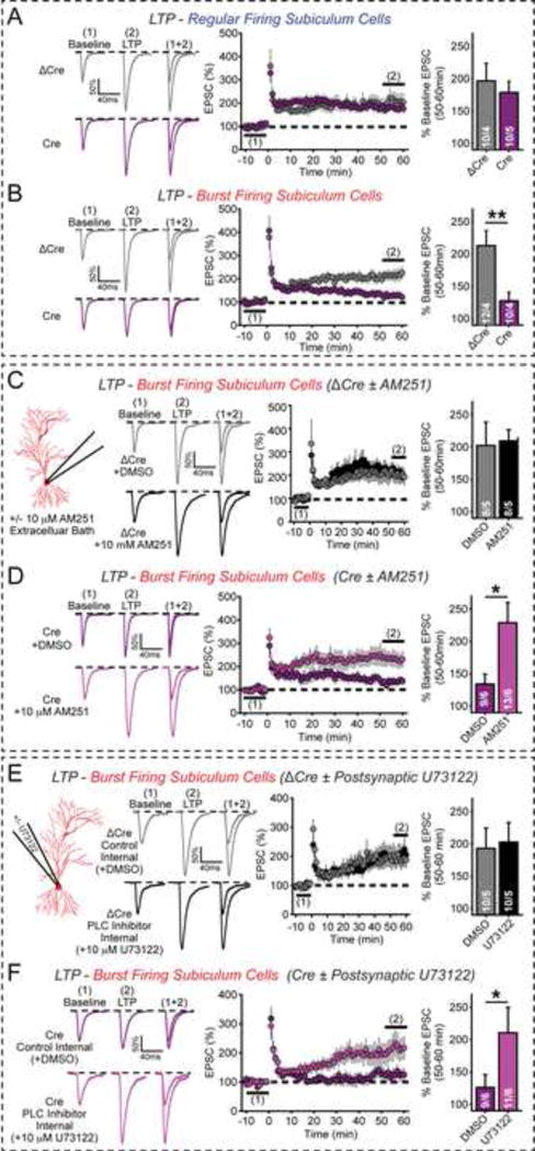
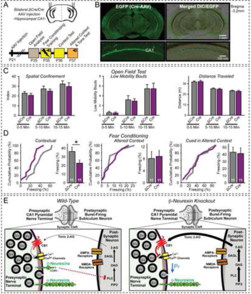
α- and β-neurexins are presynaptic cell-adhesion molecules implicated in autism and schizophrenia. We find that, although β-neurexins are expressed at much lower levels than α-neurexins, conditional knockout of β-neurexins with continued expression of α-neurexins dramatically decreased neurotransmitter release at excitatory synapses in cultured cortical neurons. The β-neurexin knockout phenotype was attenuated by CB1-receptor inhibition, which blocks presynaptic endocannabinoid signaling, or by 2-arachidonoylglycerol synthesis inhibition, which impairs postsynaptic endocannabinoid release. In synapses formed by CA1-region pyramidal neurons onto burst-firing subiculum neurons, presynaptic in vivo knockout of β-neurexins aggravated endocannabinoid-mediated inhibition of synaptic transmission and blocked LTP; presynaptic CB1-receptor antagonists or postsynaptic 2-arachidonoylglycerol synthesis inhibition again reversed this block. Moreover, conditional knockout of β-neurexins in CA1-region neurons impaired contextual fear memories. Thus, our data suggest that presynaptic β-neurexins control synaptic strength in excitatory synapses by regulating postsynaptic 2-arachidonoylglycerol synthesis, revealing an unexpected role for β-neurexins in the endocannabinoid-dependent regulation of neural circuits.


Figures

Figure 1
Figure 1. Conditional KO of β-neurexins impairs excitatory synaptic transmission
(A) β-Neurexins are expressed ~10–100 fold lower levels than α-neurexins. Data show relative α- and β-neurexin mRNA levels measured by quantitative RT-PCR (n=3 mice at P30). (B) Conditional KO (cKO) strategy for neurexin-1β (left) and neurexin-2β and -3β (right dashed box). All cKOs involve floxing the 5′ β-neurexin-specific exon and adding an N-terminal epitope tag (EGFP for neurexin-1β and -3β; HA-tag for neurexin-2β). (C) Immunoblots of tagged β-neurexins in cKO mice with antibodies to GFP (top) and to a conserved C-terminal neurexin epitope (bottom). Proteins in cortex homogenates (from 3-week old control and triple β-neurexin cKO mice; input) were immunoprecipitated with GFP antibodies (GFP-IP) to visualize the low-abundance β-neurexins in cKO mouse samples. (D) β-Neurexin KO does not alter morphological parameters in cultured cortical neurons. Left, representative images of neurons filled with Alexa Fluor 594 via the patch pipette; right, summary graphs of spine density. (E) β-Neurexin KO does not impair excitatory synapse density and size. Left, representative images; right, summary graphs of vGlut1-positive synapse density. (F–H) β-Neurexin KO in cultured cortical neurons impairs excitatory but not inhibitory synaptic transmission evoked by isolated action potentials (F, AMPAR-mediated EPSCs; G, NMDAR-mediated EPSCs; H, GABAR-mediated IPSCs). (I) β-Neurexin KO decreases the presynaptic release probability, measured via the MK-801 induced progressive block of NMDAR-mediated synaptic responses. Left, representative EPSC traces for the 1st, 10th, 25th, and 125th stimulus; center, mean ESPC amplitudes; right, summary graphs of decay constants. (J) β-Neurexin KO does not alter the readily-releasable vesicle pool as analyzed by stimulation with 0.5 M sucrose. Left, representative traces; right, total charge transfer. Data in D–J are means ± SEM (numbers of neurons/independent cultures examined are shown in graphs). Statistical analyses were performed by Student’s t-test (*p<0.05, **p<0.01, ***p<0.001). For additional data, see Figs. S1–S4.
Figure 2
Figure 2. Conditional KO of β-neurexins impairs presynaptic Ca2+-influx
(A) Design of Ca2+-imaging experiments. Left, schematic of the GCaMP5G-Syb2 fusion protein that acts as a presynaptic Ca2+-probe; right, flow diagram of experiments. (B) Representative Ca2+-imaging experiments. Left, sample images of presynaptic boutons containing GCaMP5G-Syb2 (green) that contact mCherry-containing dendritic spines (red; images were obtained before and after maximal stimulation to saturate Ca2+-transients [(ΔFsat]; white circles = regions of interest [ROIs] for quantitative analysis by line scans [arrows]). Right, summary graphs of line scans through ROIs at before (top) and after maximal stimulation (ΔFsat; bottom; green = presynaptic Ca2+-concentration; red = postsynaptic mCherry signal). (C) Presynaptic Ca2+-transients saturate after 20 stimuli and are blocked by TTX. Left, representative Ca2+-transients; right, summary graphs (-TTX, n=12 neurons; +TTX, n=2 neurons). (D) β-Neurexin KO impairs action potential-induced presynaptic Ca2+-influx in cultured cortical neurons (ΔCre = control; Cre = KO). Left, representative fluorescence traces of 10 stimuli applied at 50 Hz; right, scatter plots of individual bouton responses to 1, 5, and 10 stimuli. (E) Summary plot of mean Ca2+-transients after 1, 5, and 10 stimuli. Left, n=3 independent experiments with 2–4 boutons per neuron; right, summary graph of the mean linear slopes fitted through the 1, 5, 10 stimuli plots. (F) β-Neurexin KO does not alter levels or localization of presynaptic Ca2+-channels (representative images of cortical pyramidal neuron spines with sparse GFP expression that are stained for presynaptic P/Q-type (CaV2.1) or N-type (CaV2.2) Ca2+-channels). For quantitative analyses, see Figs. S4F–S4H. Data are means ± SEM. Statistical analysis was performed by Student’s t-test (*p<0.05, **p<0.01).
Figure 3
Figure 3. Neurexin-1β, but not neurexin-1α, and the CB1R antagonist AM251 rescue impaired spontaneous mini release in β-neurexin KO neurons
(A–D) β-Neurexin KO impairs mini release at excitatory but not inhibitory synapses (A & B, mESPCs; C & D, mIPSCs) in cortical neurons from triple β-neurexin cKO mice expressing inactive (ΔCre) or active Cre-recombinase (Cre). A & C, representative traces (left), cumulative distributions of inter-event intervals (right), and mean event frequencies (inset); B & D, average individual events (left), cumulative distributions of event amplitudes (right), and mean event amplitudes (inset). (E) Neurexin-1β without an insert in SS#4 (−SS4), but not with an insert (+SS4), rescues the decreased mEPSC frequency in β-neurexin KO neurons. (F) Neurexin-1α without an insert in SS#4 (−SS4) fails to rescue the decreased mEPSC frequency in β-neurexin KO neurons. (G) Blocking CB1Rs with AM251 reverses the decrease in mEPSC frequency in β-neurexin KO neurons. Left, representative traces; center, cumulative distributions of event frequencies with insets showing mean frequencies; right, summary graphs of AM251-induced changes. (H) Activating CB1Rs with WIN (WIN 55,212–2 mesylate) depresses mini release significantly more in control (ΔCre) than in β-neurexin KO neurons (Cre). Figure design is analogous to that of G. Data are means ± SEM; numbers of neurons/independent cultures examined are shown in the graphs. Statistical analyses were performed using Student’s t-test (*p<0.05, **p<0.01, ***p<0.001). For additional data, see Figs. S4 and S5.
Figure 4
Figure 4. Blocking postsynaptic 2-AG synthesis rescues presynaptic β-neurexin KO phenotype in cultured neurons
(A) Diagram of endocannabinoid signaling pathways. Two distinct endocannabinoids (2-arachidonoylglycerol [2-AG] and anandamide [AEA, N-arachidonoylethanolamine]) are synthesized by different postsynaptic enzymes; both act on presynaptic CB1Rs (abbreviations: PLC, phospholipase C; PIP2, phosphatidylinositol 4,5-bisphosphate; DAG, diacylglycerol; DAGL, diacylglycerol lipase; NAT, N-acyltransferase; PE, phosphatidylethanolamine; NAPE, N-arachidonoyl phosphatidylethanolamine; PLD, phospholipase D). Modes of action of the 2-AG synthesis inhibitor U73122 and the CB1R agonist WIN and antagonist AM251 are indicated. (B–D) CB1R levels are similar in cortical neurons from triple β-neurexin cKO mice expressing inactive (ΔCre) or active Cre-recombinase (Cre). B, representative immunoblots with antibodies to CB1R, CaV2.1 and CaV2.2 Ca2+-channels, GluR1, NR1 (an NMDAR subunit), and α-neurexins; C & D, immunocytochemistry quantifications of CB1R levels (C) and CB1R localization (D); note that neurons were sparsely transfected with EGFP for visualization of neuronal morphology. (E & F) 2-AG causes a larger depression of mini release in control than in β-neurexin KO neurons (E), whereas anandamide is ineffective likely because it is only a partial agonist (F). Left, representative traces; center, cumulative distributions of mEPSC inter-event intervals (insets = mean frequencies); right, summary graphs of anandamide- or 2-AG-induced changes. (G) Selective postsynaptic block of 2-AG synthesis by U73122 in the patch pipette rescues decreased mini release in β-neurexin KO neurons. Left, experimental set-up and representative mEPSC traces; right, bar diagrams of mEPSC frequencies and amplitudes. (H & I) Selective postsynaptic block of 2-AG synthesis by U73122 prevents CB1R activation in β-neurexin KO neurons as measured by blocking CB1Rs with bath applied AM251 (H) or activating CB1Rs with bath-applied 2-AG (I). Left, representative traces; center, cumulative distributions of mEPSC inter-event intervals (insets = mean frequencies); right, summary graphs of AM251- or 2-AG-induced changes in mEPSC frequencies. Data are means ± SEM; numbers of neurons/independent cultures examined are shown in graphs. Statistical analyses were with Student’s t-test (*p<0.05, **p<0.01, ***p<0.001).
Figure 5
Figure 5. Presynaptic KO of β-neurexins in CA1 pyramidal neurons decreases synaptic strength at burst-firing neuron synapses in the subiculum
(A) Experimental design. Left, diagram of stereotactic injections into the CA1 region; center, representative images of slices from stereotactically injected mice at P35 to visualize AAV infections (slices with <90% EGFP expression in the CA1 region were rejected); right, electrophysiological recording configuration in acute subiculum slices (Aoto et al., 2013). (B) Identification of regular- and burst-firing pyramidal subiculum neurons in current-clamp mode. Left and right, representative traces; center, summary graph of the initial spiking frequency. (C & D) Input/output (I/O) relations of AMPAR-mediated EPSCs elicited by stimulation of CA1-derived axons and recorded in regular- (C) or burst-firing subiculum neurons (D). Left, summary plots with representative traces on top; right, summary graphs of fitted linear input/output slopes. (E & F) Paired-pulse ratio (PPR) measurements of AMPAR EPSCs in regular- (E) and burst-firing subiculum neurons (F). Left, summary plots of PPRs vs. inter-stimulus intervals with representative traces on top; right, summary graphs of PPRs at 100 ms inter-stimulus intervals. (G) Only burst-firing but not regular-firing subiculum neurons exhibit tonic endocannabinoid signaling in wild-type mice. EPSCs were elicited at 0.1 Hz before and after bath-application of the CB1R antagonist AM251. Left, representative EPSC traces; center, plots of relative EPSC amplitudes and AM251-induced EPSC amplitude changes in individual neurons; right, summary graphs of AM251-induced EPSC amplitude changes. (H) Presynaptic β-neurexin KO increases tonic endocannabinoid signaling in burst-firing subiculum neurons. Experiments were performed as in G, except that burst-firing neurons were analyzed in slices from β-neurexin cKO mice with presynaptic expression of ΔCre- or Cre-EGFP in the CA1 region (see A). Data are means ± SEM; numbers of neurons/mice examined are shown in the summary graphs. Statistical analysis was by paired Student’s t-test for single cell plots, and unpaired Student’s t-test for comparisons in other summary graphs (*p<0.05, **p<0.01, ***p<0.001).
Figure 6
Figure 6. Presynaptic KO of β-neurexins selectively impairs LTP at burst-firing subiculum neurons by enhancing basal endocannabinoid activity
(A) KO of β-neurexins does not change LTP of CA1 EPSCs onto regular-firing subiculum neurons. LTP was induced by 4 × 100 Hz/1 s stimulation with 10 s intervals in current-clamp mode at resting potential in acute slices from CA1-region specific β-neurexin KO mice obtained as described in Fig. 5A. Left, representative traces; center, average EPSC amplitudes (1 min bins); right, summary graphs of mean LTP magnitude 50–60 min after induction. (B) Same as (A), except that burst-firing subiculum neurons were analyzed. (C & D) Same as (B), except that the effect of the CB1R antagonist AM251 on LTP was examined in slices from mice injected with inactive Cre-recombinase (C) or with active Cre-recombinase (D). (E & F) Same as (C & D), except that the effect of the phospholipase C inhibitor U73122 introduced into the postsynaptic neuron via the patch pipette was examined. Data shown are means ± SEM; numbers of neurons/mice examined are shown in the graphs. Statistical analysis was performed by Student’s t-test (* p<0.05, **p<0.01).
Figure 7
Figure 7. Conditional KO of β-neurexins in the hippocampal CA1 region impairs contextual memory: Model for β-neurexin action
(A) Design of behavioral experiments (after Xu et al., 2012). (B) Representative coronal images illustrating expression of Cre-EGFP in the CA1 region of the hippocampus after stereotactic injection (top) and zoomed CA1 image (bottom). (C & D) Analysis of ΔCre- or Cre-injected mice in open field (C) and fear-conditioning tests (D). Open field behavior (analyzed in three 5 min segments) was quantified as spatial confinement (C, left), low mobility bouts (C, center), and total distance traveled (C, right). Fear-conditioning training exposed mice to three 30 s tones ending with 2 s electrical footshocks separated by 1 min intervals (D; left graphs, cumulative distributions; right summary graphs, mean fear-conditioning memory as measured by freezing). Data are means ± SEM; numbers of mice examined are shown in the graphs. Statistical analysis was performed by Student’s t-test (* p<0.05). (E) Model for β-neurexin action. In wild-type excitatory synapses (left), presynaptic β-neurexins regulate endocannabinoid signaling by controlling postsynaptic 2-AG synthesis, possibly via trans-synaptic interaction with postsynaptic neuroligin isoforms that exclusively bind to β- but not α-neurexins lacking an insert in SS#4. In excitatory β-neurexin KO synapses (right), 2-AG synthesis is disinhibited, CB1Rs are activated, and synaptic strength is decreased; moreover, in burst-firing subiculum neurons LTP is blocked which may be responsible for the impairment in contextual memory.

Comment in

References

    1. Aoto J, Martinelli DC, Malenka RC, Tabuchi K, Südhof TC. Presynaptic neurexin-3 alternative splicing trans-synaptically controls postsynaptic AMPA receptor trafficking. Cell. 2013;154:75–88. - PMC - PubMed
    1. Bang ML, Owczarek S. A matter of balance: role of neurexin and neuroligin at the synapse. Neurochemical research. 2013;38:1174–1189. - PubMed
    1. Behr J, Wozny C, Fidzinski P, Schmitz D. Synaptic plasticity in the subiculum. Prog Neurobiol. 2009;89:334–342. - PubMed
    1. Boucard AA, Chubykin AA, Comoletti D, Taylor P, Südhof TC. A splice code for trans-synaptic cell adhesion mediated by binding of neuroligin 1 to alpha- and beta-neurexins. Neuron. 2005;48:229–236. - PubMed
    1. Brown SP, Safo PK, Regehr WG. Endocannabinoids inhibit transmission at granule cell to Purkinje cell synapses by modulating three types of presynaptic calcium channels. J Neurosci. 2004;24:5623–5631. - PMC - PubMed

Publication types