Micropatterning Alginate Substrates for in vitro Cardiovascular Muscle on a Chip
- PMID: 26213529
- PMCID: PMC4511503
- DOI: 10.1002/adfm.201203319
Micropatterning Alginate Substrates for in vitro Cardiovascular Muscle on a Chip
Abstract
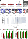
Soft hydrogels such as alginate are ideal substrates for building muscle in vitro because they have structural and mechanical properties close to the in vivo extracellular matrix (ECM) network. However, hydrogels are generally not amenable to protein adhesion and patterning. Moreover, muscle structures and their underlying ECM are highly anisotropic, and it is imperative that in vitro models recapitulate the structural anisotropy in reconstructed tissues for in vivo relevance due to the tight coupling between sturcture and function in these systems. We present two techniques to create chemical and structural heterogeneities within soft alginate substrates and employ them to engineer anisotropic muscle monolayers: (i) microcontact printing lines of extracellular matrix proteins on flat alginate substrates to guide cellular processes with chemical cues, and (ii) micromolding of alginate surface into grooves and ridges to guide cellular processes with topographical cues. Neonatal rat ventricular myocytes as well as human umbilical artery vascular smooth muscle cells successfully attach to both these micropatterned substrates leading to subsequent formation of anisotropic striated and smooth muscle tissues. Muscular thin film cantilevers cut from these constructs are then employed for functional characterization of engineered muscular tissues. Thus, micropatterned alginate is an ideal substrate for in vitro models of muscle tissue because it facilitates recapitulation of the anisotropic architecture of muscle, mimics the mechanical properties of the ECM microenvironment, and is amenable to evaluation of functional contractile properties.
Keywords: Biomedical Applications; Hydrogels; Microcontact Printing; Surface Modification; Tissue Engineering.
Figures


Similar articles
-
Microfluidic Genipin Deposition Technique for Extended Culture of Micropatterned Vascular Muscular Thin Films.J Vis Exp. 2015 Jun 26;(100):e52971. doi: 10.3791/52971. J Vis Exp. 2015. PMID: 26168271 Free PMC article.
-
Muscle on a chip: in vitro contractility assays for smooth and striated muscle.J Pharmacol Toxicol Methods. 2012 May-Jun;65(3):126-35. doi: 10.1016/j.vascn.2012.04.001. Epub 2012 Apr 12. J Pharmacol Toxicol Methods. 2012. PMID: 22521339 Free PMC article.
-
Micropatterned conductive hydrogels as multifunctional muscle-mimicking biomaterials: Graphene-incorporated hydrogels directly patterned with femtosecond laser ablation.Acta Biomater. 2019 Oct 1;97:141-153. doi: 10.1016/j.actbio.2019.07.044. Epub 2019 Jul 26. Acta Biomater. 2019. PMID: 31352108
-
Extracellular Matrix-Derived Hydrogels as Biomaterial for Different Skeletal Muscle Tissue Replacements.Materials (Basel). 2020 May 29;13(11):2483. doi: 10.3390/ma13112483. Materials (Basel). 2020. PMID: 32486040 Free PMC article. Review.
-
Design of Advanced Polymeric Hydrogels for Tissue Regenerative Medicine: Oxygen-Controllable Hydrogel Materials.Adv Exp Med Biol. 2020;1250:63-78. doi: 10.1007/978-981-15-3262-7_5. Adv Exp Med Biol. 2020. PMID: 32601938 Review.
Cited by
-
3D Bioprinted Spheroidal Droplets for Engineering the Heterocellular Coupling between Cardiomyocytes and Cardiac Fibroblasts.Cyborg Bionic Syst. 2021;2021:9864212. doi: 10.34133/2021/9864212. Epub 2021 Dec 28. Cyborg Bionic Syst. 2021. PMID: 35795473 Free PMC article.
-
Recapitulation of microtissue models connected with real-time readout systems via 3D printing technology.J Thorac Dis. 2017 Feb;9(2):233-236. doi: 10.21037/jtd.2017.02.33. J Thorac Dis. 2017. PMID: 28275467 Free PMC article. No abstract available.
-
Advancement of Sensor Integrated Organ-on-Chip Devices.Sensors (Basel). 2021 Feb 15;21(4):1367. doi: 10.3390/s21041367. Sensors (Basel). 2021. PMID: 33671996 Free PMC article. Review.
-
Dynamic display of bioactivity through host-guest chemistry.Angew Chem Int Ed Engl. 2013 Nov 11;52(46):12077-80. doi: 10.1002/anie.201306278. Epub 2013 Sep 24. Angew Chem Int Ed Engl. 2013. PMID: 24108659 Free PMC article. No abstract available.
-
Microfabrication of liver and heart tissues for drug development.Philos Trans R Soc Lond B Biol Sci. 2018 Jul 5;373(1750):20170225. doi: 10.1098/rstb.2017.0225. Philos Trans R Soc Lond B Biol Sci. 2018. PMID: 29786560 Free PMC article. Review.
References
Grants and funding
LinkOut - more resources
Full Text Sources
Other Literature Sources
