. 2014 Nov 26;24(44):7053-7062.
doi: 10.1002/adfm.201401483.

Delivery of iPS-NPCs to the Stroke Cavity within a Hyaluronic Acid Matrix Promotes the Differentiation of Transplanted Cells

Affiliations

Delivery of iPS-NPCs to the Stroke Cavity within a Hyaluronic Acid Matrix Promotes the Differentiation of Transplanted Cells

Jonathan Lam et al. Adv Funct Mater. .

Abstract

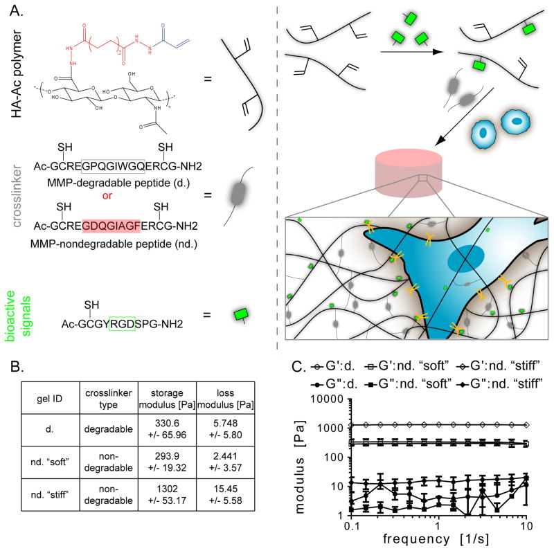
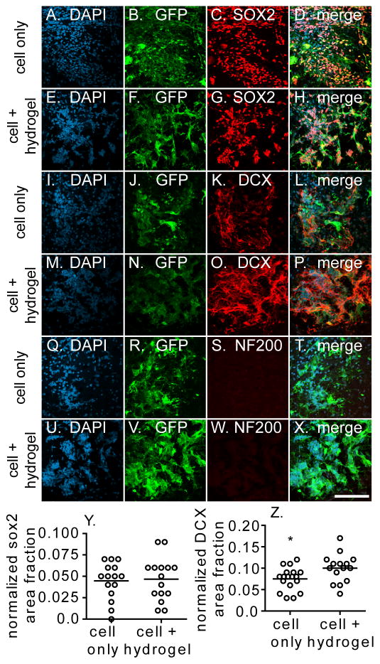
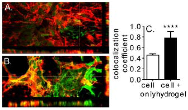
Stroke is the leading cause of adult disability with ~80% being ischemic. Stem cell transplantation has been shown to improve functional recovery. However, the overall survival and differentiation of these cells is still low. The infarct cavity is an ideal location for transplantation as it is directly adjacent to the highly plastic peri-infarct region. Direct transplantation of cells near the infarct cavity has resulted in low cell viability. Here we deliver neural progenitor cells derived from induce pluripotent stem cells (iPS-NPC) to the infarct cavity of stroked mice encapsulated in a hyaluronic acid hydrogel matrix to protect the cells. To improve the overall viability of transplanted cells, each step of the transplantation process was optimized. Hydrogel mechanics and cell injection parameters were investigated to determine their effects on the inflammatory response of the brain and cell viability, respectively. Using parameters that balanced the desire to keep surgery invasiveness minimal and cell viability high, iPS-NPCs were transplanted to the stroke cavity of mice encapsulated in buffer or the hydrogel. While the hydrogel did not promote stem cell survival one week post-transplantation, it did promote differentiation of the neural progenitor cells to neuroblasts.

Keywords: Hyaluronic acid; Hydrogel; Induced pluripotent stem cells; Stroke.


Figures

Fig. 1
Fig. 1
(A) The hydrogel contains acrylated hyaluronic acid, peptide crosslinker, and an RGD-containing peptide. The crosslinker is MMP-degradable (d.) or MMP-nondegradable (nd.). The hydrogel is made by pre-reacting the HA with the RGD peptide and then mixing in the cross-linker and cells. (B) Three hydrogel formulations were made with (C) rheology confirming their mechanical properties.
Fig. 2
Fig. 2
Four different conditions were injected into the striatum of naïve or stroked mice: HEPES buffer, d. hydrogel, nd. “soft” hydrogel and nd. “stiff” hydrogel. (A) Overview of entire brain section showing contralateral and ipsilateral regions of interest. (B–E) Images were taken on the contralateral side of the brain as well as the area (B′–E′) directly adjacent to the injection site (ipsilateral) in naïve mice. (J) GFAP and (K) IBA1 signal was quantified. The same four conditions were then injected into the infarct cavity of stroked mice. Images were taken on the (F–I) contralateral and (F′–I′) ipsilateral of the brain for (L) GFAP and (M) IBA1 quantification. Scale bar = 50 μm
Fig. 3
Fig. 3
(A) The effect of the injection process on iPS-NPC viability was tested using the same setup used on in vivo experiments. Viability of cells encapsulated in media or hydrogel “d.” were tested at different (B) infusion speeds, (C) needle gauge, and (D) cell concentration. +: between w/gel, * between w/o gel, # between w/ and w/o gel
Fig. 4
Fig. 4
100,000 iPS-NPCs injected into the infarct cavity of stroked NSG mice suspended in buffer or hydrogel “d.” (A–B) The inflammatory response of the peri-infarct tissue was quantified and normalized to the contralateral side for (C) GFAP and (D) IBA1. (E–G) Human nuclei were stained and (H) quantified for both conditions. Scale bar = 50 μm
Fig. 5
Fig. 5
Transplantation zone was stained for markers of the GFP-labeled (A–H) iPS-NPCs (SOX2), (I–P) neuroblasts (DOUBLECORTIN, DCX), and (Q–X) mature neurons (NF200). (Y) SOX2 and (Z) DCX signal was quantified and with an increased DOUBLECORTIN signal seen in the cell + hydrogel condition. Scale bar = 100 μm
Fig. 6
Fig. 6
3-dimensional reconstruction of (A) cell only and (B) cell + hydrogel sections stained for GFP-labeled transplanted cells and DOUBLECORTIN. (C) Colocalization analysis shows that the majority of DCX positive signal seen in cell + hydrogel condition is from transplanted cell differentiation.

References

    1. Taylor TN, Davis PH, Torner JC, Holmes J, Meyer JW, Jacobson MF. Stroke. 1996;27:1459. - PubMed
    1. Broderick JP. Stroke. 2004;35:205. - PubMed
    1. Donnan GA, Fisher M, Macleod M, Davis SM. Lancet. 2008;371:1612. - PubMed
    1. Buhnemann C, Scholz A, Bernreuther C, Malik CY, Braun H, Schachner M, Reymann KG, Dihne M. Brain. 2006;129:3238. - PubMed
    1. Hicks AU, Lappalainen RS, Narkilahti S, Suuronen R, Corbett D, Sivenius J, Hovatta O, Jolkkonen J. European Journal of Neuroscience. 2009;29:562. - PubMed