

Electrostatically Directed Self-Assembly of Ultrathin Supramolecular Polymer Microcapsules
- PMID: 26213532
- PMCID: PMC4511391
- DOI: 10.1002/adfm.201501079
Electrostatically Directed Self-Assembly of Ultrathin Supramolecular Polymer Microcapsules
Abstract
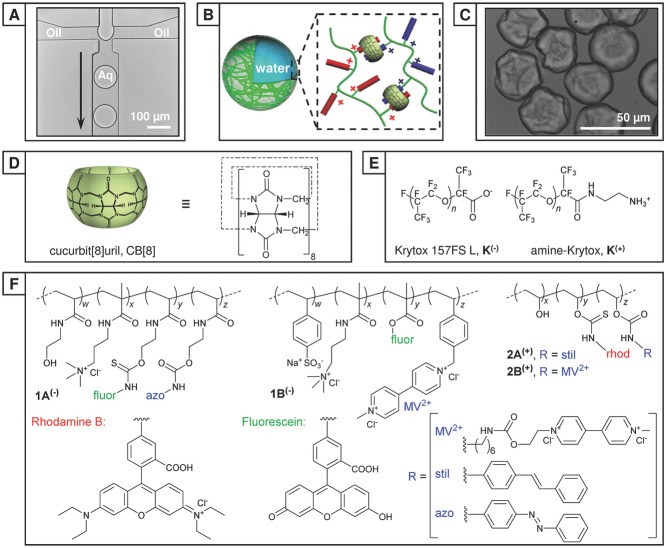
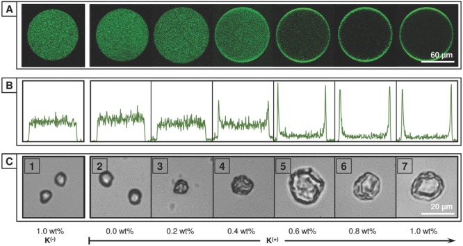
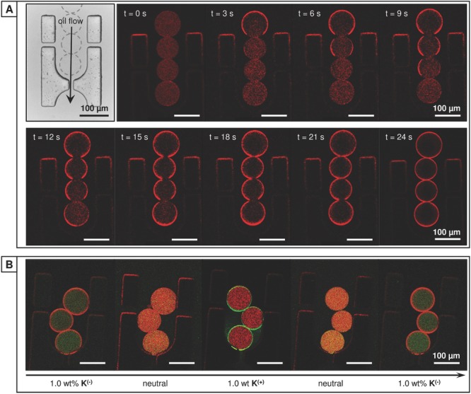
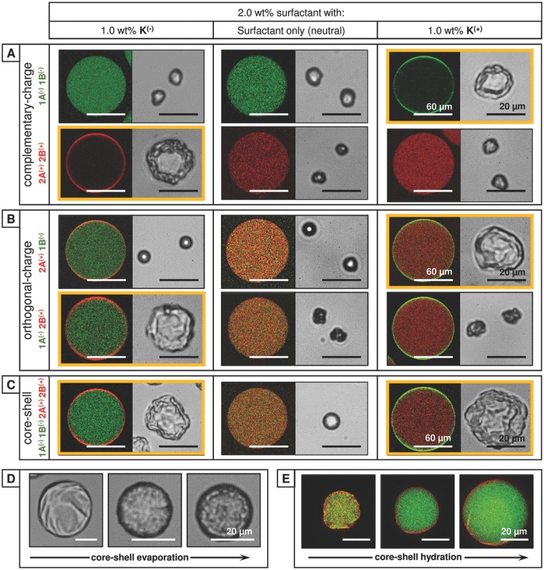
Supramolecular self-assembly offers routes to challenging architectures on the molecular and macroscopic scale. Coupled with microfluidics it has been used to make microcapsules-where a 2D sheet is shaped in 3D, encapsulating the volume within. In this paper, a versatile methodology to direct the accumulation of capsule-forming components to the droplet interface using electrostatic interactions is described. In this approach, charged copolymers are selectively partitioned to the microdroplet interface by a complementary charged surfactant for subsequent supramolecular cross-linking via cucurbit[8]uril. This dynamic assembly process is employed to selectively form both hollow, ultrathin microcapsules and solid microparticles from a single solution. The ability to dictate the distribution of a mixture of charged copolymers within the microdroplet, as demonstrated by the single-step fabrication of distinct core-shell microcapsules, gives access to a new generation of innovative self-assembled constructs.
Keywords: microcapsules; microfluidics; microstructures; self-assembly; supramolecular materials.
Figures
Similar articles
-
Cucurbit[n]uril-Based Microcapsules Self-Assembled within Microfluidic Droplets: A Versatile Approach for Supramolecular Architectures and Materials.Acc Chem Res. 2017 Feb 21;50(2):208-217. doi: 10.1021/acs.accounts.6b00429. Epub 2017 Jan 11. Acc Chem Res. 2017. PMID: 28075551 Free PMC article.
-
Supramolecular hydrogel microcapsules via cucurbit[8]uril host-guest interactions with triggered and UV-controlled molecular permeability.Chem Sci. 2015 Aug 1;6(8):4929-4933. doi: 10.1039/c5sc01440a. Epub 2015 Jun 11. Chem Sci. 2015. PMID: 28717496 Free PMC article.
-
Microfluidic Droplet-Facilitated Hierarchical Assembly for Dual Cargo Loading and Synergistic Delivery.ACS Appl Mater Interfaces. 2016 Apr 6;8(13):8811-20. doi: 10.1021/acsami.6b00661. Epub 2016 Mar 25. ACS Appl Mater Interfaces. 2016. PMID: 26982167 Free PMC article.
-
Cucurbit[8]uril-Based Polymers and Polymer Materials.Small. 2018 Nov;14(46):e1802234. doi: 10.1002/smll.201802234. Epub 2018 Aug 31. Small. 2018. PMID: 30168673 Review.
-
Molecular Recognition in the Colloidal World.Acc Chem Res. 2017 Nov 21;50(11):2756-2766. doi: 10.1021/acs.accounts.7b00370. Epub 2017 Oct 6. Acc Chem Res. 2017. PMID: 28984441 Review.
Cited by
-
Cucurbit[n]uril-Based Microcapsules Self-Assembled within Microfluidic Droplets: A Versatile Approach for Supramolecular Architectures and Materials.Acc Chem Res. 2017 Feb 21;50(2):208-217. doi: 10.1021/acs.accounts.6b00429. Epub 2017 Jan 11. Acc Chem Res. 2017. PMID: 28075551 Free PMC article.
-
Enabling Technology for Supramolecular Chemistry.Front Chem. 2021 Nov 15;9:774987. doi: 10.3389/fchem.2021.774987. eCollection 2021. Front Chem. 2021. PMID: 34869224 Free PMC article. Review.
-
Aqueous interfacial gels assembled from small molecule supramolecular polymers.Chem Sci. 2017 Feb 1;8(2):1350-1355. doi: 10.1039/c6sc04103e. Epub 2016 Oct 11. Chem Sci. 2017. PMID: 29780448 Free PMC article.
-
Supramolecular hydrogel microcapsules via cucurbit[8]uril host-guest interactions with triggered and UV-controlled molecular permeability.Chem Sci. 2015 Aug 1;6(8):4929-4933. doi: 10.1039/c5sc01440a. Epub 2015 Jun 11. Chem Sci. 2015. PMID: 28717496 Free PMC article.
-
Charge controlled interactions between DNA-modified silica nanoparticles and fluorosurfactants in microfluidic water-in-oil droplets.Nanoscale Adv. 2023 Jul 5;5(15):3914-3923. doi: 10.1039/d3na00124e. eCollection 2023 Jul 25. Nanoscale Adv. 2023. PMID: 37496619 Free PMC article.
References
-
- She Z, Wang C, Li J, Sukhorukov GB, Antipina MN. Biomacromolecules. 2012;13:2174. - PubMed
-
- Fattahi P, Borhan A, Abidian MR. Adv. Mater. 2013;25:4555. - PubMed
-
- Amali AJ, Sharma B, Rana RK. Chem. - A Eur. J. 2014;20:12239. - PubMed
-
- Horecha Marta, Kaul E, Horechyy A, Stamm M. J. Mater. Chem. A. 2014;2:7431.
-
- Augustin MA, Hemar Y. Chem. Soc. Rev. 2009;38:902. - PubMed
LinkOut - more resources
Full Text Sources
Other Literature Sources
Molecular Biology Databases