

The Effect of Preparation Size on Efficacy of Smear Layer Removal; A Scanning Electron Microscopic Study
- PMID: 26213539
- PMCID: PMC4509124
- DOI: 10.7508/iej.2015.03.005
The Effect of Preparation Size on Efficacy of Smear Layer Removal; A Scanning Electron Microscopic Study
Abstract
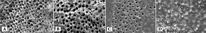
Introduction: Enlargement of the root canal may potentially affect efficient smear layer (SL) removal. The aim of the present in vitro study was to compare SL removal following canal preparation with two different sizes/tapers by means of scanning electron microscopy (SEM).
Methods and materials: A total of 50 extracted human mandibular premolars were decoronated. The teeth were randomly divided into two experimental groups (n=20) and two negative control groups. In groups 1 and 2 the sizes of master apical file (MAF) were #25 and 40, respectively. Coronal part of the canals were flared with #2 Piezo drills in group 1 and sizes #2 to 6 in group 2. Finally FlexMaster NiTi rotary instruments were used to complete canal preparation (25/0.04 and 35/0.06 in groups 1 and 2, respectively). The irrigation protocol consisted of 10 mL of 17% ethylenediaminetetraacetic acid (EDTA) for 1 min followed by 10 mL of 5.25% NaOCl for 3 min. The patency of dentinal tubules was evaluated under SEM with Hülsmann scores. Data were analyzed with the Kruskal-Wallis and Mann-Whitney U tests.
Results: The number of patent dentinal tubules in coronal third of the group 1 was significantly more than group 2 (P<0.001). However, this difference was not significant for the middle and apical segments. There was a significant difference in the number of patent dentinal tubules between coronal, middle and apical thirds (P<0.05).
Conclusion: Increasing the canal preparation size did not lead to better cleanliness of the canal walls and more efficient smear layer removal.
Keywords: Dentinal Tubules; EDTA; Ethylenediaminetetraacetic Acid; Root Canal Preparation; Scanning Electron Microscopy; Smear Layer.
Figures

Similar articles
-
[In vitro evaluation of the effectiveness of XP-endo Finisher file on smear layer removal after root canal instrumentation].Hua Xi Kou Qiang Yi Xue Za Zhi. 2019 Feb 1;37(1):48-52. doi: 10.7518/hxkq.2019.01.009. Hua Xi Kou Qiang Yi Xue Za Zhi. 2019. PMID: 30854818 Free PMC article. Chinese.
-
Comparative Evaluation of Smear Layer and Debris on the Canal Walls prepared with a Combination of Hand and Rotary ProTaper Technique using Scanning Electron Microscope.J Contemp Dent Pract. 2016 Jul 1;17(7):574-81. J Contemp Dent Pract. 2016. PMID: 27595725
-
Effects of NiTi rotary and reciprocating instruments on debris and smear layer scores: an SEM evaluation.J Appl Biomater Funct Mater. 2014 Dec 30;12(3):256-62. doi: 10.5301/jabfm.5000161. J Appl Biomater Funct Mater. 2014. PMID: 24425380
-
A comparative scanning electron microscopy evaluation of smear layer removal with chitosan and MTAD.Niger J Clin Pract. 2018 Jan;21(1):76-80. doi: 10.4103/1119-3077.224798. Niger J Clin Pract. 2018. PMID: 29411728
-
A comparative study of the removal of smear layer by three endodontic irrigants and two types of laser.Int Endod J. 1999 Jan;32(1):32-9. doi: 10.1046/j.1365-2591.1999.00182.x. Int Endod J. 1999. PMID: 10356467
Cited by
-
To Evaluate the Efficacy of an Innovative Irrigant on Smear Layer Removal - SEM Analysis.J Clin Diagn Res. 2016 Apr;10(4):ZC104-6. doi: 10.7860/JCDR/2016/17200.7685. Epub 2016 Apr 1. J Clin Diagn Res. 2016. PMID: 27190941 Free PMC article.
-
Effect of Apical Size and Taper on the Efficacy of Root Canal Disinfection With LED Photodynamic Therapy as an Adjunct to Irrigation With Sodium Hypochlorite.J Lasers Med Sci. 2021 Oct 9;12:e58. doi: 10.34172/jlms.2021.58. eCollection 2021. J Lasers Med Sci. 2021. PMID: 35155143 Free PMC article.
-
Influence of Final Apical Width on Smear Layer Removal Efficacy of Xp Endo Finisher and Endodontic Needle: An Ex Vivo Study.Eur Endod J. 2020 Mar 17;5(1):18-22. doi: 10.14744/eej.2019.58076. eCollection 2020. Eur Endod J. 2020. PMID: 32342033 Free PMC article.
-
Removal of Composite Restoration from the Root Surface in the Cervical Region Using Er: YAG Laser and Drill-In Vitro Study.Materials (Basel). 2020 Jul 7;13(13):3027. doi: 10.3390/ma13133027. Materials (Basel). 2020. PMID: 32645864 Free PMC article.
References
-
- Silva LA, Novaes AB, de Oliveira RR, Nelson-Filho P, Santamaria M, Silva RA. Antimicrobial photodynamic therapy for the treatment of teeth with apical periodontitis: a histopathological evaluation. J Endod. 2012;38(3):360–6. - PubMed
-
- Paranjpe A, de Gregorio C, Gonzalez AM, Gomez A, Silva Herzog D, Pina AA, Cohenca N. Efficacy of the self-adjusting file system on cleaning and shaping oval canals: a microbiological and microscopic evaluation. J Endod. 2012;38(2):226–31. - PubMed
-
- Young GR, Parashos P, Messer HH. The principles of techniques for cleaning root canals. Aust Dent J. 2007;52(1 Suppl):S52–63. - PubMed
-
- Schilder H. Cleaning and shaping the root canal. Dent Clin North Am. 1974;18(2):269–96. - PubMed
-
- Bartha T, Kalwitzki M, Lost C, Weiger R. Extended apical enlargement with hand files versus rotary NiTi files. Part II. Oral Surg Oral Med Oral Pathol Oral Radiol Endod. 2006;102(5):692–7. - PubMed
LinkOut - more resources
Full Text Sources