Case Reports
. 2015 Jul 26:8:54.
doi: 10.1186/s13039-015-0162-3. eCollection 2015.

Periventricular heterotopia and white matter abnormalities in a girl with mosaic ring chromosome 6

Affiliations
Case Reports

Periventricular heterotopia and white matter abnormalities in a girl with mosaic ring chromosome 6

Satsuki Nishigaki et al. Mol Cytogenet. .

Abstract

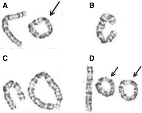
Ring chromosome 6 is a rare chromosome abnormality that arises typically de novo. The phenotypes can be highly variable, ranging from almost normal to severe malformations and neurological defects. We report a case of a 3-year-old girl with mosaic ring chromosome 6 who presented with being small for gestational age and intellectual disability, and whose brain MRI later revealed periventricular heterotopia and white matter abnormalities. Mosaicism was identified in peripheral blood cells examined by standard G-bands, mos 46,XX,r(6)(p25q27)[67]/45,XX,-6[25]/46,XX,dic r(6:6)(p25q27:p25q27)[6]/47,XX,r(6)(p25q27) × 2[2]. Using array-comparative genomic hybridization, we identified terminal deletion of 6q27 (1.5 Mb) and no deletion on 6p. To our knowledge, this is the first report of periventricular heterotopia and white matter abnormalities manifested in a patient with ring chromosome 6. These central nervous system malformations are further discussed in relation to molecular genetics.

Keywords: Array-comparative genomic hybridization; Brain MRI; Periventricular heterotopia; Ring chromosome 6; Small for gestational age; White matter abnormality.


Figures

Fig. 1
Fig. 1
Chromosome G-banding result from the patient. The total number of cells counted was hundred. a Ring chromosome (67 %). b Monosomy 6 (25 %). c Dicentric ring chromosome (6 %). d Double ring chromosome (2 %)
Fig. 2
Fig. 2
a-f: MRI imaging of the brain of this patient. a and b Axial T1W and T2W image, showing left parietal lobe. c Coronal T2W section showing white matter abnormalities in the bilateral parietal lobe. d Coronal T2W section showing white matter abnormalities in the bilateral occipital lobe. e and f Axial and Coronal FLAIR image, showing periventricular heterotopia on the bilateral posterior horn of the lateral ventricle
Fig. 3
Fig. 3
The result of array-CGH. Vertical and horizontal axes indicate the genomic location and signal log2 ratio, respectively. Dots indicate the location and the signal log2 ratio of the microarray probes. a Chromosomal view of the chromosome 6 shows a genomic copy number loss at q27. b Gene view expands the aberration region. The blue rectangle indicates and the blue bars indicate the aberration region and the locations of the genes. Two genes discussed in the text are marked by red oblongs. The mean log2 ratio of the aberration region is −0.780567, indicating mosaic deletion

References

    1. Ahzad HA, Ramli SF, Loong TM, Salahshourifar I, Zilfalil BA, Yusoff NM. De novo ring chromosome 6 in a child with multiple congenital anomalies. Kobe J Med Sci. 2010;56(2):E79–84. - PubMed
    1. Lee SJ, Han DK, Cho HJ, Cho YK, Ma JS. Mosaic ring chromosome 6 in an infant with significant patent ductus arteriosus and multiple congenital anomalies. J Korean Med Sci. 2012;27:948–952. doi: 10.3346/jkms.2012.27.8.948. - DOI - PMC - PubMed
    1. Kini KR, Van Dyke DL, Weiss L, Logan MS. Ring chromosome 6: case report and review of literature. Hum Genet. 1979;50(2):145–9. doi: 10.1007/BF00390235. - DOI - PubMed
    1. Urban M, Bommer C, Tennstedt C, Lehmann K, Thiel G, Wegner RD, Bollmann R, Becker R, Schulzke I, Körner H. Ring chromosome 6 in three fetuses: case reports, literature review, and implications for prenatal diagnosis. Am J Med Genet. 2002;108:97–104. doi: 10.1002/ajmg.10215. - DOI - PubMed
    1. Zhang HZ, Li P, Wang D, Huff S, Nimmakayalu M, Qumsiyeh M, Pober BR. FOXC1 gene deletion is associated with eye anomalies in ring chromosome 6. Am J Med Genet A. 2004;124A(3):280–7. doi: 10.1002/ajmg.a.20413. - DOI - PubMed

Publication types