

Fast degradable citrate-based bone scaffold promotes spinal fusion
- PMID: 26213625
- PMCID: PMC4511467
- DOI: 10.1039/C5TB00607D
Fast degradable citrate-based bone scaffold promotes spinal fusion
Abstract
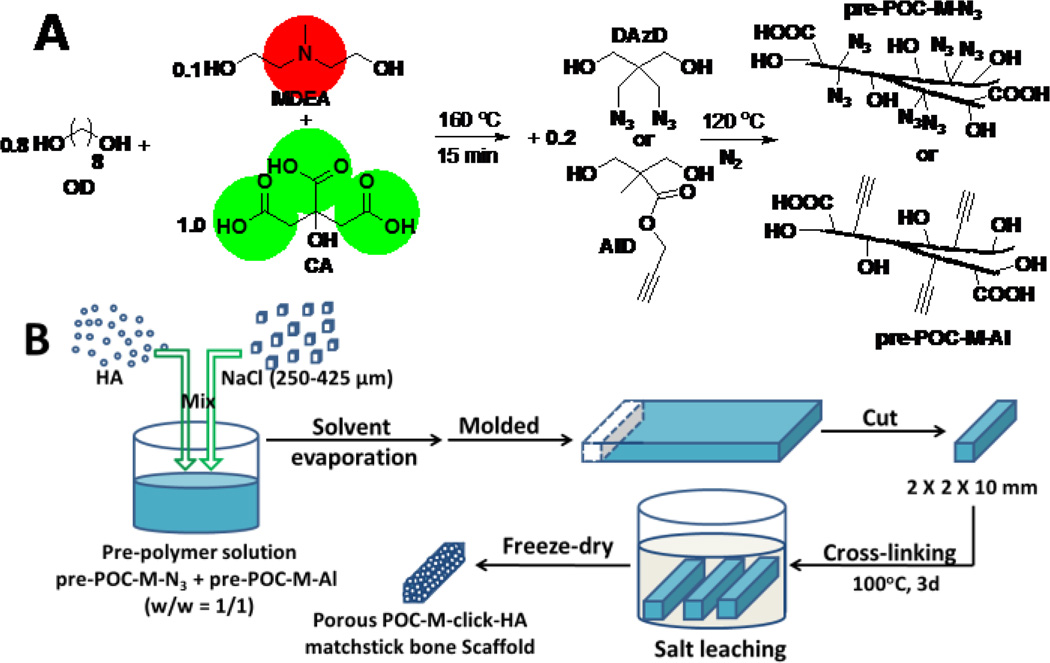
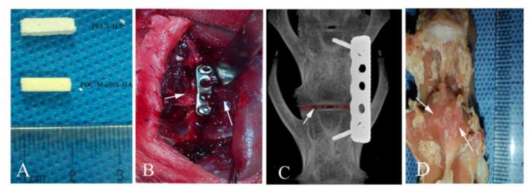
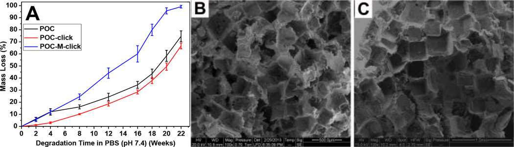
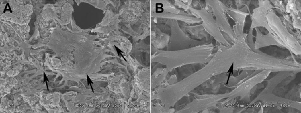
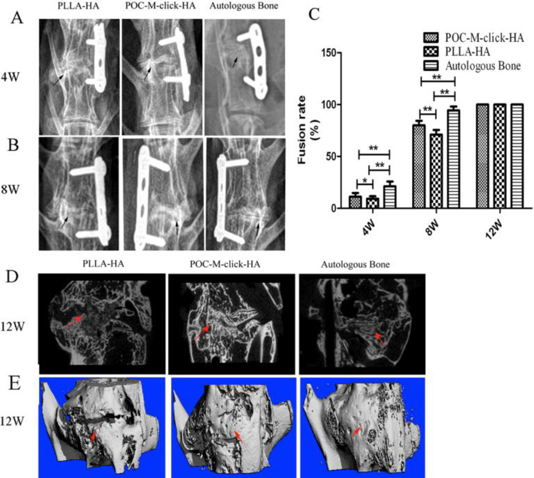
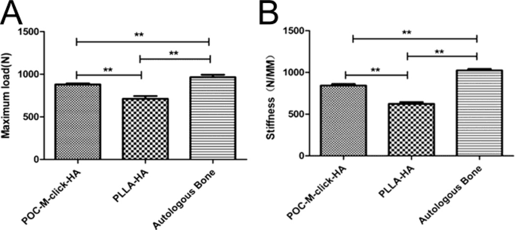
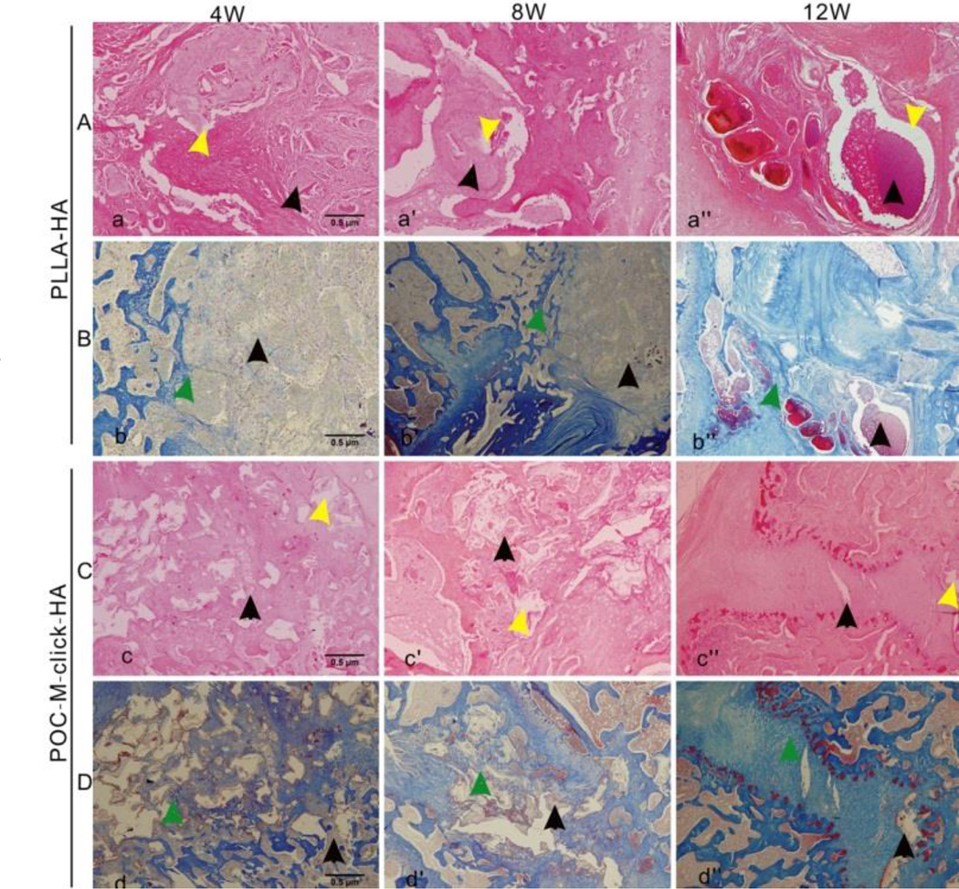
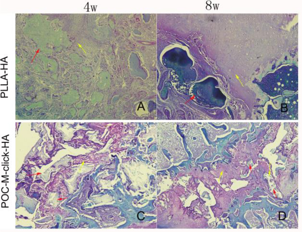
It is well known that high rates of fusion failure and pseudoarthrosis development (5~35%) are concomitant in spinal fusion surgery, which was ascribed to the shortage of suitable materials for bone regeneration. Citrate was recently recognized to play an indispensable role in enhancing osteconductivity and osteoinductivity, and promoting bone formation. To address the material challenges in spinal fusion surgery, we have synthesized mechanically robust and fast degrading citrate-based polymers by incorporating N-methyldiethanolamine (MDEA) into clickable poly(1, 8-octanediol citrates) (POC-click), referred to as POC-M-click. The obtained POC-M-click were fabricated into POC-M-click-HA matchstick scaffolds by compositing with hydroxyapatite (HA) for interbody spinal fusion in a rabbit model. Spinal fusion was analyzed by radiography, manual palpation, biomechanical testing, and histological evaluation. At 4 and 8 weeks post surgery, POC-M-click-HA scaffolds presented optimal degradation rates that facilitated faster new bone formation and higher spinal fusion rates (11.2±3.7, 80±4.5 at week 4 and 8, respectively) than the poly(L-lactic acid)-HA (PLLA-HA) control group (9.3±2.4 and 71.1±4.4) (p<0.05). The POC-M-click-HA scaffold-fused vertebrates possessed a maximum load and stiffness of 880.8±14.5 N and 843.2±22.4 N/mm, respectively, which were also much higher than those of the PLLA-HA group (maximum: 712.0±37.5 N, stiffness: 622.5±28.4 N/mm, p<0.05). Overall, the results suggest that POC-M-click-HA scaffolds could potentially serve as promising bone grafts for spinal fusion applications.
Figures
Similar articles
-
Development of osteopromotive poly (octamethylene citrate glycerophosphate) for enhanced bone regeneration.Acta Biomater. 2019 Jul 15;93:180-191. doi: 10.1016/j.actbio.2019.03.050. Epub 2019 Mar 27. Acta Biomater. 2019. PMID: 30926580 Free PMC article.
-
Citric acid-based hydroxyapatite composite scaffolds enhance calvarial regeneration.Sci Rep. 2014 Nov 5;4:6912. doi: 10.1038/srep06912. Sci Rep. 2014. PMID: 25372769 Free PMC article.
-
Osteoinductivity and biomechanical assessment of a 3D printed demineralized bone matrix-ceramic composite in a rat spine fusion model.Acta Biomater. 2021 Jun;127:146-158. doi: 10.1016/j.actbio.2021.03.060. Epub 2021 Apr 6. Acta Biomater. 2021. PMID: 33831576 Free PMC article.
-
Citrate-based biphasic scaffolds for the repair of large segmental bone defects.J Biomed Mater Res A. 2015 Feb;103(2):772-81. doi: 10.1002/jbm.a.35228. Epub 2014 May 29. J Biomed Mater Res A. 2015. PMID: 24829094 Free PMC article.
-
Low-pressure foaming: a novel method for the fabrication of porous scaffolds for tissue engineering.Tissue Eng Part C Methods. 2012 Feb;18(2):113-21. doi: 10.1089/ten.TEC.2011.0289. Epub 2011 Dec 22. Tissue Eng Part C Methods. 2012. PMID: 21933018
Cited by
-
Citric Acid: A Nexus Between Cellular Mechanisms and Biomaterial Innovations.Adv Mater. 2024 Aug;36(32):e2402871. doi: 10.1002/adma.202402871. Epub 2024 Jun 11. Adv Mater. 2024. PMID: 38801111 Free PMC article. Review.
-
Flexible biodegradable citrate-based polymeric step-index optical fiber.Biomaterials. 2017 Oct;143:142-148. doi: 10.1016/j.biomaterials.2017.08.003. Epub 2017 Aug 4. Biomaterials. 2017. PMID: 28802101 Free PMC article.
-
Injectable Self-Setting Ternary Calcium-Based Bone Cement Promotes Bone Repair.ACS Omega. 2023 May 1;8(19):16809-16823. doi: 10.1021/acsomega.3c00331. eCollection 2023 May 16. ACS Omega. 2023. PMID: 37214722 Free PMC article.
-
Citrate-based materials fuel human stem cells by metabonegenic regulation.Proc Natl Acad Sci U S A. 2018 Dec 11;115(50):E11741-E11750. doi: 10.1073/pnas.1813000115. Epub 2018 Nov 26. Proc Natl Acad Sci U S A. 2018. PMID: 30478052 Free PMC article.
-
Engineering multifunctional bioactive citrate-based biomaterials for tissue engineering.Bioact Mater. 2022 May 7;19:511-537. doi: 10.1016/j.bioactmat.2022.04.027. eCollection 2023 Jan. Bioact Mater. 2022. PMID: 35600971 Free PMC article. Review.
References
-
- Reid JJ, Johnson JS, Wang JC. J. Biomech. 2011;44:213. - PubMed
-
- Ghogawala 1Z, Whitemore RG, Watters WC, 3rd, Sharan A, Mummaneni PV, Dailey AT, Choudhri TF, Eck JC, Groff MW, Wang JC, Resnick DK, Dhall SS, Kaiser MG. J. Neurosurg Spine. 2014;21:14. - PubMed
-
- Ransom BR, Neale E, Henkart M, Bullock PN, Nelson PG. J Neurophysiol. 1977;40:1132. - PubMed
-
- Dimitriou R, Mataliotakis GI, Angoules AG, Kanakaris NK, Giannoudis PV. Injury. 2011;42:S3. - PubMed
Grants and funding
LinkOut - more resources
Full Text Sources
Other Literature Sources