

STAT1 is Constitutively Activated in the T/C28a2 Immortalized Juvenile Human Chondrocyte Line and Stimulated by IL-6 Plus Soluble IL-6R
- PMID: 26213636
- PMCID: PMC4512305
- DOI: 10.4172/2155-9899.1000307
STAT1 is Constitutively Activated in the T/C28a2 Immortalized Juvenile Human Chondrocyte Line and Stimulated by IL-6 Plus Soluble IL-6R
Abstract
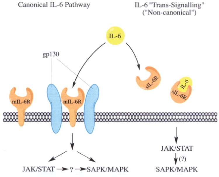
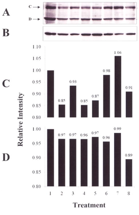
T/C28a2 immortalized juvenile human chondrocytes were employed to determine the extent to which activation of Signal Transducers and Activators of Transcription-1 (STAT1) occurred in response to recombinant human interleukin-6 (rhIL-6) or rhIL-6 in combination with the soluble IL-6 receptor (sIL-6R). Two forms of STAT1, STAT1A and STAT1B, were identified on SDS-PAGE and western blotting with anti-STAT1 antibody. Western blotting revealed that STAT1 was constitutively phosphorylated (p-STAT1). Although incubation of T/C28a2 chondrocytes with rhIL-6 (50 ng/ml) increased p-STAT1A by Δ=22.3% after 30 min, this percent difference failed to reach significance by Chi-square analysis. Similarly, no effect of rhIL-6 (Δ=+10.7%) on p-STAT1B was seen at 30 min. In contrast, although the combination of rhIL-6 plus sIL-6R had no effect on p-STAT1A, rhIL-6 plus sIL-6R increased p-STAT1B by Δ=73.3% (p<0.0001) after 30 min compared to the control group and by Δ=56.7% (p<0.0001) compared to rhIL-6 alone. Janex-1, a Janus kinase-3-specific inhibitor (100 μM) partially reduced the effect of rhIL-6 on p-STAT1B by Δ=27.7% (p<0.05). The results of this study showed that STAT1A/STAT1B was constitutively activated in T/C28a2 chondrocytes. Although rhIL-6 increased p-STAT1B to a small extent, the combination of rhIL-6 plus sIL-6R was far more effective in stimulating STAT1B phosphorylation compared to controls or rhIL-6 alone. These data support the likelihood that although JAK3-mediated activation of STAT1 in T/C28a2 chondrocytes may involve the IL-6/IL-6R/gp130 pathway, these results indicated that STAT1 activation in response to IL-6 preferentially involved IL-6 trans-signaling via sIL-6R.
Keywords: Chondrocyte; Cytokine; Rheumatoid arthritis.
Figures

References
-
- Murray PJ. The JAK-STAT signaling pathway: input and output integration. J Immunol. 2007;178:2623–2629. - PubMed
-
- Malemud CJ, Pearlman E. Targeting JAK/STAT signaling pathway in inflammatory disorders. Curr Signal Transduct Ther. 2009;4:201–221.
-
- Hanada T, Yoshimura A. Regulation of cytokine signaling and inflammation. Cytokine Growth Factor Rev. 2002;13:413–421. - PubMed
-
- Decker T, Kovarik P. Serine phosphorylation of STATs. Oncogene. 2000;19:2628–2637. - PubMed
Grants and funding
LinkOut - more resources
Full Text Sources
Other Literature Sources
Research Materials
Miscellaneous