. 2015 Jul 9:3:e1093.
doi: 10.7717/peerj.1093. eCollection 2015.

Retrodeformation and muscular reconstruction of ornithomimosaurian dinosaur crania

Affiliations

Retrodeformation and muscular reconstruction of ornithomimosaurian dinosaur crania

Andrew R Cuff et al. PeerJ. .

Abstract

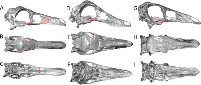
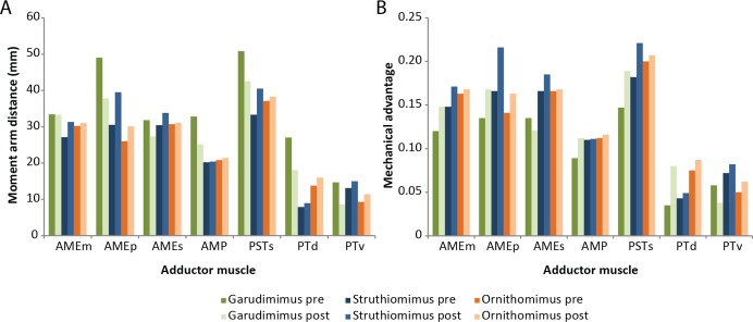
Ornithomimosaur dinosaurs evolved lightweight, edentulous skulls that possessed keratinous rhamphothecae. Understanding the anatomy of these taxa allows for a greater understanding of "ostrich-mimic" dinosaurs and character change during theropod dinosaur evolution. However, taphonomic processes during fossilisation often distort fossil remains. Retrodeformation offers a means by which to recover a hypothesis of the original anatomy of the specimen, and 3D scanning technologies present a way to constrain and document the retrodeformation process. Using computed tomography (CT) scan data, specimen specific retrodeformations were performed on three-dimensionally preserved but taphonomically distorted skulls of the deinocheirid Garudimimus brevipesBarsbold, 1981 and the ornithomimids Struthiomimus altusLambe, 1902 and Ornithomimus edmontonicusSternberg, 1933. This allowed for a reconstruction of the adductor musculature, which was then mapped onto the crania, from which muscle mechanical advantage and bite forces were calculated pre- and post-retrodeformation. The extent of the rhamphotheca was varied in each taxon to represent morphologies found within modern Aves. Well constrained retrodeformation allows for increased confidence in anatomical and functional analysis of fossil specimens and offers an opportunity to more fully understand the soft tissue anatomy of extinct taxa.

Keywords: Bite forces; Myology; Ornithomimosaurs; Retrodeformation; Rhamphotheca; Skull.


Conflict of interest statement

The authors declare there are no competing interests.

Figures

Figure 1
Figure 1. Foramina and rugosities in the rostra of certain taxa.
(A) Anterior, right mandible of Struthiomimus altus (RTMP 1990.026.0001); (B) Dorsal view of anterior premaxilla of ostrich and mandible (ROM R1080); (C) Anterior dentary of a tyrannosaur (Daspletosaurus?) RTMP (1967.009.0164). Scale bars = 1 cm.
Figure 2
Figure 2. Garudimimus brevipes reconstruction (GIN 100/13).
(A), (C), (E), original skull, (B), (D), (F), retrodeformed skulls. (A), (B), right lateral; (C), (D) dorsal; (E), (F), ventral views. Scale bar = 5 cm. See Video S1 and Video S2 showing video of the skull before and after retrodeformation.
Figure 3
Figure 3. Struthiomimus altus reconstruction (RTMP 1990.026.0001).
Note the dorsoventral expansion of the skull after retrodeformation, particularly of the orbital region. (A), (C), (E), original skull, (B), (D), (F), retrodeformed skulls. (A),(B), right lateral; (C), (D) dorsal; (E), (F), ventral views. Scale bar = 5 cm. See Videos S3 and S4 showing video of the skull before and after retrodeformation.
Figure 4
Figure 4. Ornithomimus edmontonicus reconstruction (RTMP 1995.110.0001) showing the effect of the mediolateral expansion after separating the taphonomically deformed bones of the palate.
(A), (C), (E), original skull, (B), (D), (F), retrodeformed skulls. (A), (B), right lateral; (C), (D) dorsal; (E), (F), ventral views. Scale bar = 5 cm. See Videos S5 and S6 showing video of the skull before and after retrodeformation.
Figure 5
Figure 5. Reconstructions showing the regions where material was added using the paintbrush region-selecting tool within Avizo.
Regions in red showing the areas where new material was added. (A)–(C) Garudimimus, (D)–(F) Struthiomimus, (G)–(I) Ornithomimus.
Figure 6
Figure 6. Full cranial reconstruction including musculature of the jaw.
(A) Garudimimus, (B) Struthiomimus, (C) Ornithomimus. Scale bars = 5 cm. Pink, PSTs; purple, AMEp; red, AMEm; blue, AMEs; green, AMP; yellow, PTd; orange, PTv.
Figure 7
Figure 7. Ornithomimosaur beaks.
(A) Small and (B) big beak morphs on Garudimimus; (C) small and (D) big beak morphs on Ornithomimus; (E) small and (F) big beak morphs on Struthiomimus. Scale bars = 5 cm. Triangles represent bite locations for mid-beak and tip of the beak bites (Table 6).
Figure 8
Figure 8. Effects of retrodeformation on myological reconstructions.
(A) Moment arm distances, (B) Mechanical advantages. ‘Pre’ and ‘Post’ refer to pre- and post-retrodeformation.

References

    1. Adams LA. A memoir of the phylogeny of the jaw muscles in recent and fossil vertebrates. Annals of the New York Academy of Science. 1919;58:51–166.
    1. Angielczyk KD, Sheets HD. Investigation of simulated tectonic deformation in fossils using geometric morphometrics. Paleobiology. 2007;33:125–148. doi: 10.1666/06007.1. - DOI
    1. Arbour VM, Currie PJ. Analyzing taphonomic deformation of ankylosaur skulls using retrodeformation and finite element analysis. PLoS ONE. 2012;7(6):e1093. doi: 10.1371/journal.pone.0039323. - DOI - PMC - PubMed
    1. Barsbold R. Toothless carnivorous dinosaurs of Mongolia. Transactions, Joint Soviet-Mongolian Palaeontological Expedition. 1981;15:28–39.
    1. Barsbold R, Perle A. On first new find of a primitive ornithomimosaur from the Cretaceous of the MPR. Paleontologicheskii Zhurnal. 1984;2:121–123.

LinkOut - more resources