Review
. 2015 Jul 23;6(3):685-713.
doi: 10.3390/genes6030685.

The Structural Determinants behind the Epigenetic Role of Histone Variants

Affiliations
Review

The Structural Determinants behind the Epigenetic Role of Histone Variants

Manjinder S Cheema et al. Genes (Basel). .

Abstract

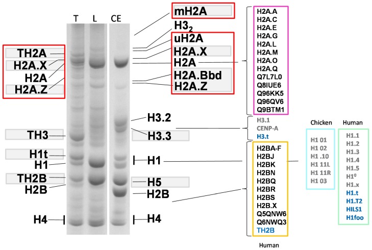
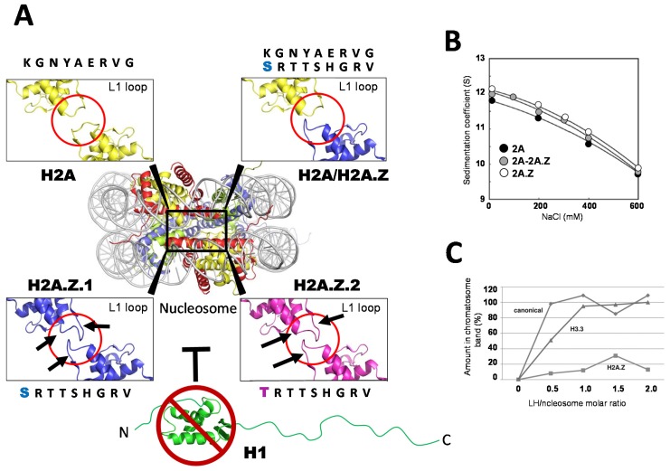
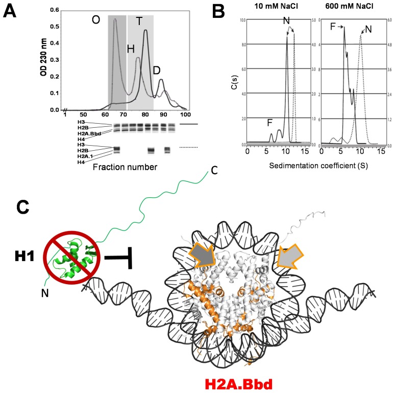
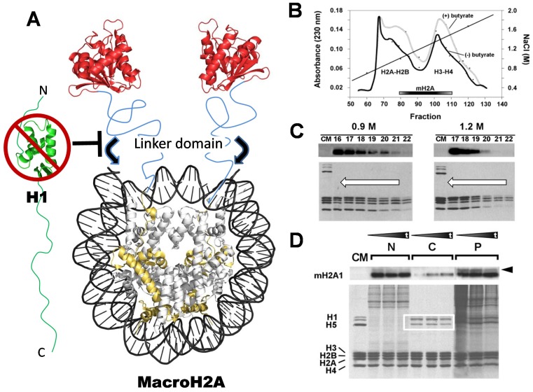
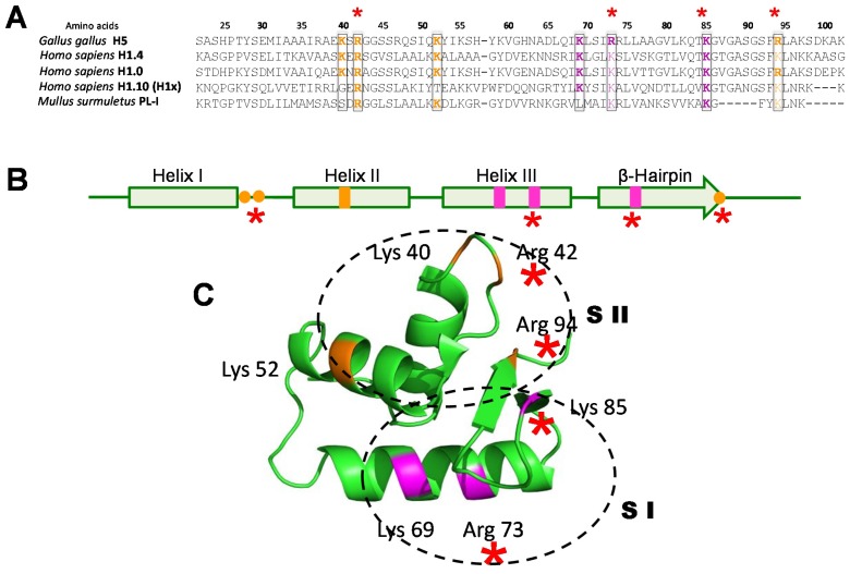
Histone variants are an important part of the histone contribution to chromatin epigenetics. In this review, we describe how the known structural differences of these variants from their canonical histone counterparts impart a chromatin signature ultimately responsible for their epigenetic contribution. In terms of the core histones, H2A histone variants are major players while H3 variant CenH3, with a controversial role in the nucleosome conformation, remains the genuine epigenetic histone variant. Linker histone variants (histone H1 family) haven't often been studied for their role in epigenetics. However, the micro-heterogeneity of the somatic canonical forms of linker histones appears to play an important role in maintaining the cell-differentiated states, while the cell cycle independent linker histone variants are involved in development. A picture starts to emerge in which histone H2A variants, in addition to their individual specific contributions to the nucleosome structure and dynamics, globally impair the accessibility of linker histones to defined chromatin locations and may have important consequences for determining different states of chromatin metabolism.

Keywords: chromatin; epigenetics; histone variants; histones; nucleosome.


Figures

Figure 1
Figure 1
Acetic acid (5%) urea (5M) -triton (0.3%) polyacrylanmide gel electrophoresis of HCl-extracted histones from nuclei of: CE (chicken erythrocyte); L (rat liver) and T (rat testes). The replacement histone variants are highlighted with a grey background, and the different types for the canonical variants are also indicated. Members of the histone H2A family are highlighted by red squares to underscore the large number of variants within this family. Names in light blue on the right hand side of the image correspond to the germline variants. For clarity, the old histone nomenclature has been used in this figure. For equivalence to the newly unified phylogeny-based nomenclature, the reader is referred to [36].
Figure 2
Figure 2
(A) Contacting regions, involving loop 1, of the histone fold for the two H2A histones present in the nucleosome. The details are shown for a homotypic nucleosome consisting of two canonical H2A histones (H2A), a heterotypic nucleosome consisting of a canonical H2A and an H2A.Z histone variant (H2A/H2A.Z), and two homotypic nucleosomes consisting of two H2A.Z.1 (H2A.Z.1) or two H2A.Z.2 (H2A.Z.2) variants. The loop 1 regions are highlighted in red and the black arrows to indicate the sites where differences are observed. The images were prepared from the crystallographic structures of the canonical histone nucleosome [78], the H2A.Z-containing nucleosome [79], and the H2A.Z.1 and H2A.Z.2 containing nucleosomes [67]. The inability of histone H1 to bind to the H2A.Z-containing nucleosome (see (C)) is indicated (┬); (B) Dependence of the sedimentation coefficient of reconstituted canonical nucleosomes (2A), homotypic nucleosomes consisting of H2A.Z (2A.Z), and heterotypic nucleosomes consisting of H2A and H2A.Z (2A-2A.Z), in Svedberg units (S) as a function of the ionic strength concentration (NaCl) in a 20 mM Tris-HCl (pH7.5) 0.1 mM EDTA buffer [64]; (C) Binding of histone H1 (linker histone, (LH)) to the nucleosome (chromatosome formation) as a function of the molar amount of linker histone (LH) per mol of nucleosome [68]. The titration was carried out using reconstituted nucleosomes consisting of either canonical histones, H3.3, or H2A.Z.
Figure 3
Figure 3
(A) Stability of the histone core octamer consisting of canonical H2A (solid line) or histone H2A.Bbd (dotted line) in the presence of 2M NaCl, as determined by gel filtration chromatography on a Sephacryl S-300 HR resin. O: octamer ((H3-H4)2.2(H2A-H2B)), H: hexamer ((H3-H4)2. (H2A-H2B)), T: tetramer ((H3-H4)2.), D: dimer ((H2A-H2B)/(H2A.Bbd-H2B)). The histones eluting with the different elution peaks are visualized in the SDS-PAGE shown underneath; (B) Ionic-strength-dependent stability of reconstituted NCPs containing H2A.Bbd (dotted line), in comparison to NCPs consisting of canonical H2A (solid line) as determined by sedimentation velocity analysis in the analytical ultracentrifuge. F: free (dissociated) DNA, N: nucleosomes; (C) Model for the H2A.Bbd (orange) structure of the H2A.Bbd-containing NCP, based on the crystallographic image of the nucleosome [78,98] and on the hydrodynamic characteristics [99,101] of the particle. The wide arrows indicate the region corresponding to the C-terminal domain of canonical H2A, which is missing in H2A.Bbd. The inability of histone H1 to bind to the H2A.Bbd-containing nucleosome [102] is indicated.
Figure 4
Figure 4
(A) Schematic representation of the macroH2A-containing nucleosome, based on the crystallographic structure of the N-terminal histone domain of macroH2A (gold) and on the crystallographic structure of the C-terminus of the macro domain (red) [111]. The tertiary structure of the linker domain region (depicted in blue) is not known, but it likely corresponds to an intrinsically disordered domain. The black arrows indicate the additional protection of DNA (approximately 10 bp at the entry and exit site of the DNA into the NCP by this linker region [112]). The inability of histone H1 to bind to the macroH2A-containing nucleosome (see (D)) is indicated; (B) Hydroxyapatite chromatography salt (NaCl) elution profiles of macroH2A, from chromatin extracts obtained from HeLa cells treated with or without sodium butyrate (to enhance global levels of histone acetylation). The elution of the H2A–H2B dimers, the H3-H4 tetramers, and also that of macroH2A, are indicated [113]; (C) MacroH2A western blot analysis of fractions (numbers) obtained from nucleosomes run on (5%–20%) sucrose gradients in the presence of 0.9 and 1.2 M NaCl. The SDS-PAGE analysis of the fractions is shown underneath. The arrows indicate the direction of the sedimentation. CM is chicken erythrocyte histone standard [110]; (D) MacroH2A western and SDS-PAGE analysis of chicken liver chromatin digested at increasing times, with micrococcal nuclease (black triangles). The digested chromatin thus obtained was fractionated according to a method initially described by Ada Olins et al. [114], which allows its separation into histone H1 depleted (N), histone H1-containing (white square) (C), and an insoluble (P) fractions [110].
Figure 5
Figure 5
(A) Figures were obtained from the crystallographic structures of the H3.1 [134], H3.2, and H3.3-containing nucleosomes [128]. The amino acid residues where the sequences differ in the α2 helix of the histone fold are shown in fuchsia. The generic image of the nucleosome is from the early crystallographic analysis by Luger et al. (1997) [78]; (B) Ionic strength (NaCl)-dependent stability of reconstituted nucleosome core particles containing H2A.Z and H3.3 histones, as visualized in the analytical ultracentrifuge [68]. D: free DNA, N: nucleosomes, s20,w: sedimentation coefficient corrected for standard water and 20 °C conditions. The DNA used in the nucleosome reconstitutions was 155 ± 5 bp random sequence DNA fragments purified from chicken erythrocyte nucleosome core particles [135].
Figure 6
Figure 6
(A) Amino acid sequence alignment of the WHD regions of several linker histones. The amino acids corresponding to the first and second sites of interaction of this domain with DNA [154] in the chromatosome are highlighted in orange and magenta (respectively). The amino acid numbers refer to their position in the histone H5 sequence; (B) Schematic representation of the secondary structure of the WHD. The sites corresponding to the first and the second histone—DNA interacting domains are in the same colours as in (A); (C) The tertiary structure of the WHD of chicken erythrocyte histone H5 as determined by crystallographic analysis [155], showing the regions and amino acids corresponding to the first (SI) and second (SII) sites of interaction with DNA. The red asterisks highlight the minimal ionic interaction sites that appear to be indispensable for this domain to perform its function in vertebrate linker histones.
Figure 7
Figure 7
Histone occupancy profiles around TSS. (A) Averaged H3.3/H2A.Z nucleosome positioning [70]; (B) Profile of histone H2A.Z distribution in high and medium transcriptionally active genes [167]; (C) Histone H1 subtypes distribution at TSS for transcribed and non-transcribed genes. H1.1 corresponds to the distribution observed for this subtype as compared to H1m, corresponding to the average for H1.2-H1.3-H1.4-H1.5 [162]; (D) Depletion of histone H1-tagged subtypes at promoters. The asterisks indicate analysis conducted using the endogenous histones [163]; (E). Schematic representation of histone variants H3.3 and H2A.Z, and histone H1 distribution around gene promoters.

References

    1. Kossel A. Uber einen peptonartigen bestandteil des zellkerns. Z. Physiol. Chem. 1884;8:511–515.
    1. Shechter D., Dormann H.L., Allis C.D., Hake S.B. Extraction, purification and analysis of histones. Nat. Protocols. 2007;2:1445–1457. doi: 10.1038/nprot.2007.202. - DOI - PubMed
    1. Ausió J., Moore S.C. Reconstitution of chromatin complexes from high-performance liquid chromatography-purified histones. Methods. 1998;15:333–342. doi: 10.1006/meth.1998.0637. - DOI - PubMed
    1. Arents G., Moudrianakis E.N. The histone fold: A ubiquitous architectural motif utilized in DNA compaction and protein dimerization. Proc. Natl. Acad. Sci. USA. 1995;92:11170–11174. doi: 10.1073/pnas.92.24.11170. - DOI - PMC - PubMed
    1. Oudet P., Gross-Bellard M., Chambon P. Electron microscopic and biochemical evidence that chromatin structure is a repeating unit. Cell. 1975;4:281–300. doi: 10.1016/0092-8674(75)90149-X. - DOI - PubMed

LinkOut - more resources