. 2015 Jul 27;10(7):e0131518.
doi: 10.1371/journal.pone.0131518. eCollection 2015.

Photodynamic Treatment of Tumor with Bacteria Expressing KillerRed

Affiliations

Photodynamic Treatment of Tumor with Bacteria Expressing KillerRed

Libo Yan et al. PLoS One. .

Abstract

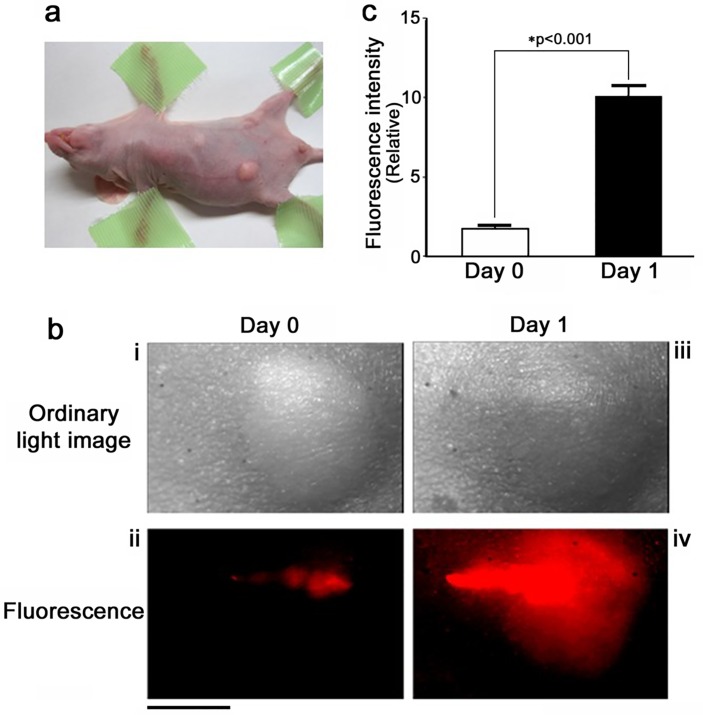
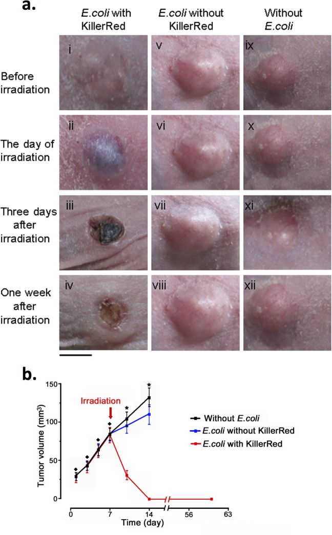
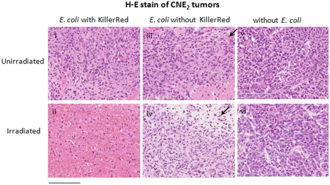
Photodynamic therapy (PDT) is a cancer treatment modality in which a photosensitizing dye is administered and exposed to light to kill tumor cells via the production of reactive oxygen species (ROS). A fundamental obstacle for PDT is the low specificity for staining solid tumors with dyes. Recently, a tumor targeting system guided by anaerobic bacteria was proposed for tumor imaging and treatment. Here, we explore the feasibility of the genetically encoded photosensitizer KillerRed, which is expressed in Escherichia coli, to treat tumors. Using nitroblue tetrazolium (NBT), we detected a lengthy ROS diffusion from the bodies of KillerRed-expressing bacteria in vitro, which demonstrated the feasibility of using bacteria to eradicate cells in their surroundings. In nude mice, Escherichia coli (E. coli) expressing KillerRed (KR-E. coli) were subcutaneously injected into xenografts comprising CNE2 cells, a human nasopharyngeal carcinoma cell line, and HeLa cells, a human cervical carcinoma cell line. KR-E. coli seemed to proliferate rapidly in the tumors as observed under an imaging system. When the intensity of fluorescence increased and the fluorescent area became as large as the tumor one day after KR-E. coli injection, the KR-E. coli-bearing tumor was irradiated with an orange light (λ = 540-580 nm). In all cases, the tumors became necrotic the next day and were completely eliminated in a few days. No necrosis was observed after the irradiation of tumors injected with a vehicle solution or a vehicle carrying the E. coli without KillerRed. In successfully treated mice, no tumor recurrence was observed for more than two months. E. coli genetically engineered for KillerRed expression are highly promising for the diagnosis and treatment of tumors when the use of bacteria in patients is cleared for infection safety.


Conflict of interest statement

Competing Interests: The authors have declared that no competing interests exist.

Figures

Fig 1
Fig 1. Superoxide is released from KR-E. coli upon orange light irradiation.
a, Ordinary light image (left) and fluorescence image (right) of a KR-E. coli suspension observed under a microscope. Bar, 10 m. b, The production of superoxide by the irradiation of KR-E. coli detected with nitroblue tetrazolium (NBT) in an agarose gel. The top panels demonstrate a change in the E. coli with KillerRed before and after irradiation. A magnified portion (right) of the agarose gel near the KR-E. coli containing central pool indicates a formazan precipitation due to the diffusion of superoxide from the pool. Bar, 5 mm. The bottom panels demonstrate change in the color of the E. coli without KillerRed expression before and after irradiation.
Fig 2
Fig 2. Absence of singlet oxygen in the phototoxic effect of illuminated KR-E. coli.
To quantitatively measure the singlet oxygen produced by KillerRed, the intensity of spontaneous luminescence was measured at wavelengths in a near infrared region. (1) KillerRed in PBS. (2) KillerRed in an O2-saturated solution. (3) KillerRed in a deuterium oxide (D2O) solution. (4) Methylene blue solution used as a control. In reference to methylene blue, KillerRed did not produce any singlet oxygen upon irradiation.
Fig 3
Fig 3. Intratumoral proliferation of KR-E. coli.
a, NPC tumor subcutaneously formed in a nude mouse. b, The distribution of KR-E. coli immediately after intratumoral injection (left), and one day after the injection (right). Scale bar, 5 mm. c, Quantification of the fluorescence intensity of KR-E. coli immediately after the intratumoral injection and one day after the injection (n = 10). *p<0.001 (t-test). Error bars indicate standard error of the mean (SEM). The fluorescence intensity was calculated by summating the digital values of all points in the field of view, and expressed in a relative manner.
Fig 4
Fig 4. Effect of PDT with KR-E. coli on CNE2 tumors.
a, The left column represents the tumor (CNE2) injected with KR-E. coli and irradiated with the orange light. The middle column represents the tumor injected with wild type E. coli and irradiated with the orange light. The right column represents the tumor irradiated without E. coli injection (n = 12). Bar, 5 mm. b, Growth curves of tumors in the presence and absence of KillerRed-E. coli. The black line indicates the volume change of tumors irradiated without E. coli. The blue line indicates the volume change of tumors injected with E. coli that expressed no KillerRed. The red line indicates the volume change of tumors injected with E. coli that expressed KillerRed. All tumors were irradiated at the same dose on the day 7 after tumor cell transplantation (Day 1 after bacterial injection). Before treatment: p > 0.05 (rank sum test). After treatment: *p < 0.01 (rank sum test). Error bars indicate SEM.
Fig 5
Fig 5. Growth curves of the HeLa tumors and CNE2 tumors of a large size treated with KR-E. coli.
The black line indicates the volume change of tumors injected with E. coli that expressed no KillerRed. The red dotted line indicates the volume change of CNE2 tumors (volume = 150 mm3) injected with E. coli that expressed KillerRed. The red full line indicates the volume change of HeLa tumors injected with E. coli that expressed KillerRed. All tumors were irradiated at the same dose on the day 7. Before treatment, p > 0.05 (rank sum test), error bars indicate SEM. After treatment, *P< 0.01 (rank sum test), error bars indicate SEM.
Fig 6
Fig 6. Histological sections of tissue showing the phototoxicity induced by KR-E. coli on CNE2 tumors.
The top panels demonstrate sections of CNE2 tumors that were injected with KR-E. coli, E. coli without KillerRed expression, and a physiological saline, and fixed before irradiation. The bottom panels demonstrate the preparations treated in the same way but fixed after irradiation with the orange light. All sections were H&E stained. The arrows indicate blood vessels of the tumor. Bar, 200 m.

References

    1. Dolmans DE, Fukumura D, Jain RK. Photodynamic therapy for cancer. Nat Rev Cancer. 2003; 3: 380–387. - PubMed
    1. Dougherty TJ, Gomer CJ, Henderson BW, Jori G, Kessel D, Korbelik M, et al. Photodynamic therapy. J Natl Cancer Inst. 1998; 90: 889–905. - PMC - PubMed
    1. Brown SB, Brown EA and Walker I. The present and future role of photodynamic therapy in cancer treatment. Lancet Oncol. 2004; 5: 497–508. - PubMed
    1. Chen J, Keltner L, Christophersen J, Zheng F, Krouse M, Singhal A, et al. New technology for deep light distribution in tissue for phototherapy. Cancer J. 2002; 8: 154–163. - PubMed
    1. Ethan DS, David D. Porphyrin-based photosensitizers for use in photodynamic therapy. Tetrahedron. 1998; 54: 4151–4202.

Publication types

LinkOut - more resources