Review
. 2015 Aug 18;54(32):4953-68.
doi: 10.1021/acs.biochem.5b00565. Epub 2015 Aug 5.

Dynamics and Membrane Interactions of Protein Kinase C

Review

Dynamics and Membrane Interactions of Protein Kinase C

Tatyana I Igumenova. Biochemistry. .

Abstract

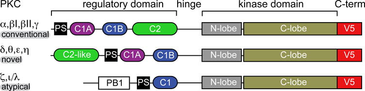
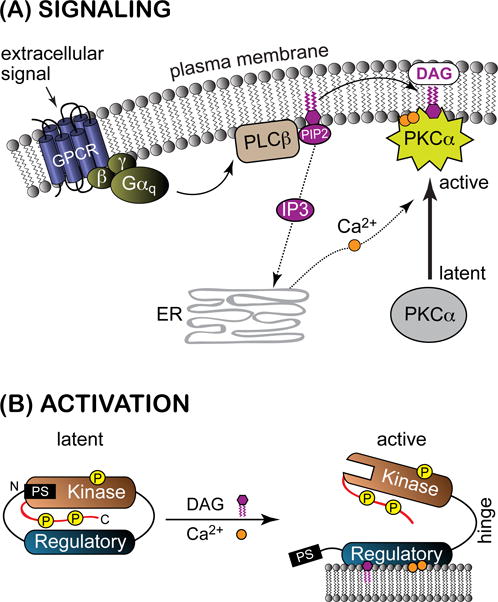
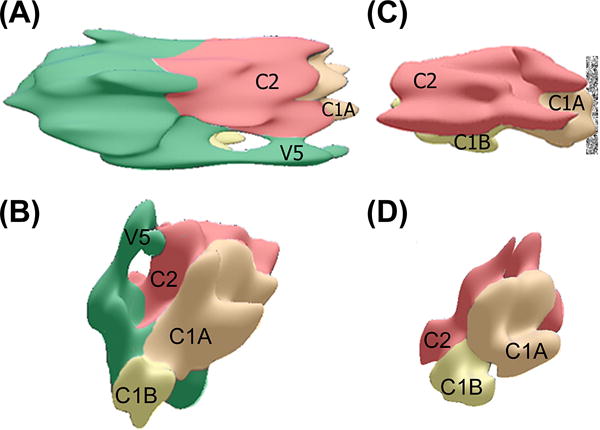
Protein kinase C (PKC) is a family of Ser/Thr kinases that regulate a multitude of cellular processes through participation in the phosphoinositide signaling pathway. Significant research efforts have been directed at understanding the structure, function, and regulatory modes of the enzyme since its discovery and identification as the first receptor for tumor-promoting phorbol esters. The activation of PKC involves a transition from the cytosolic autoinhibited latent form to the membrane-associated active form. The membrane recruitment step is accompanied by the conformational rearrangement of the enzyme, which relieves autoinhibitory interactions and thereby allows PKC to phosphorylate its targets. The multidomain structure and intrinsic flexibility of PKC present remarkable challenges and opportunities for the biophysical and structural biology studies of this class of enzymes and their interactions with membranes, the major focus of this Current Topic. I will highlight the recent advances in the field, outline the current challenges, and identify areas where biophysics and structural biology approaches can provide insight into the isoenzyme-specific regulation of PKC activity.


Figures

Figure 1
Figure 1
Multi-modular structure of PKC isoenzymes. C1 and C2 are conserved region-1 and conserved region-2 domains, respectively; PS is a pseudo-substrate region; and PB1 is a Phox and Bem1 domain. The most variable PKC regions are the N-terminal regulatory and the C-terminal V5 domains.
Figure 2
Figure 2
Signaling pathway (A) and activation (B) of PKCα. The abbreviations are given in the text. The C-terminal V5 domain is shown in red. DAG-dependent PKCs are constitutively phosphorylated as part of the maturation process, with one site on the kinase activation loop and two sites on the V5 domain.
Figure 3
Figure 3
3D reconstructions of PKCδ (A, B) and its regulatory domain RDδ (C, D) on lipid monolayers, showing the proposed assignments of individual domains (adapted from). (A) and (C) represent the views from the membrane surface, which is shown with a textured bar. The catalytic domain is colored green.
Figure 4
Figure 4
(A) The crystal structure of C2 domain from PKCα complexed to Ca2+ and PtdIns(4,5)P2. CMBL regions and the LRC lysine residues, K197, K199, K209, and K211, are highlighted with orange and blue, respectively. Adapted from: coordination geometries of Pb2+ (B), Ca2+ (C), and Cd2+ (D) ions bound to the C2 domain from PKCα. The ligands are the sidechain oxygens of aspartates, with the exception of W247 and M186, where it is the carbonyl oxygen. The coordination sphere of Pb2 is hemi-directed: all eight ligands are located in one coordination hemisphere that is facing the viewer. The top axial ligands of Ca1 in 1DSY are the phosphoryl oxygens of the short-chain PtdSer analog.
Figure 5
Figure 5
Probing the C2-membrane interactions with EPR and NMR. (A) Adapted from: depth parameters Φ are mapped onto the structure of the Ca2+-complexed C2α (PDB ID 1DSY). The spheres correspond to the backbone N-H groups, to facilitate a comparison with the NMR data. The dashed line represents the bilayer phosphate plane. (B) The CSP analysis of C2α in mixed DPS/DPC micelles. The N-H groups are color-coded according to the deviation SD from the mean CSP value. Loop residues that enter an intermediate-exchange regime upon micelle binding (N189, R249, T250, T251, R252) are colored red, with no sphere representation. Prolines and residues whose N-H groups are not spectrally resolved are in black. The CSP values Δ were calculated as described previously using 15N-1H HSQC spectra, two expansions of which are given in (C). The data in (C) illustrate the changes in chemical shifts experienced by C2α upon interactions with mixed micelles; the cross-peaks of Nε1-Hε1 groups of the Trp side-chains in the micelle-complexed form are shown in green.
Figure 6
Figure 6
C1 domains and their ligands (adapted from). (A) Primary and tertiary structure of C1Bδ complexed to phorbol-13 acetate, P13A (PDB ID 1PTR). The loop regions β12 (residues 237–242) and β34 (residues 252–257) are highlighted in green; α1 is a short α-helix comprising residues 270–275. W252 and Zn2+-coordinating residues are highlighted with orange and yellow, respectively, in the primary structure. (B) Expansion of the C1Bδ ligand-binding site showing hydrogen bonds between P13A and the backbone atoms of C1Bδ. The sidechain of W252 is not involved in direct interactions with the ligand. (C)(E) Chemical structures of representative C1 ligands: (C) 1,2-dioctanoyl-sn-glycerol, (D) phorbol-12,13-dibutyrate; and (E) Bryostatin-1. Red boxes mark the chemical groups that according to the molecular modeling studies are involved in hydrogen-bonding interactions with the protein.
Figure 7
Figure 7
Dynamics of C1 loops and their interaction with mixed micelles. (A) Differences in the μs-timescale dynamics of the wt (solid circles) and Y123W C1Bα (empty circles), illustrated using relaxation-dispersion curves for the N-H backbone groups of L122, Y123, Q128, and G129. (B) Intra- and inter-loop hydrogen bonds that stabilize β12 and β34 loops (top) and summary of conformational dynamics of loop hinges (bottom). Residues with quantifiable dispersion in both wt and mutant are underlined; residues with quantifiable dispersion in either wt or mutant are shown with regular and bold fonts, respectively (adapted from). (C) Probing the depth of C1Bδ insertion into micellar environment using PRE. The intensity ratios Ipara/Idia of the N-H resonances of W252Y C1Bδ, complexed to paramagnetic and diamagnetic preparations of mixed DPS/DPC micelles, are plotted as a function of amino acid sequence. Data in the absence and presence of DOG are shown with black and green bars, respectively. Residues whose broadening in the micelle-bound state is unrelated to PRE are labeled with “B”. The ratios were normalized to G281, the most C-terminal residue. Δ(Ipara/Idia) is the difference in Ipara/Idia ratios between the DOG-bound and micelle-only W252Y (adapted from).
Figure 8
Figure 8
Properties of V5 domain. (A) Alignment of the V5 primary structures of PKC isoenzymes from M. musculus. The conserved motifs are boxed. Acidic residues implicated in the intra-molecular interactions with the C2 domain are shown in green. (B) Catalytic domain (residues 339–679) taken from the partial crystal structure of PKCβII, PDB ID 3PFQ. The B-factors of backbone Cα atoms are mapped onto the structure as a color gradient. The N-/C-lobes of the kinase domain and V5 are shown with transparent and opaque representations, respectively. (C) SSP scores calculated using NMR chemical shift data for the free (black) and micelle-bound V5α (red). Upon binding to DPC micelles, V5α acquires a partial α-helical structure, most notably in the NFD region and the most C-terminal residues (adapted from).

References

    1. Inoue M, Kishimoto A, Takai Y, Nishizuka Y. Studies on a cyclic nucleotide-independent protein kinase and its proenzyme in mammalian tissues. 2. Proenzyme and its actiavtion by calcium-dependent protease from rat brain. J Biol Chem. 1977;252:7610–7616. - PubMed
    1. Takai Y, Kishimoto A, Inoue M, Nishizuka Y. Studies on a cyclic nucleotide-independent protein kinase and its proenzyme in mammalian tissues. 1. Purification and characterization of an active enzyme from bovine cerebellum. J Biol Chem. 1977;252:7603–7609. - PubMed
    1. Castagna M, Takai Y, Kaibuchi K, Sano K, Kikkawa U, Nishizuka Y. Direct Activation of Calcium-Activated, Phospholipid-Dependent Protein-Kinase by Tumor-Promoting Phorbol Esters. J Biol Chem. 1982;257:7847–7851. - PubMed
    1. Newton AC. Protein kinase C: Structural and spatial regulation by phosphorylation, cofactors, and macromolecular interactions. Chem Rev. 2001;101:2353–2364. - PubMed
    1. Rosse C, Linch M, Kermorgant S, Cameron AJM, Boeckeler K, Parker PJ. PKC and the control of localized signal dynamics. Nature Reviews Molecular Cell Biology. 2010;11:103–112. - PubMed

Publication types