

doi: 10.1038/nn.4068.
Epub 2015 Jul 27.
Coding the direct/indirect pathways by D1 and D2 receptors is not valid for accumbens projections
Affiliations
- PMID: 26214370
- PMCID: PMC4551610
- DOI: 10.1038/nn.4068
Item in Clipboard
Coding the direct/indirect pathways by D1 and D2 receptors is not valid for accumbens projections
Nat Neurosci.
2015 Sep.
Abstract
It is widely accepted that D1 dopamine receptor-expressing striatal neurons convey their information directly to the output nuclei of the basal ganglia, whereas D2-expressing neurons do so indirectly via pallidal neurons. Combining optogenetics and electrophysiology, we found that this architecture does not apply to mouse nucleus accumbens projections to the ventral pallidum. Thus, current thinking attributing D1 and D2 selectivity to accumbens projections akin to dorsal striatal pathways needs to be reconsidered.
Figures
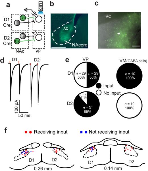
(a) Schematic of the experimental protocol. Cre-dependent ChR2 was injected into the NAcore of D1-Cre (top) or D2-Cre (bottom) mice. Terminals of the infected MSNs (green) were optogenetically activated and responses were recorded in VP neurons. (b) Representative image of injection sites (see Supplementary Figure 1a for composite of injection sites) of Cre-dependent ChR2 into the NAcore (D1-Cre mouse). Calibration bar - 500 μm. AC - anterior commissure. (c) Representative micrograph of Cre-dependent ChR2 expression surrounding the recording pipette (arrowhead) in the dVP. ChR2 expression in dVP was used to define recording site for all experiments. Calibration bar - 150 μm. (d) Representative responses to optogenetic stimulation (red dots) of D1-MSN or D2-MSN input to the dVP, obtained in all experiments involving dVP recordings. (e) Half of the neurons in the dVP received input from NAcore D1-MSNs. In the VM only D1-MSN input from the NAcore was detected. n represents number of cells. n in figure is cells recorded, number of animals was: D1-VP n= 12, D2-VP n= 10, D1-VM n= 3, D2-VM n=3. (f) Location of patched cells exhibiting (red) or not (blue) eIPSCs after stimulation of D1-MSN (squares) or D2-MSN (circles) terminals.
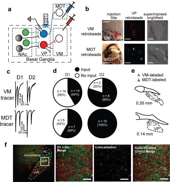
(a) Schematic of experimental protocol. Cre-dependent ChR2 was injected into the NAcore of D1-Cre or D2-Cre mice while retrobeads were injected into the VM or MDT. Infected terminals (green) were optogenetically activated and retrogradely-labeled VP projection neurons were recorded from. (b) Representative images of retrobeads in the injection sites and in retrogradely-labeled VP neurons. Red MDT retrobeads are shown in cyan pseudo-color. Injection site scale bars - 1 mm. Retrobeads scalebars - 5 μm. (c) Representative traces of optogenetically-generated IPSCs recorded from retrogradely-labeled VP neurons in D1-Cre and D2-Cre mice. (d) Approximately half of the VM-projecting VP neurons received D1-MSN input from the NAcore while 80% received D2-MSN input. All MDT-projecting VP neurons responded to D2-MSN activation while 58% received D1-MSN input from the NAcore. n in figure represents number of cells, number mice was: D1-VM n= 6, D2-VM n= 3, D1-MDT n= 3, D2-MDT n= 3. (e) In the more rostral VP VM-projecting (red) neurons tended to be dorsolateral to the MDT-projecting (blue) neurons while at the caudal VP both projection cells were intermingled. (f) 2.4% (n= 3) co-localization of D1- and D2-expression in fibers in the dVP was quantified in D1-tomato X D2-GFP BAC transgenic mice. The left panel shows low magnification of the location in the dVP where we quantified fiber co-expression shown in the next 3 panels. Bar= 50 μm.
References
Publication types
MeSH terms
Substances
Grants and funding
LinkOut - more resources
Full Text Sources
Other Literature Sources
Molecular Biology Databases
