. 2015 Aug 3;125(8):2995-3007.
doi: 10.1172/JCI79386. Epub 2015 Jul 27.

Lymph flow regulates collecting lymphatic vessel maturation in vivo

Lymph flow regulates collecting lymphatic vessel maturation in vivo

Daniel T Sweet et al. J Clin Invest. .

Abstract

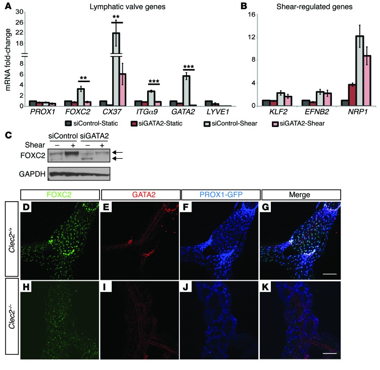
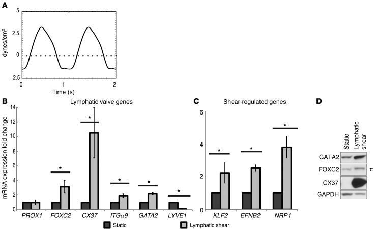
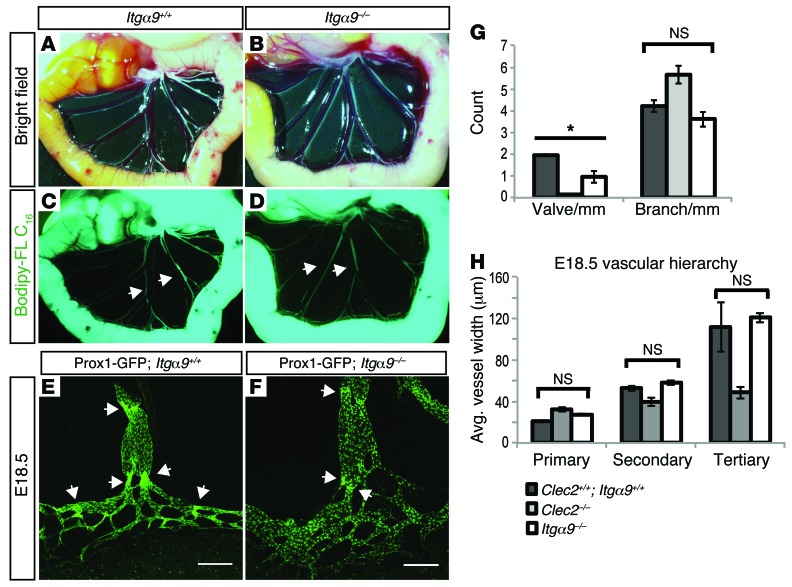
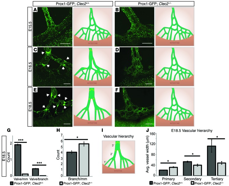
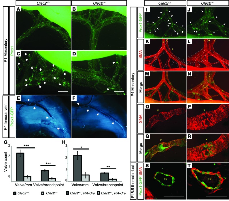
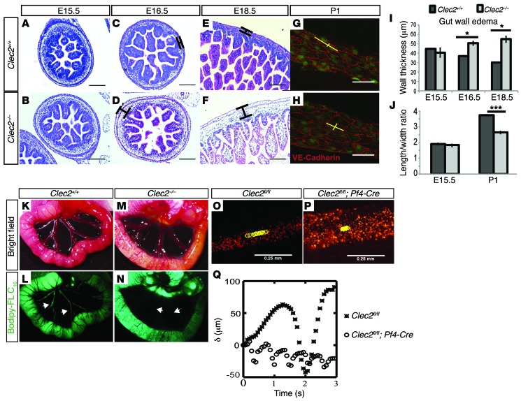
Fluid shear forces have established roles in blood vascular development and function, but whether such forces similarly influence the low-flow lymphatic system is unknown. It has been difficult to test the contribution of fluid forces in vivo because mechanical or genetic perturbations that alter flow often have direct effects on vessel growth. Here, we investigated the functional role of flow in lymphatic vessel development using mice deficient for the platelet-specific receptor C-type lectin-like receptor 2 (CLEC2) as blood backfills the lymphatic network and blocks lymph flow in these animals. CLEC2-deficient animals exhibited normal growth of the primary mesenteric lymphatic plexus but failed to form valves in these vessels or remodel them into a structured, hierarchical network. Smooth muscle cell coverage (SMC coverage) of CLEC2-deficient lymphatic vessels was both premature and excessive, a phenotype identical to that observed with loss of the lymphatic endothelial transcription factor FOXC2. In vitro evaluation of lymphatic endothelial cells (LECs) revealed that low, reversing shear stress is sufficient to induce expression of genes required for lymphatic valve development and identified GATA2 as an upstream transcriptional regulator of FOXC2 and the lymphatic valve genetic program. These studies reveal that lymph flow initiates and regulates many of the key steps in collecting lymphatic vessel maturation and development.


Figures

Figure 6
Figure 6. GATA2 is required for flow-induced expression of lymphatic valve genes.
(A and B) Changes in LEC valve gene expression induced by lymphatic fluid shear following siRNA knockdown of GATA2. Gene expression was measured following transfection with siGATA2 or control siRNA (siControl) in static LEC or LEC exposed to lymphatic flow for 24 hours. n = 4 independent experiments. All values are means ± SEM. **P < 0.01, ***P < 0.001, calculated by Student’s t test. (C) Western blot of FOXC2 molecular weight shift in siControl vs. siGATA2 LEC exposed to lymphatic shear for 24 hours or kept static. n = 3 experiments. (DK) Whole-mount staining for FOXC2 (green), GATA2 (red), and PROX1-GFP (blue) in E17.5 mesenteric lymphatic vessels of Clec2+/+ and Clec2–/– embryos. Scale bars: 100 μm.
Figure 5
Figure 5. Lymphatic shear stress induces the expression of most genes required for lymphatic valve development.
(A) The reversing flow regimen shown was used to expose human LECs to fluid shear forces known to exist in lymphatic collecting vessels in vivo for 48 hours. LECs were exposed to a maximum of 3.25 dynes/cm2, a minimum of –1.25 dynes/cm2, and an average 0.67 dynes/cm2 of shear stress. (B and C) Changes in the expression of genes associated with lymphatic valve development (B) and genes known to be induced by laminar shear in blood ECs (C) were measured as fold-change compared with static control and normalized to GAPDH following LEC exposure to the lymphatic flow regimen shown in (A). n = 4 experiments. All values expressed as fold-change means ± SEM. *P < 0.05, calculated by Student’s t test. (D) Western blot showing upregulation of protein expression in response to 48-hour lymphatic shear. Blots were probed for GATA2, FOXC2, and CX37, with GAPDH as a loading control. Arrows indicate slight molecular weight shift of FOXC2 after flow. n = 3 experiments.
Figure 4
Figure 4. Embryonic vessel remodeling is preserved in Itga9–/– animals that lack lymphatic valves.
(AD) Oral lymphangiography in P1 Itga9–/– neonates and littermate controls. Images of the intestine and mesentery were acquired 2 hours after feeding of Bodipy-FL C16 (green). White arrows indicate contrast-filled mesenteric lymphatic vessels. Images are representative of 3 mice per genotype. (E and F) Mesenteric lymphatic vessel morphology and patterning is preserved in E18.5 Itga9–/– embryos. Mesenteric lymphatic vasculature was visualized in E18.5 Itga9–/– embryos and littermate controls on a Prox1-GFP reporter background. White arrows indicate nascent lymphatic valves. Scale bars: 200 μm. (G) Quantitation of mesenteric lymphatic valve number and vessel branchpoint number in E18.5 control, Itga9–/–, and Clec2–/– embryos. Control embryos were a combination of the littermates of Itga9–/– and Clec2–/– embryos. n = 4 or more embryos per genotype. (H) Quantitation of lymphatic vascular hierarchy in E18.5 control, Itga9–/–, and Clec2–/– embryos. Lymphatic vessel width was measured at 3 levels of the lymphatic vascular tree: primary (most proximal to the gut), secondary, and tertiary (most distal to the gut). n = 4 embryos per genotype. All values are means ± SEM. *P < 0.05, calculated by Student’s t test.
Figure 3
Figure 3. Failure of mesenteric lymphatic vessel remodeling and valve initiation in Clec2–/– mice.
(AF) Analysis of mesenteric lymphatic vessel morphology and patterning at E15.5 (A and B), E16.5 (C and D), and E18.5 (E and F) in Prox1-GFP BAC transgenic embryos. White arrows indicate sites of lymphatic valve formation. Vascular architecture and valve formation are shown diagrammed on the right. Scale bars: 200 μm. (G and H) Quantitation of valve number (G) and vessel branchpoint number (H) in E18.5 Clec2–/– and Clec2+/+ mesenteric lymphatics. n = 9 embryos per genotype. (I) Schematic of the late gestation WT mesenteric lymphatic network demonstrating vessels denoted as primary, secondary, and tertiary based on branching and distance from the intestine. (J) Quantitation of lymphatic vascular hierarchy in E18.5 Clec2–/– and Clec2+/+ embryos. Lymphatic vessel width was measured at the 3 levels of the lymphatic vascular tree indicated in I. n = 4 embryos per genotype. At least 6 vessels at each level were measured per embryo. All values are means ± SEM. *P < 0.05, ***P < 0.001, calculated by Student’s t test.
Figure 2
Figure 2. Lymphatic valve development is blocked in Clec2–/– mesenteric lymphatics.
(AD) Whole-mount staining for PROX1 in P1 neonatal mesentery was used to identify lymphatic valves. White arrows indicate PROX1HI lymphatic valves. Scale bars: 200 μm. Representative images shown from 6 mice per genotype. (E and F) Analysis of venous valves in the femoral vein of P6 pups by visualization of PROX1-GFP, which is expressed in venous valve BECs. White arrows indicate venous valves. Representative images shown from 4 mice per genotype. (G) Quantitation of lymphatic valve number in neonatal Clec2–/– and Clec2+/+ littermates. n = 6 mice per genotype. (H) Quantitation of lymphatic valve number in neonatal platelet-specific conditional Clec2fl/–; Pf4-Cre and Clec2 fl/+; Pf4-Cre littermates. n = 3 mice per genotype. All values are means ± SEM. *P < 0.05, **P < 0.01, ***P < 0.001, calculated by Student’s t test. (KT) Analysis of SMC coverage of Clec2–/– and Clec2+/+ littermates. (IN) Staining of mesenteric vessels for smooth muscle actin (red) and lymphatic ECs (PROX1-GFP, green) in P4 neonates. V, vein; A, artery; L, lymphatic. White dotted line in KL outlines lymphatic vessels. White arrows indicate lymphatic valves. Scale bars: 200 μm. Representative images shown from 3 mice per genotype. (OR) High-magnification confocal microscopy of P4 lymphatic valve regions (smooth muscle actin, red) and lymphatic ECs (PROX1-GFP, green). V, valve. Scale bars: 100 μm. (S and T) Histology of E18.5 thoracic duct (TD) for smooth muscle actin (red) and lymphatic ECs (PROX1-GFP, green). Scale bars: 100 μm. Representative images shown from 2 mice per genotype.
Figure 1
Figure 1. Lack of detectable lymph flow in developing mesenteric lymphatics of Clec2–/– mice.
(AF) Clec2–/– mice exhibit edema in the intestine wall after E15.5. H&E staining of cross sections of embryonic intestine is shown at E15.5, E16.5, and E18.5. Scale bars: 100 μm. (G and H) LEC elongation in P1 mesenteric lymphatics was measured by staining for PROX1 (green) and VE-cadherin (red). Yellow lines indicate representative measurements of cell length and width. Scale bars: 50 μm. (I) Quantitation of intestine wall thickness. n = 2 mice per timepoint for each genotype and 2 sections per mouse with at least 3 wall thickness measurements per section. (J) Quantitation of LEC elongation using length/width ratio. n = 4 animals at E15.5 and 5 animals at P1 per genotype. Over 100 LECs were measured per mouse. All values are means ± SEM. *P < 0.05, ***P < 0.001. P value calculated by Student’s t test. (KN) Oral lymphangiogram 2 hours after neonatal ingestion of Bodipy-FL C16. White arrows indicate lymphatic vessels in the mesentery. Images are representative of 5 mice per genotype. (O and P) In vivo measurement of lymphatic flow by bead tracking in surviving 4-week-old in Clec2fl/fl vs. Clec2fl/fl; Pf4-Cre mice with blood-filled lymphatic phenotype. Yellow circles indicate position of bead in each frame of video over one contraction cycle. (Q) Quantitation of bead displacement (μm) over time relative to starting point in Clec2fl/fl (black asterisks) vs. Clec2fl/fl; Pf4-Cre (white circles). Representative images shown from bead tracking in 3 mice per genotype.

Comment in

Similar articles

Cited by

References

    1. Tammela T, Alitalo K. Lymphangiogenesis: Molecular mechanisms and future promise. Cell. 2010;140(4):460–476. doi: 10.1016/j.cell.2010.01.045. - DOI - PubMed
    1. Baluk P, et al. Functionally specialized junctions between endothelial cells of lymphatic vessels. J Exp Med. 2007;204(10):2349–2362. doi: 10.1084/jem.20062596. - DOI - PMC - PubMed
    1. Wang Y, Simons M. Flow-regulated lymphatic vasculature development and signaling. Vasc Cell. 2014;6:14. doi: 10.1186/2045-824X-6-14. - DOI - PMC - PubMed
    1. Makinen T, Norrmen C, Petrova TV. Molecular mechanisms of lymphatic vascular development. Cell Mol Life Sci. 2007;64(15):1915–1929. doi: 10.1007/s00018-007-7040-z. - DOI - PMC - PubMed
    1. Bazigou E, Makinen T. Flow control in our vessels: vascular valves make sure there is no way back. Cell Mol Life Sci. 2013;70(6):1055–1066. doi: 10.1007/s00018-012-1110-6. - DOI - PMC - PubMed

Publication types

MeSH terms