. 2015 Aug 3;125(8):2979-94.
doi: 10.1172/JCI78888. Epub 2015 Jul 27.

GATA2 is required for lymphatic vessel valve development and maintenance

GATA2 is required for lymphatic vessel valve development and maintenance

Jan Kazenwadel et al. J Clin Invest. .

Abstract

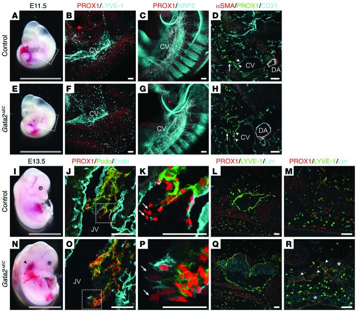
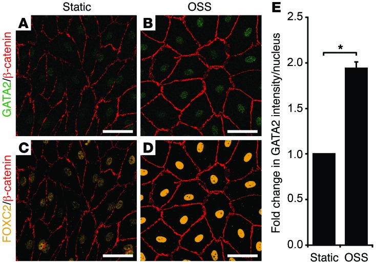
Heterozygous germline mutations in the zinc finger transcription factor GATA2 have recently been shown to underlie a range of clinical phenotypes, including Emberger syndrome, a disorder characterized by lymphedema and predisposition to myelodysplastic syndrome/acute myeloid leukemia (MDS/AML). Despite well-defined roles in hematopoiesis, the functions of GATA2 in the lymphatic vasculature and the mechanisms by which GATA2 mutations result in lymphedema have not been characterized. Here, we have provided a molecular explanation for lymphedema predisposition in a subset of patients with germline GATA2 mutations. Specifically, we demonstrated that Emberger-associated GATA2 missense mutations result in complete loss of GATA2 function, with respect to the capacity to regulate the transcription of genes that are important for lymphatic vessel valve development. We identified a putative enhancer element upstream of the key lymphatic transcriptional regulator PROX1 that is bound by GATA2, and the transcription factors FOXC2 and NFATC1. Emberger GATA2 missense mutants had a profoundly reduced capacity to bind this element. Conditional Gata2 deletion in mice revealed that GATA2 is required for both development and maintenance of lymphovenous and lymphatic vessel valves. Together, our data unveil essential roles for GATA2 in the lymphatic vasculature and explain why a select catalogue of human GATA2 mutations results in lymphedema.


Figures

Figure 13
Figure 13. Model depicting mechanisms by which Gata2 regulates lymphatic vascular development.
GATA2 is elevated in valve-forming territories at the onset of lymphatic vessel valve development and regulates the levels of PROX1 and FOXC2 in valve-forming endothelial cells. Deletion of Gata2 in the lymphatic vasculature prior to the initiation of lymphatic vessel valve development results in reduced FOXC2 levels throughout the lymphatic vasculature, ectopic recruitment of vascular SMCs to dermal lymphatic vessels, dilated lymphatic vessels, and arrested valve development. Postnatal deletion of Gata2 in the lymphatic vasculature following valve assembly results in diminished levels of PROX1 in the endothelial cells comprising lymphatic vessel valves and consequent valve disorganization.
Figure 12
Figure 12. Lymphatic vascular defects in adult Gata2ΔEC mice.
Adult heterozygous Gata2ΔEC/+ mice injected with Evans Blue dye exhibited collecting lymphatic vessels of substantially larger caliber (BD) than controls (A). Thoracic duct area was measured using ImageJ in control (n = 5) and heterozygous Gata2ΔEC/+ (n = 6) adult mice (D). *P < 0.05, by 2-tailed Student’s t-test. Reduced transport of Evans Blue dye to the thoracic duct and blood within the thoracic duct (C; arrow) were also observed in Gata2ΔEC/+ mice. Scale bars: 1 mm. TD, thoracic duct.
Figure 11
Figure 11. Gata2 deletion results in degeneration of lymphatic vessel valves and lymphatic vessel distension.
Gata2ΔLEC and littermate control (Cre-negative; Gata2fl/fl) pups were injected with tamoxifen at P4. Whole-mount immunostaining of mesenteries at P10 with PROX1 (cyan) and CD31 (green) demonstrated severely dysmorphic lymphatic vessel valves and distended lymphatic vessels in Gata2ΔLEC mesenteries (CH, JL, and NP), compared with littermate controls (A, B, I, and M). Scale bars: 100 μm (AH) or 50 μm (IP).
Figure 10
Figure 10. Gata2 is required for lymphatic vessel valve maintenance.
Gata2ΔLEC and littermate control (Cre-negative;Gata2fl/fl) pups were injected with tamoxifen at P4. Whole-mount immunostaining of mesenteries at P8 with PROX1 (cyan) and CD31 (green) demonstrated reduced PROX1 levels in lymphatic vessel valves of Gata2ΔLEC mice (C, E, H, and I), compared with littermate controls (A and G). Blood vascular patterning was indistinguishable from littermate controls. Scale bars: 100 μm (AF) or 50 μm (GL).
Figure 9
Figure 9. Lymphatic vessel valve development is perturbed in Gata2ΔLEC embryos.
Prox1-CreERT2 Gata2fl/+ male mice were crossed with Gata2fl/fl female mice, and tamoxifen was administered at E12.5, E13.5, and E14.5. E16.5 Gata2ΔLEC embryos exhibit blood-filled dermal lymphatic vessels and pooling of blood in the jugular region (F), phenotypes not observed in control littermates (Cre-negative;Gata2fl/fl, A). Whole-mount immunostaining of skin from E16.5 Gata2ΔLEC embryos revealed enlarged, blood-filled dermal lymphatic vessels (GJ) that were ectopically associated with vascular SMCs (H, arrows), phenotypes not observed in control littermates (BE). Reduced numbers of PROX1-high valve territories (I, arrowheads) were obvious in E16.5 Gata2ΔLEC embryos compared with littermate controls (D, arrowheads). Gata2ΔLEC embryos appeared indistinguishable from control littermates at E18.5 (K and O). Whole-mount immunostaining of E18.5 mesenteries demonstrated bulbous mesenteric lymphatic vessels in Gata2ΔLEC embryos (PR), lacking organized valve forming territories (LN). Single-channel images of boxed regions in L and P are shown in S and T, and U and V, respectively. Scale bars: 5 mm (A, F, K, and O) or 100 μm (BE, GJ, LN, and PR). Lin+; APC mouse lineage antibody cocktail used to detect hematopoietic cells.
Figure 8
Figure 8. Lymphatic vascular phenotypes in E13.5 and E14.5 Gata2ΔLEC embryos.
Prox1-CreERT2 Gata2fl/+ male mice were crossed with Gata2fl/fl female mice, and tamoxifen was administered at E10.5, E11.5, and E12.5, followed by analysis at E13.5 (AD), or E10.5 and 11.5, followed by analysis at E14.5 (EL). E13.5 Gata2ΔLEC embryos exhibit striking edema (C, arrow) and greatly enlarged, blood-filled jugular lymph sacs (arrowhead, C and D), compared with control littermates (Cre-negative; Gata2fl/fl, A and B). At E14.5, Gata2ΔLEC embryos exhibit edema (I, arrow). Immunostaining of coronal E14.5 tissue sections demonstrated no obvious morphological abnormalities in LVVs of Gata2ΔLEC mice (JL) compared with control littermates (FH). Scale bars: 5 mm (A, C, E, and I) or 100 μm (B, D, FH, and JL). JLS, jugular lymph sac; JV, jugular vein.
Figure 7
Figure 7. Lymphatic vessel hyperplasia in Gata2ΔEC embryos.
E11.5 Gata2ΔEC embryos appear phenotypically normal (A and E). Whole-mount immunostaining for PROX1 (red) and LYVE-1 or NRP2 (cyan) demonstrated that PROX1-positive lymphatic endothelial progenitor cells are specified and migrate away from the cardinal veins of Gata2ΔEC embryos (F and G). Immunostaining of transverse sections for α smooth muscle actin (αSMA; red), PROX1 (green), and CD31 (cyan) confirmed PROX1 expression in lymphatic endothelial progenitor cells of the cardinal veins of Gata2ΔEC embryos (D and H; arrowheads) and migration of these cells from the veins (D and H, arrows). Boxed regions in AE correspond to areas shown in B, C, F, and G. E13.5 Gata2ΔEC embryos exhibit severe edema, pooling of blood in the jugular region (N, arrowhead), and focal dermal hemorrhages (R, asterisks), compared with control littermates (Cre-negative;Gata2fl/fl; I and M). LVVs appeared multi-layered (O) with reduced PROX1 levels in the venous leaflets (Podoplanin-negative, Endomucin-low) of Gata2ΔEC embryos (P, arrows) compared with controls (J and K). PROX1 levels in lymphatic leaflets (Podoplanin-positive, Endomucin-negative) of Gata2ΔEC embryos were not substantially altered (O and P). Jugular lymph sacs (Q) and dermal lymphatic vessels (R, arrowheads) were enlarged and blood-filled in Gata2ΔEC embryos, phenotypes not observed in control embryos (L and M). Magnified images of boxed regions in J and O are shown in K and P, respectively. Gray scale bars: 5 mm. White scale bars: 50 μm (K and P) or 100 μm (all panels excluding K and P). CV, cardinal vein; DA, dorsal aorta; JV, jugular vein; Lin+, APC mouse lineage antibody cocktail used to detect hematopoietic cells.
Figure 6
Figure 6. OSS induced elevation of FOXC2 is dependent on GATA2.
hLECs were transfected with control or GATA2 siRNA and cultured under static (A, D, G, and J) conditions, or subjected to laminar shear stress (LSS; B, E, H, and K) or OSS (C, F, I, and L) for 48 hours (4 dyn/cm2, 1/4 Hz). Immunostaining revealed reduced nuclear FOXC2 levels in GATA2 siRNA–treated hLECs in response to OSS (L compared with I). Scale bars: 50 μm.
Figure 5
Figure 5. In vitro OSS increases GATA2 levels in hLECs.
hLECs were cultured under static conditions (A and C) or subjected to OSS (4 dyn/cm2, 1/4 Hz) (B and D) for 48 hours. Immunostaining for GATA2 and FOXC2 demonstrated elevated levels of nuclear GATA2 (A, B, and E) and FOXC2 (C and D) following exposure of hLECs to OSS. Error bars correspond to ± SEM, n = 4 independent experiments. *P < 0.00001, by 2-tailed Student’s t test. Scale bars: 50 μm.
Figure 4
Figure 4. Localization of GATA2 in valves and arteries.
(AE) Immunostaining of E13.5 WT mouse tissue sections demonstrated that GATA2 is present at higher levels in the endothelial cells comprising LVV leaflets (E, arrows) compared with endothelial cells lining the jugular veins and jugular lymph sacs (E, arrowheads). GATA2 is also present in cardiac valves (FI) and arterial endothelial cells (JM). Boxed region in A is shown at a higher magnification in BE. Scale bars: 50 μm. JLS, jugular lymph sac; JV, jugular vein; VA, vertebral artery.
Figure 3
Figure 3. Occupancy of chromatin at the PROX1 –11 kb enhancer element.
(A) PROX1 locus as viewed in UCSC Human Genome Browser (http://genome.ucsc.edu/). Red boxed area indicates approximate location of the PROX1 –11 kb enhancer element. (B) ChIP demonstrates that GATA2, FOXC2, and NFATC1 ChIP at the PROX1 –11 kb enhancer in LECs and BECs, but not K562 cells. Where error bars are shown, error bars represent ± SEM, n ≥ 3 independent experiments. Where error bars are not shown, data represents an average of 2 independent experiments. (C) ChIP-seq profile illustrating occupancy of the PROX1 locus by GATA2 in lymphatic endothelial cells. (D) ChIP with markers of active (H3K4Me1) and inactive/repressed (H3K27Me3) chromatin at the PROX1 –11 kb element demonstrates that PROX1 –11 kb is active in LECs and repressed in BECs and K562. Data are representative of 2 independent experiments.
Figure 2
Figure 2. GATA2 Emberger mutants exhibit reduced DNA-binding affinity.
(A) EMSA analyses using recombinant, purified GATA2 WT C-terminal zinc finger (CF) and mutants showing that Emberger mutants (red) exhibit negligible binding to the PROX1 –11 kb element. (B) Far-UV CD spectra and (C) 1-D H1 NMR spectra (amide region) of recombinant purified proteins at 25°C, pH 7.4 in 20 mM sodium phosphate, 50 mM NaCl, 1 mM DTT showing C373R is largely disordered and that T354M is partly disordered. (D) Structure of the C-terminal finger of GATA3 (cyan) bound to DNA (gray; Protein Data Bank accession code 4hc7 [http://www.rcsb.org/]) showing positions of mutated residues. Emberger mutations are shown as red sticks, residues that moderately affect DNA binding by EMSA are shown as magenta sticks, and those that show WT binding are shown as pink sticks. Other zinc-coordinating residues are shown as yellow sticks, and zinc as a gray sphere.
Figure 1
Figure 1. GATA2 Emberger mutants have reduced capacity to bind and transactivate a novel PROX1 –11 kb enhancer element.
(A) Schematic demonstrating location of PROX1 –11 kb enhancer element relative to the PROX1 gene and arrangement of consensus transcription factor binding sites. (B) Treatment of hLECs with GATA2 siRNA results in substantial reduction in PROX1 levels. (C) Mutation of the GATA binding site (BS) in the PROX1 –11 kb element reduced luciferase activity mediated by WT GATA2. EV, empty vector control. Error bars correspond to ± SEM, n = 5 independent experiments. *P < 0.05, by 2-tailed Student’s t test. (D) The PROX1 –11 kb and +4.5 kb elements cooperatively enhance luciferase activity. Fold-change is shown relative to EV controls. Error bars correspond to ± SEM, n = 3 independent experiments. *P < 0.05, by 2-tailed Student’s t test. (E) HEK293 cells were cotransfected with GATA2 (WT or mutant) expression constructs together with a PROX1 –11 kb luciferase reporter construct. Luciferase activity was measured after 24 hours. Error bars correspond to ± SD (n = 6) from 2 independent experiments (1-way ANOVA, *P < 0.05 relative to WT GATA2) Emberger mutants (here and in subsequent panels) are labeled red. (F) Binding of WT and mutant GATA2 proteins to the PROX1 –11 kb element, assessed by WEMSA.

Comment in

Similar articles

Cited by

References

    1. Mansour S, et al. Emberger syndrome-primary lymphedema with myelodysplasia: report of seven new cases. Am J Med Genet A. 2010;152A(9):2287–2296. doi: 10.1002/ajmg.a.33445. - DOI - PubMed
    1. Ostergaard P, et al. Mutations in GATA2 cause primary lymphedema associated with a predisposition to acute myeloid leukemia (Emberger syndrome). Nat Genet. 2011;43(10):929–931. doi: 10.1038/ng.923. - DOI - PubMed
    1. Kazenwadel J, et al. Loss-of-function germline GATA2 mutations in patients with MDS/AML or MonoMAC syndrome and primary lymphedema reveal a key role for GATA2 in the lymphatic vasculature. Blood. 2012;119(5):1283–1291. doi: 10.1182/blood-2011-08-374363. - DOI - PMC - PubMed
    1. Ishida H, et al. GATA-2 anomaly and clinical phenotype of a sporadic case of lymphedema, dendritic cell, monocyte, B- and NK-cell (DCML) deficiency, and myelodysplasia. Eur J Pediatr. 2012;171(8):1273–1276. doi: 10.1007/s00431-012-1715-7. - DOI - PubMed
    1. Khandekar M, et al. A Gata2 intronic enhancer confers its pan-endothelia-specific regulation. Development. 2007;134(9):1703–1712. doi: 10.1242/dev.001297. - DOI - PubMed

Publication types

MeSH terms

Associated data