. 2015 Aug 3;125(8):3117-31.
doi: 10.1172/JCI80313. Epub 2015 Jul 27.

Fibrinolysis is essential for fracture repair and prevention of heterotopic ossification

Fibrinolysis is essential for fracture repair and prevention of heterotopic ossification

Masato Yuasa et al. J Clin Invest. .

Erratum in

Abstract

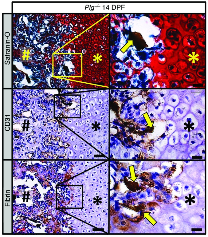
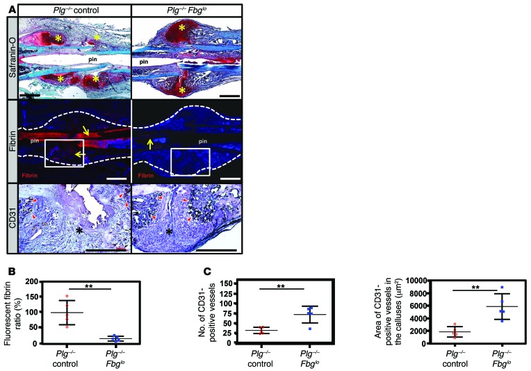
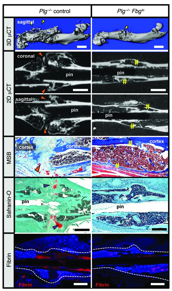
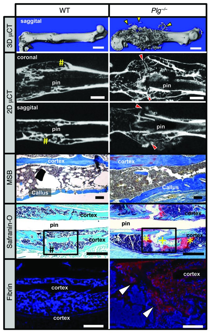
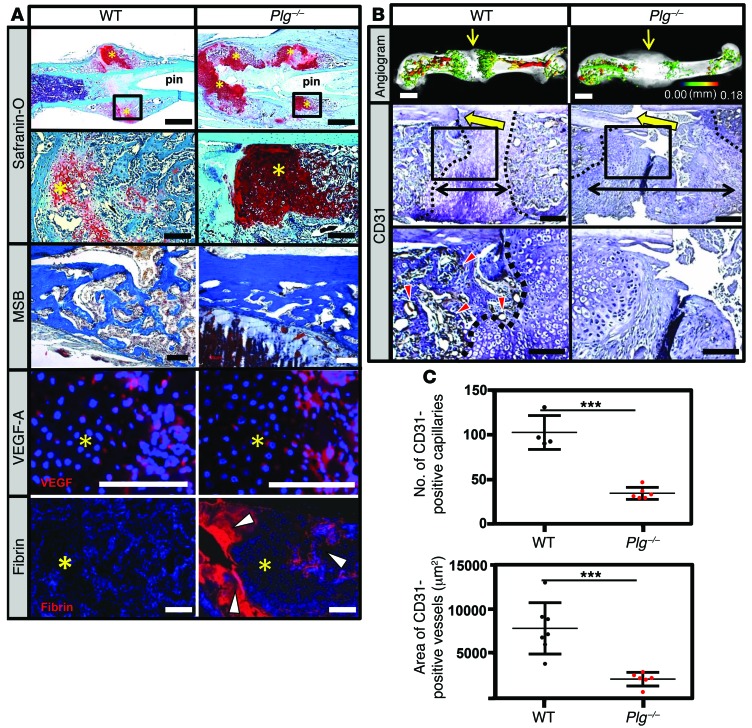
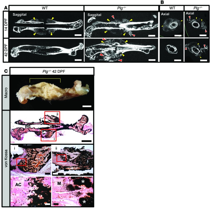
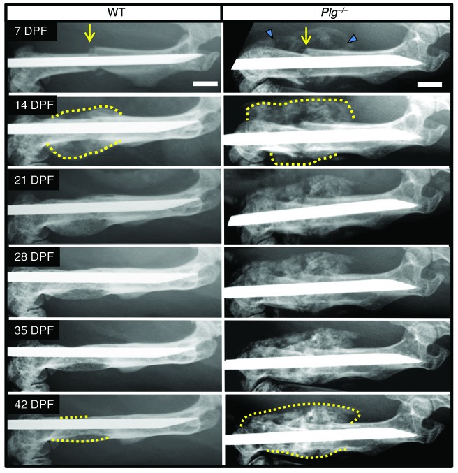
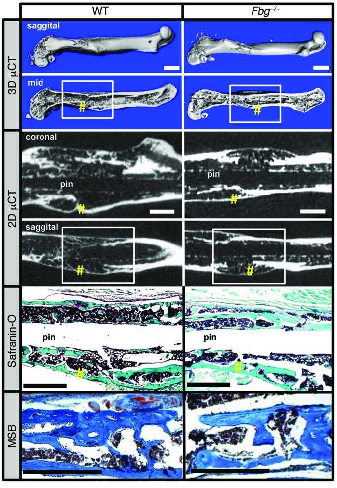
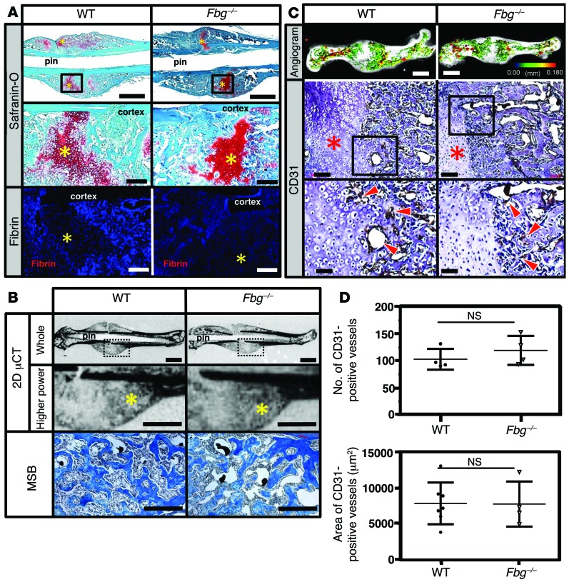
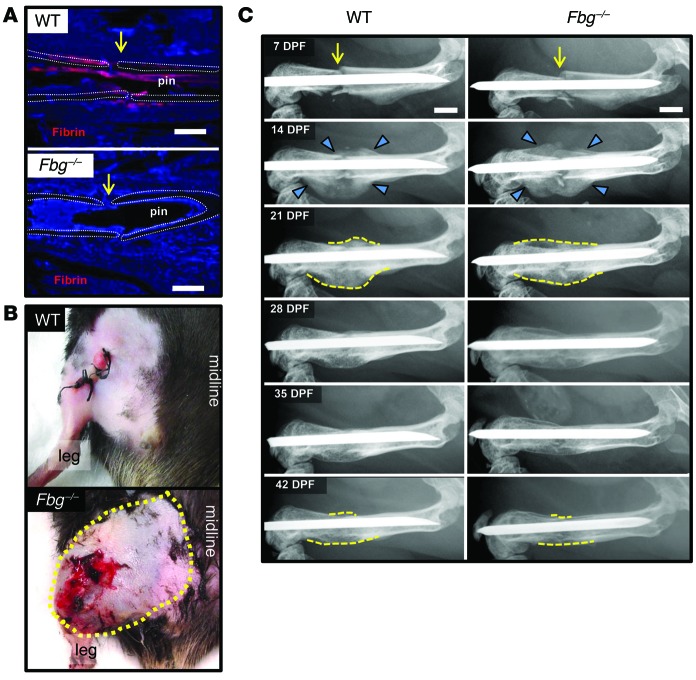
Bone formation during fracture repair inevitably initiates within or around extravascular deposits of a fibrin-rich matrix. In addition to a central role in hemostasis, fibrin is thought to enhance bone repair by supporting inflammatory and mesenchymal progenitor egress into the zone of injury. However, given that a failure of efficient fibrin clearance can impede normal wound repair, the precise contribution of fibrin to bone fracture repair, whether supportive or detrimental, is unknown. Here, we employed mice with genetically and pharmacologically imposed deficits in the fibrin precursor fibrinogen and fibrin-degrading plasminogen to explore the hypothesis that fibrin is vital to the initiation of fracture repair, but impaired fibrin clearance results in derangements in bone fracture repair. In contrast to our hypothesis, fibrin was entirely dispensable for long-bone fracture repair, as healing fractures in fibrinogen-deficient mice were indistinguishable from those in control animals. However, failure to clear fibrin from the fracture site in plasminogen-deficient mice severely impaired fracture vascularization, precluded bone union, and resulted in robust heterotopic ossification. Pharmacological fibrinogen depletion in plasminogen-deficient animals restored a normal pattern of fracture repair and substantially limited heterotopic ossification. Fibrin is therefore not essential for fracture repair, but inefficient fibrinolysis decreases endochondral angiogenesis and ossification, thereby inhibiting fracture repair.


Figures

Figure 10
Figure 10. Fibrin is deposited at the interface between avascular soft-tissue callus and vascularized hard-tissue callus.
Histological evaluation of the fracture callus of Plg–/– mice at 14 DPF discloses angiogram contrast–perfused blood vessels (safranin O, yellow arrow) at the interface of the soft-tissue callus (asterisks) and hard-tissue callus (pound symbols). Immunohistochemical staining for CD31 (brown staining) highlights thin-walled blood vessels filled with angiogram contrast material (CD31, yellow arrows) at the interface of soft- and hard-tissue callus. Immunohistochemistry also identifies fibrin (brown staining) at this interface surrounding angiogram contrast–perfused blood vessels (fibrin, yellow arrows). Immunohistochemistry slides were counterstained with hematoxylin. Scale bars: 50 μm (left column); 20 μm (right column). Representative of n = 5.
Figure 9
Figure 9. Fibrinogen knockdown rescues impaired callus formation and revascularization.
(A) Representative histological sections of femoral fracture sites from Plg–/– mice treated with ASO as a control and Plg–/– mice (Plg–/– control; n = 5) treated with an ASO against fibrinogen (Plg–/– Fbglo; n = 10) at 14 DPF. Sections were stained with safranin O and assessed by immunofluorescence for fibrin and by immunohistochemistry for CD31. Whereas the fracture callus of Plg–/– mice is somewhat disorganized, with multiple foci of chondroid soft-tissue callus (safranin O, yellow asterisks), the callus in Plg–/– Fbglo mice is more organized, with a central soft-tissue callus bordered proximally and distally by hard-tissue callus. There is markedly reduced fibrin deposition in the fracture callus of Plg–/– Fbglo mice compared with Plg–/– control mice (yellow arrows; white box denotes area shown for CD31 staining). CD31 highlights numerous CD31-positive cells lining blood vessels in the hard-tissue callus of Plg–/– Fbglo mice (red arrowheads), with fewer vessels in Plg–/– mice (black asterisks denote areas of soft-tissue callus in fracture gap). Scale bars: 1 mm. Statistical comparison (Student’s t test) of (B) fibrin deposition at fracture site, (C) number of CD31-positive vessels in the callus (left), and total area of CD31-positive vessels (right). n ≥ 5 for each group. Four sections in each sample were averaged to constitute a single replicate (1 n). **P < 0.01. Error bars represent SEM.
Figure 8
Figure 8. Fibrinogen knockdown rescues hard-tissue callus union.
Representative 3D and 2D μCT images, safranin O–stained sections, and immunofluorescence microscopy for fibrin in Plg–/– and Plg–/– Fbglo mice. 3D μCT images demonstrate heterotopic ossification (yellow arrowheads) and poorly macroscopically remodeled hard-tissue fracture callus in Plg–/– mice for 42 DPF formation. 2D μCT of the fracture sites in Plg–/– Fbglo mice shows cortical bridging by hard-tissue callus (yellow sharps), whereas this is not shown in Plg–/– mice (orange arrowheads). Histological sections confirmed united fractures with the presence of reactive woven bone in Plg–/– Fbglo mice in contrast to the disorganized callus with residual soft-tissue callus in Plg–/– mice (purple arrowheads). Immunofluorescence for fibrin confirms markedly less fibrin in the fracture site in Plg–/– Fbglo mice (red stain, fibrin, with DAPI blue counterstain). White dotted line denotes border of the femur and fracture callus. Scale bars: 1 mm. n ≥ 5 for each genotype.
Figure 7
Figure 7. Plasmin is required for hard-tissue callus union.
3D μCT reconstruction of a displaced fracture at 42 DPF shows evidence of soft-tissue mineralization away from the fracture callus in Plg–/– mice (yellow arrowheads). Coronal and sagittal 2D μCT slices of WT and Plg–/– mouse femurs show cortical bridging by hard fracture callus (yellow pound symbols) in WT mice. In contrast, a failure to unite the proximal and distal hard-tissue callus was observed in Plg–/– mice (red arrowheads). MSB stains demonstrate the presence of organized woven bone formation within the callus, and safranin O–stained sections confirm complete cortical bridging in WT mice (black pound symbol), whereas the fracture gap in Plg–/– mice remains composed of chondroid soft-tissue callus (yellow asterisks) and fibrous tissue (yellow plus sign) without evidence of cortical bridging. Immunofluorescence microscopy for fibrin shows abundant fibrin deposition at the fracture site in Plg–/– mice (red stain with DAPI blue counterstain, white arrowheads). Black boxes in safranin O sections indicate region displayed for fibrin immunofluorescence. n = 15 for each genotype. White scale bars: 1 mm (top two rows), 500 μm (bottom row); black scale bars: 1 mm.
Figure 6
Figure 6. Plasmin is not required for soft-tissue callus formation, but is essential for vascularization of fracture callus.
(A) Safranin O– and MSB-stained sections and immunofluorescence for VEGF-A and fibrin in the fracture callus at 14 DPF. Safranin O staining reveals the presence of the soft-tissue callus, while MSB-stained sections demonstrate that, while reduced in quantity compared with WT mice, the newly formed bone is histologically indistinguishable from newly formed bone in WT mice. VEGF immunofluorescence staining reveals VEGF expression within the hypertrophic chondrocytes (avascular soft-tissue callus, yellow asterisks) of WT and Plg–/– mice. Fibrin immunofluorescence reveals excessive fibrin deposition (white arrowheads) at the fracture site and in the hard-tissue callus of Plg–/– mice at 14 DPF compared with WT mice. Scale bars: 1 mm (top row); 100 μm (second, third, and fourth rows). (B) Representative 3D μCT reconstructions of angiogram contrast–perfused femurs show reduced revascularization of the fracture callus in Plg–/– mice at 14 DPF (color scale bar indicates vessel size range: black = 0.00 mm, red = 0.18 mm). Immunohistochemistry for CD31-positive vessels (red arrowheads) in the fracture site (yellow arrows). The interface of avascular soft-tissue callus and vascularized hard-tissue callus is marked by a black dashed line. The fracture gap is defined as the region between hard-tissue calluses (black double arrows). Scale bars: 1 mm (top row); 100 μm (middle and bottom rows). (C) Statistical analysis (Student’s t test) comparing number (top) and total area (bottom) of CD31-positive blood vessels in the callus. Four sections in each sample were averaged to constitute a single replicate (1 n). ***P < 0.01. Error bars represent SD. n ≥ 4 for each genotype.
Figure 5
Figure 5. Heterotopic ossification in plasminogen-deficient mice.
(A) 2D μCT sagittal slices of WT and Plg–/– mice at 14 and 42 DPF. 14 DPF images show hard-tissue callus formation in both WT and Plg–/– mice (yellow arrowheads). In addition, separate foci of mineralization in soft tissue away from the fracture callus seen only in Plg–/– mice are suggestive of heterotopic ossification (red arrowheads). By 42 DPF, the hard-tissue fracture callus has largely remodeled in WT mice. In contrast, the fracture callus in Plg–/– mice is not only persistent, but also hypertrophic and disorganized, with an irregular pattern of mineralization. Scale bars: 1 mm. n ≥ 13 for each genotype. (B) 2D μCT axial images of fractured femurs at 14 and 42 DPF in Plg–/– mice show that the areas of soft-tissue mineralization (red arrowheads) are distinct from the femoral fracture callus (yellow arrowheads), consistent with heterotopic ossification. Scale bars: 1 mm. n ≥ 13 for each genotype. (C) Macroscopic photograph of lateral aspect of a fractured femur of a Plg–/– mouse at 42 DPF (macro, yellow bracket denotes fracture callus). Nondemineralized tissue sections from the fracture callus of Plg–/– mice were stained with von Kossa and van Gieson solution to identify regions of hard-tissue callus formation (box i) and heterotopic ossification (box ii). Red boxes denote regions shown at higher power magnification. Foci of heterotopic ossification are denoted by white asterisks. Scale bars: 1 mm (top and second row); 200 μm (third row); 20 μm (bottom row). AC, avascular cartilage; M, muscle.
Figure 4
Figure 4. Plasminogen-deficient mice show abnormal hard-tissue callus formation.
Serial radiographic analysis of fractured femurs (yellow arrows) revealing marked differences in the formation (blue arrowheads) and macroscopic remodeling of the hard-tissue fracture callus (dashed yellow lines) in WT and Plg–/– mice. Note that relative to WT mice, the fracture callus in Plg–/– mice was hypertrophic and failed to undergo remodeling of the macroscopic hard-tissue callus over the first 6 weeks after fracture. In addition, both appreciable and persistent soft-tissue mineralization were noted adjacent to the injury site (blue triangles). Representative images of n = 15 for each genotype; see Table 1 and Supplemental Figure 4 for quantification. Scale bars: 1 mm.
Figure 3
Figure 3. Fibrin is not essential for hard-tissue callus union.
Top panels show 3D μCT reconstructions of femurs from WT and Fbg–/– mice at 42 DPF. Note there are no clear structural differences in animals of each genotype (white boxes denote site of fracture; yellow pound symbols denote areas of cortical bridging). Middle panels show coronal and sagittal 2D μCT images, further demonstrating cortical bridging by hard-tissue callus in both WT and Fbg–/– mice (yellow pound symbols). Bottom panels show safranin O and MSB staining establishing cortical bone union in both WT and Fbg–/– mice (yellow pound symbols). n ≥ 10 for each genotype. Scale bars: 1 mm.
Figure 2
Figure 2. Fibrin is not essential for soft-tissue callus formation and vascularization of fracture callus.
(A) WT and Fbg–/– mice stained with safranin O or immunofluorescence for fibrin at 14 DPF. Other than the distinct absence of local fibrin deposition in Fbg–/– mice, no obvious differences were detected in fracture callus formation (chondroid soft-tissue callus, yellow asterisks). Scale bars: 1 mm (top panels); 100 μm (middle and bottom panels). n ≥ 5 for each genotype. (B) 2D μCT images of mineralized bone (yellow asterisks) and MSB-stained histologic sections of tissues in WT and Fbg–/– mice. Note that regardless of fibrinogen status, these areas contain reactive hypercellular trabecular woven bone. Scale bars: 1 mm (whole, μCT); 500 μm (higher power, μCT); 100 μm (MSB). (C) Comparative analyses of vasculature in WT and Fbg–/– mice. Note that both 3D μCT reconstructions of contrast-perfused femurs and immunofluorescence for CD31 revealed no clear differences in the macro- and microvasculature of the fracture callus at 14 DPF in WT and Fbg–/– mice (soft-tissue callus, red asterisks; CD31-positive vessels, red arrowheads). Scale bars: 1 mm (top panels); 100 μm (middle panels); 50 μm (bottom panels). n ≥ 4 for each genotype. In the angiographic images, the color scale bar denotes vessel size: black (0.00 mm); red (0.18 mm). Black boxes denote areas shown at higher magnification. (D) Quantitative analysis of CD31-positive blood vessels in the callus in tissue sections. No significant differences (Student’s t test) were observed in the either the number (top) or total area of CD31-positive blood vessels (bottom). n ≥ 4 for each genotype. Four sections in each sample were averaged to constitute a single replicate (1 n). Error bars indicate SD.
Figure 1
Figure 1. Fibrin is essential for limiting hemorrhage, but not for fracture callus formation.
(A) Immunofluorescence-based detection of fibrin (red) at fracture site (yellow arrow) in WT and fibrinogen-deficient (Fbg–/–) mice. Note that fibrin was prominent in WT mice proximal to the fracture site (approximate location of the femora cortical bone outlined with white dotted lines), whereas fibrin was completely absent in Fbg–/– animals at 1 DPF. (B) Gross photographs of WT and Fbg–/– mouse extremities at 1 DPF showing local excessive bleeding and hematoma formation in Fbg–/– mice (outlined by yellow dashes) compared with WT mice. (C) Temporal radiographic analysis of fractured femurs (yellow arrows) in WT and Fbg–/– mice. Note that the formation (blue arrowheads) of the fracture callus and macroscopic hard-tissue callus remodeling (dashed yellow lines) of the fracture callus were effectively indistinguishable in WT and Fbg–/– mice. n ≥ 10 for each genotype. Scale bars: 1 mm.

Similar articles

Cited by

References

    1. Yuasa M, et al. The temporal and spatial development of vascularity in a healing displaced fracture. Bone. 2014;67:208–221. doi: 10.1016/j.bone.2014.07.002. - DOI - PubMed
    1. Gerstenfeld LC, Cullinane DM, Barnes GL, Graves DT, Einhorn TA. Fracture healing as a post-natal developmental process: molecular, spatial, and temporal aspects of its regulation. J Cell Biochem. 2003;88(5):873–884. doi: 10.1002/jcb.10435. - DOI - PubMed
    1. Rockwood CA, Green DP, Bucholz RW. Rockwood and Green’s Fractures In Adults. Philadelphia, Pennsylvania, USA: Wolters Kluwer Health/Lippincott Williams & Wilkins; 2010.
    1. Anitua E, Andia I, Ardanza B, Nurden P, Nurden AT. Autologous platelets as a source of proteins for healing and tissue regeneration. Thromb Haemost. 2004;91(1):4–15. - PubMed
    1. Martino MM, Briquez PS, Ranga A, Lutolf MP, Hubbell JA. Heparin-binding domain of fibrin(ogen) binds growth factors and promotes tissue repair when incorporated within a synthetic matrix. Proc Natl Acad Sci U S A. 2013;110(12):4563–4568. doi: 10.1073/pnas.1221602110. - DOI - PMC - PubMed

Publication types