. 2015 Sep;47(9):996-1002.
doi: 10.1038/ng.3361. Epub 2015 Jul 27.

Exome sequencing identifies recurrent mutations in NF1 and RASopathy genes in sun-exposed melanomas

Affiliations

Exome sequencing identifies recurrent mutations in NF1 and RASopathy genes in sun-exposed melanomas

Michael Krauthammer et al. Nat Genet. 2015 Sep.

Abstract

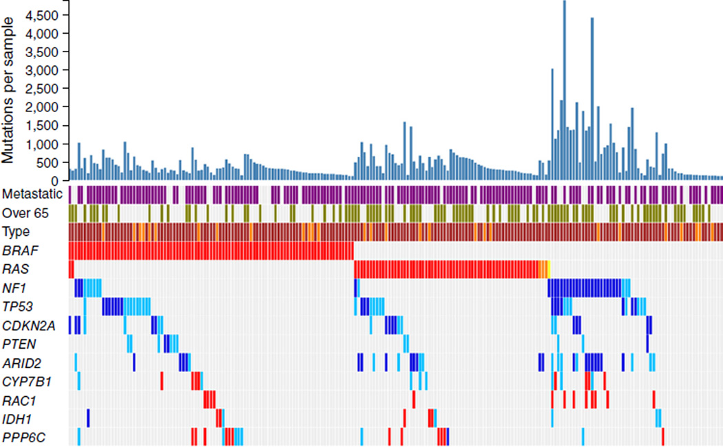
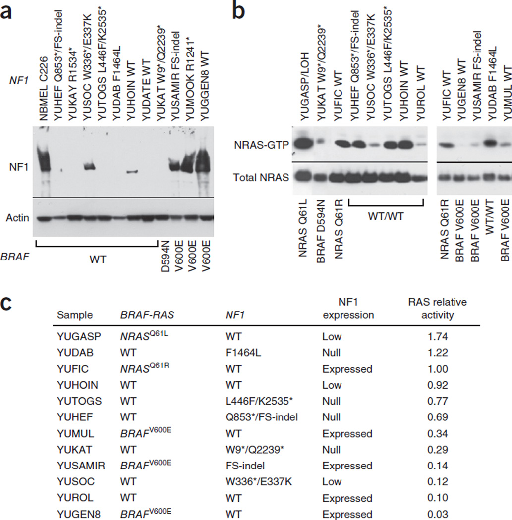
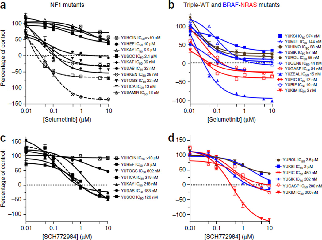
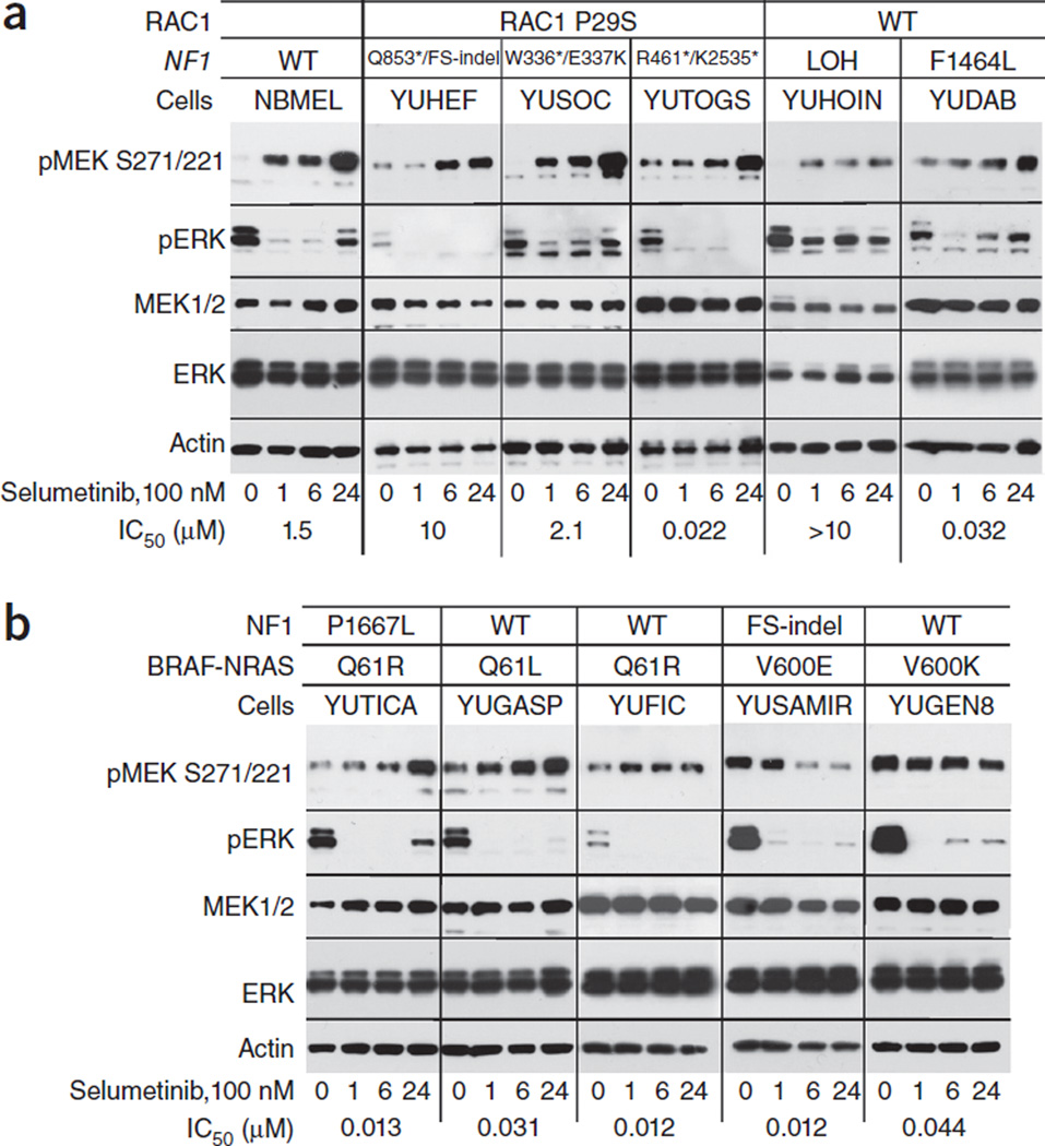
We report on whole-exome sequencing (WES) of 213 melanomas. Our analysis established NF1, encoding a negative regulator of RAS, as the third most frequently mutated gene in melanoma, after BRAF and NRAS. Inactivating NF1 mutations were present in 46% of melanomas expressing wild-type BRAF and RAS, occurred in older patients and showed a distinct pattern of co-mutation with other RASopathy genes, particularly RASA2. Functional studies showed that NF1 suppression led to increased RAS activation in most, but not all, melanoma cases. In addition, loss of NF1 did not predict sensitivity to MEK or ERK inhibitors. The rebound pathway, as seen by the induction of phosphorylated MEK, occurred in cells both sensitive and resistant to the studied drugs. We conclude that NF1 is a key tumor suppressor lost in melanomas, and that concurrent RASopathy gene mutations may enhance its role in melanomagenesis.


Figures

Figure 1
Figure 1
Melanoma mutational landscape (n = 213). Top 11 melanoma-driver genes that reach genome-wide significance according to background mutation-frequency estimation. Purple, metastatic melanoma; green, patients over 65 years old; red, mutations at recurrent positions; dark blue, inactivating mutations (nonsense, splice, indel); light blue, predicted harmful mutations. Brown and darker orange represent sun-exposed tumors and tumors of unknown origin, respectively. Mutations in HRAS and KRAS are marked in light orange and yellow, respectively. Mutation counts correspond to novel mutations that are not found in repositories of common human variants.
Figure 2
Figure 2
NF1 expression and NRAS activity. (a) Protein blot showing NF1 expression in melanoma cells (YU designation) relative to that in normal human melanocytes (NBMEL) derived from a single newborn foreskin; β-actin was used as a loading control. NF1 mutations are indicated at the top, and BRAF mutations at the bottom. The wild-type NF1 (WT) melanomas YUHOIN and YUDATE displayed LOH. All the melanoma cell lines represented in this panel are NRAS wild type. The reproducibility data and protein expression in additional representative melanoma cell lines are presented in Supplementary Figure 4. (b) NRAS activity as determined by NRAS-GTP pulldown assay showing active NRAS-GTP and total NRAS detected in lysates. The same results were obtained with pan-RAS antibodies (data not shown). (c) Ranking of NRAS activity with BRAF, NRAS and NF1 mutation and expression status. We derived numbers by scanning the bands and quantitating the bands’ intensity relative to input NRAS using ImageJ and normalizing against the NRASQ61R YUFIC melanoma. FS, frame shift.
Figure 3
Figure 3
Growth responses to selumetinib and SCH772984. (a–d) Growth of NF1-mutant (a,c) and of triple–wild-type (brown), BRAFV600-mutant (blue) and NRAFSQ61-mutant (red) melanoma cell lines (b,d). In a, the NF1 mutants YURKEN, YUTICA and YUSAMIR (dashed lines) are double-mutant cell lines (BRAFV600E, NRASQ61R and BRAFV600E, respectively) that express normal levels of NF1. The rest of the lines are null for NF1 (Fig. 2a). The selumetinib experiments were repeated at least twice. The data represent cell viability (CellTiter-Glo luminescent assay) as a percentage of the control at the end of 72 h of treatment. Each measurement is the average of triplicate or quadruplet wells. Error bars denote s.e.m.
Figure 4
Figure 4
Changes in MEK1/2 and ERK phosphorylation in response to selumetinib. Normal human melanocytes (NBMEL) and melanoma cell lines were untreated (0) or treated with 100 nM selumetinib for 1, 6 and 24 h. The panels show protein blots probed with the indicated antibodies. (a) Normal melanocytes and NF1-mutant melanoma cell lines that are wild type for BRAF and NRAS. (b) Melanoma cell lines that are NF1, NRAS or BRAF mutants, as indicated. Numbers on the bottom show the IC50 in response to selumetinib (Fig. 3). The types of NF1 and RAC1 mutations are indicated for each cell line at the top. All the cell lines in b express wild-type RAC1. Figure 2a and supplementary Figure 4 show the corresponding levels of NF1 expression. The results represent one of two similar experiments.

References

    1. Davis MJ, et al. RAC1P29S is a spontaneously activating cancer-associated GTPase. Proc. Natl. Acad. Sci. USA. 2013;110:912–917. - PMC - PubMed
    1. Hodis E, et al. A landscape of driver mutations in melanoma. Cell. 2012;150:251–263. - PMC - PubMed
    1. Krauthammer M, et al. Exome sequencing identifies recurrent somatic RAC1 mutations in melanoma. Nat. Genet. 2012;44:1006–1014. - PMC - PubMed
    1. Gold HL, et al. PP6C hotspot mutations in melanoma display sensitivity to Aurora kinase inhibition. Mol. Cancer Res. 2014;12:433–439. - PMC - PubMed
    1. Hammond D, et al. Melanoma-associated mutations in protein phosphatase 6 cause chromosome instability and DNA damage owing to dysregulated Aurora-A. J. Cell Sci. 2013;126:3429–3440. - PubMed

Publication types

MeSH terms