. 2015 Jul 27;10(7):e0134157.
doi: 10.1371/journal.pone.0134157. eCollection 2015.

Expression Profiles of Neuropeptides, Neurotransmitters, and Their Receptors in Human Keratocytes In Vitro and In Situ

Affiliations

Expression Profiles of Neuropeptides, Neurotransmitters, and Their Receptors in Human Keratocytes In Vitro and In Situ

Marta Słoniecka et al. PLoS One. .

Abstract

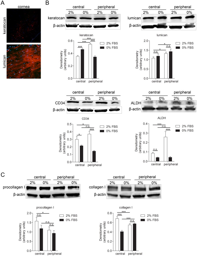
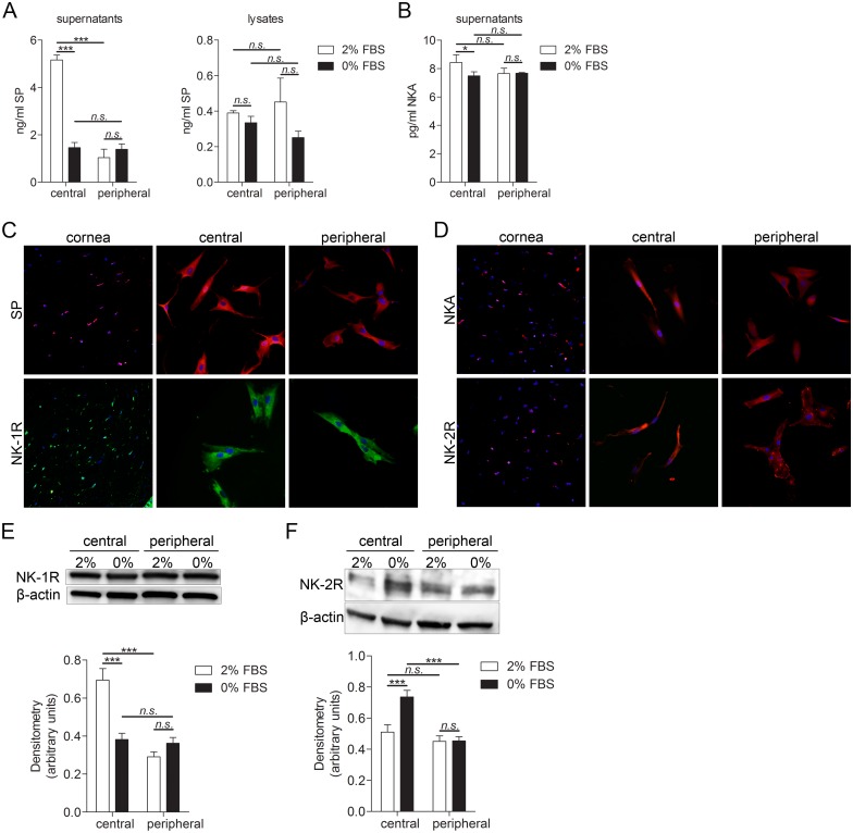
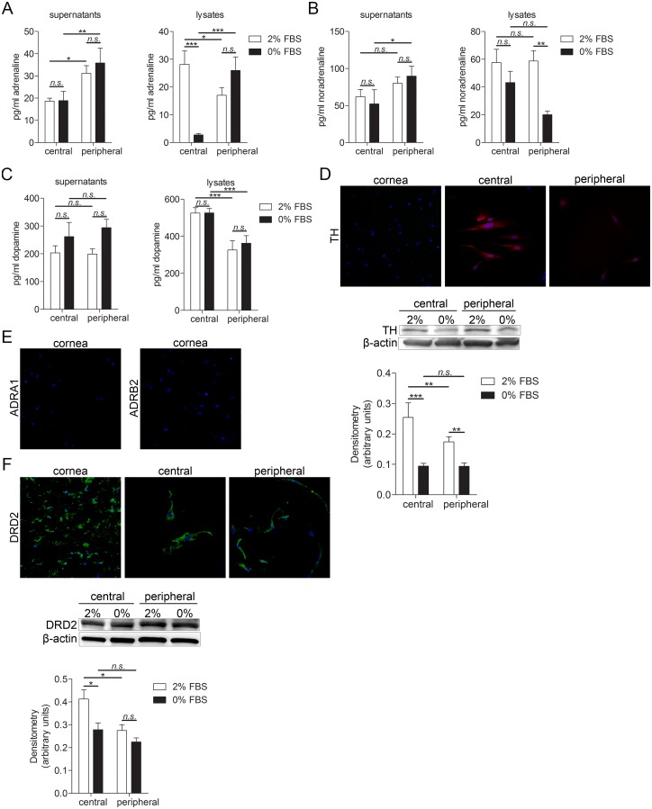
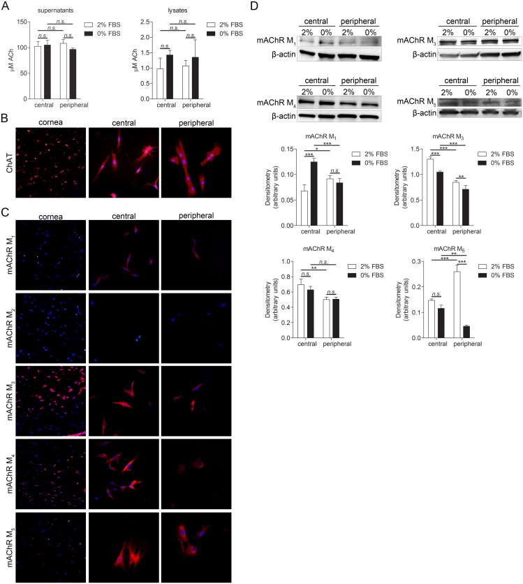
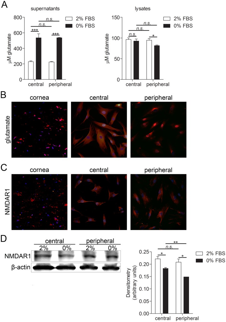
Keratocytes, the quiescent cells of the corneal stroma, play a crucial role in corneal wound healing. Neuropeptides and neurotransmitters are usually associated with neuronal signaling, but have recently been shown to be produced also by non-neuronal cells and to be involved in many cellular processes. The aim of this study was to assess the endogenous intracellular and secreted levels of the neuropeptides substance P (SP) and neurokinin A (NKA), and of the neurotransmitters acetylcholine (ACh), catecholamines (adrenaline, noradrenaline and dopamine), and glutamate, as well as the expression profiles of their receptors, in human primary keratocytes in vitro and in keratocytes of human corneal tissue sections in situ. Cultured keratocytes expressed genes encoding for SP and NKA, and for catecholamine and glutamate synthesizing enzymes, as well as genes for neuropeptide, adrenergic and ACh (muscarinic) receptors. Keratocytes in culture produced SP, NKA, catecholamines, ACh, and glutamate, and expressed neurokinin-1 and -2 receptors (NK-1R and NK-2R), dopamine receptor D2, muscarinic ACh receptors, and NDMAR1 glutamate receptor. Human corneal sections expressed SP, NKA, NK-1R, NK-2R, receptor D2, choline acetyl transferase (ChAT), M3, M4 and M5 muscarinic ACh receptors, glutamate, and NMDAR1, but not catecholamine synthesizing enzyme or the α1 and β2 adrenoreceptors, nor M1 receptor. In addition, expression profiles assumed significant differences between keratocytes from the peripheral cornea as compared to those from the central cornea, as well as differences between keratocytes cultured under various serum concentrations. In conclusion, human keratocytes express an array of neuropeptides and neurotransmitters. The cells furthermore express receptors for neuropeptides/neurotransmitters, which suggests that they are susceptible to stimulation by these substances in the cornea, whether of neuronal or non-neuronal origin. As it has been shown that neuropeptides/neurotransmitters are involved in cell proliferation, migration, and angiogenesis, it is possible that they play a role in corneal wound healing.


Conflict of interest statement

Competing Interests: The authors have declared that no competing interests exist.

Figures

Fig 1
Fig 1. Characterization of isolated keratocytes and keratocytes in cornea tissue sections.
(A) Human corneal sections were characterized by immunohistochemistry. Sections were double stained for the nuclei (DAPI; blue) together with keratocan (top panel) or lumican (bottom panel) (both TRITC; red). Both keratocan and lumican were expressed by keratocytes in corneal sections. (B-C) 250,000 cells were grown for 24h in 6 well plates in DMEM/F12 supplemented with either 2% or 0% FBS and subjected to western blot analysis. (B) Four keratocyte markers were analyzed: keratocan (50 kDa), lumican (46 kDa), CD34 (90–120 kDa) and ALDH (55 kDa). Keratocan was expressed in cultured cells. Its expression was significantly higher in peripheral keratocytes cultured in 2% FBS than in central keratocytes grown in 2% FBS (***p<0.001). Central keratocytes grown in 0% FBS expressed more keratocan than central keratocytes grown in 2% FBS (***p<0.001) and peripheral keratocytes grown in 0% FBS (***p<0.001). However, peripheral keratocytes grown in 0% FBS had significantly lower expression of keratocan than peripheral keratocytes grown in 2% FBS (***p<0.001), Lumican was expressed in cultured cells. Peripheral keratocytes expressed significantly more lumican than central keratocytes (*p<0.05 for 2% FBS, *p<0.05 for 0% FBS). CD34 was expressed in cultured keratocytes. Its expression was significantly higher in peripheral keratocytes grown in 2% FBS than in central keratocytes grown in 2% FBS (*p<0.05) and peripheral keratocytes grown in 0% FBS (***p<0.001). CD34 expression was also significantly higher in central keratocytes grown in 2% FBS than in central keratocytes grown in 0% FBS (*p<0.05). ALDH was expressed in cultured keratocytes but at low levels. Peripheral keratocytes grown in 2% FBS expressed significantly more ALDH than central keratocytes grown in 2% FBS (***p<0.001) and peripheral keratocytes grown in 0% FBS (***p<0.001). (C) Expression of procollagen I (140–210 kDa) and collagen I (139 kDa) was analyzed in cultured keratocytes. Procollagen I was expressed by cultured keratocytes. Its expression was significantly higher in central keratocytes grown in 2% FBS than in peripheral keratocytes grown in same conditions (*p<0.05). Collagen I expression was significantly higher in central keratocytes grown in 2% FBS than in central keratocytes grown in 0% FBS (***p<0.001) and peripheral keratocytes grown in 2% FBS (***p<0.001). Also, collagen I expression was significantly lower in central keratocytes grown in 0% FBS than in peripheral keratocytes grown in same conditions (***p<0.001). β-actin (42 kDa) served as a loading control. Values are means± SD.
Fig 2
Fig 2. Substance P and neurokinin A expression in keratocytes of cornea tissue sections and in cultured primary keratocytes.
(A-B) Substance P (A) and neurokinin A (B) EIA of central and peripheral cultured keratocytes. 250,000 cells were grown for 24h in 6 well plates in DMEM/F12 supplemented with either 2% or 0% FBS. Supernatant was collected, cells were lysed and subjected to EIA. (A) SP levels were significantly higher in supernatants of central keratocytes grown in medium supplemented with 2% FBS than in peripheral keratocytes grown in medium supplemented with 2% FBS (***p<0.001). SP secretion was significantly higher in central keratocytes grown in 2% FBS than central keratocytes grown in 0% FBS (***p<0.001). There were no significant differences in SP levels in lysates of keratocytes. (B) NKA secretion was significantly higher in central keratocytes grown in 2% FBS than in central keratocytes grown in 0% FBS (*p<0.05). (C) Keratocytes in sections (left panel), as well as cultured central (middle panel) and peripheral (right panel) keratocytes, were labeled with an antibody targeting SP (top row; TRITC; red) and double stained with DAPI to visualize the nuclei (blue). Sections and cultured cells were also labeled with an antibody targeting full-length NK-1R (bottom row; Alexa Fluor 488; green) and double stained with DAPI. Both the keratocytes in tissue sections and the cultured keratocytes expressed SP and a full-length version of NK-1R. (D) Keratocytes in sections (left panel), as well as cultured central (middle panel) and peripheral (right panel) keratocytes, were labeled with an antibody targeting NKA (top row; TRITC; red) and NK-2R (bottom row; TRITC; red) and double stained with DAPI. Both the keratocytes in tissue sections and the cultured keratocytes expressed NKA and NK-2R. (E-F) 250,000 central keratocytes or peripheral keratocytes grown in DMEM/F12 supplemented with either 2% or 0% FBS were subjected to western blot analysis for NK-1R (46 kDa) and NK-2R (48 kDa) expression. (E) Expression of NK-1R was significantly higher in central keratocytes grown in 2% FBS than in peripheral keratocytes grown in same conditions (***p<0.001). Expression of NK-1R was also significantly higher in central keratocytes grown in 2% FBS than in central keratocytes grown in 0% FBS (***p<0.001). (F) Expression of NK-2R was significantly higher in central keratocytes grown in 0% FBS than in peripheral keratocytes grown in same conditions (***p<0.001). NK-2R expression was significantly higher in central keratocytes grown in 0% FBS than in central keratocytes grown in 2% (***p<0.001). n.s. (non significant; p>0.05). β-actin (42 kDa) served as a loading control. Values are means ± SD.
Fig 3
Fig 3. Catecholamines and their receptors expression in keratocytes of cornea tissue sections and cultured primary keratocytes.
(A-C) Adrenaline, noradrenaline and dopamine ELISAs of lysates and supernatants of central and peripheral cultured keratocytes. 250,000 cells were grown for 24h in 6 well plates in DMEM/F12 supplemented with either 2% or 0% FBS. Supernatant was collected, cells were lysed and subjected to ELISA. (A) Levels of secreted adrenaline were significantly higher in peripheral keratocytes grown in both 2% and 0% FBS than in central keratocytes grown in same conditions (*p<0.05 for 2% FBS and **p<0.01 for 0% FBS). Levels of intracellular adrenaline were significantly higher in central keratocytes grown in 2% FBS than in peripheral keratocytes grown in 2% FBS (*p<0.05). Central keratocytes grown in 2% FBS had higher levels of adrenaline than central keratocytes grown in 0% FBS (***p<0.001). Moreover, peripheral keratocytes grown in 0% FBS had higher levels of adrenaline than central keratocytes grown in same the same condition (***p<0.001). (B) Noradrenaline was secreted from both central and peripheral keratocytes and was also present in cell lysates. Secreted noradrenaline levels were significantly higher in peripheral keratocytes grown in 0% FBS than in central keratocytes grown in same conditions (*p<0.05). Intracellular levels of noradrenaline were significantly higher in peripheral keratocytes grown in 2% FBS than peripheral keratocytes grown in 0% FBS. (C) Dopamine was secreted from both central and peripheral keratocytes. Dopamine levels were significantly higher in lysates of central keratocytes than peripheral keratocytes (***p<0.001 for both 2% FBS and 0% FBS). (D) Keratocytes in sections (left panel), as well as cultured central (middle panel) and peripheral (right panel) keratocytes, were labeled with an antibody targeting tyrosine hydroxylase (TH; TRITC; red) and double stained with DAPI to visualize the nuclei (blue). Cultured keratocytes expressed TH whereas keratocytes in tissue sections did not. 250,000 central or peripheral keratocytes grown in either 2% FBS or 0% FBS were lysed and expression of TH (60 kDa) was analyzed by western blot. Both central and peripheral keratocytes grown in 2% FBS expressed more TH than cells grown in 0% FBS (***p<0.001 for central keratocytes, **p<0.01 for peripheral keratocytes). Moreover, central keratocytes grown in 2% FBS expressed more TH than peripheral keratocytes grown in same conditions (**p<0.01). β-actin (42 kDa) served as a loading control. (E) Sections were labeled with adrenergic receptor antibodies, ADRA1 (left; TRITC, red) and ADRB2 (right; TRITC, red) and double stained with DAPI (blue). Keratocytes in tissue sections did not express either of the adrenergic receptors. (F) Keratocytes in sections (left panel), as well as cultured central (middle panel) and peripheral (right panel) keratocytes, were labeled with an antibody targeting dopamine receptor DRD2 (Alexa Fluor 488; green) and double stained with DAPI to visualize the nuclei (blue). DRD2 was expressed in both keratocytes in tissue sections and in cultured keratocytes. 250,000 central or peripheral keratocytes grown in either 2% FBS or 0% FBS were lysed and expression of DRD2 (51 kDa) was analyzed by western blot. Central keratocytes grown in 2% FBS had significantly higher expression of DRD2 than both central keratocytes grown in 0% FBS (*p<0.05) and peripheral cells grown in 2% FBS (*p<0.05). β-actin (42 kDa) served as a loading control. Values are means ± SD.
Fig 4
Fig 4. Acetylcholine and its receptors expression in keratocytes.
(A) 250,000 cells were grown for 24h in 6 well plates in DMEM/F12 supplemented with either 2% or 0% FBS. Supernatant was collected and subjected to acetylcholine (ACh) assay and cells were lysed. Both central and peripheral keratocytes secreted ACh (n.s.; p≥0.05). ACh was also present in both central and peripheral keratocyte lysates (n.s.; p≥0.05). (B) Keratocytes in sections (left panel), as well as cultured central (middle panel) and peripheral (right panel) keratocytes, were labeled with an antibody targeting choline acetyltransferase (ChAT; TRITC; red), and double stained with DAPI to visualize the nuclei (blue). Both the keratocytes in tissue sections and the cultured keratocytes expressed ChAT. (C) Keratocytes in sections (left panel), as well as cultured central (middle panel) and peripheral (right panel) keratocytes, were labeled with muscarinic acetylcholine receptors (mAChR M1, mAChR M2, mAChR M3, mAChR M4, and mAChR5; TRITC; red) and double stained with DAPI (blue). mAChR M1 was present in cultured cells but not in tissue sections, mAChR M2 was absent in both cultured cells and tissue sections. Remaining receptors (mAChR M3, mAChR M4, and mAChR M5) were expressed in both tissue sections and cultured cells. (D) 250,000 central or peripheral keratocytes grown in either 2% FBS or 0% FBS were lysed and expression of muscarinic acetylcholine receptors (mAChR M1 [52 kDa], mAChR M3 [75 kDa], mAChR M4 [74 kDa], and mAChR5 [60 kDa]; mAChR M2 was not expressed) was analyzed by western blot. mAChR M1 expression was significantly higher in central keratocytes grown in 0% FBS than in central keratocytes grown in 2% FBS (***p<0.001), significantly higher in peripheral keratocytes grown in 2% FBS than in central keratocytes grown in same the condition (*p<0.05), and significantly higher in central keratocytes grown in 0% FBS than in peripheral keratocytes grown in the same condition (***p<0.001). mAChR M3 expression was significantly higher in central keratocytes grown in 2% FBS than in both central keratocytes grown in 0% FBS (***p<0.001) and peripheral keratocytes grown in 2% FBS (***p<0.001). Central keratocytes grown in 0% FBS expressed significantly more mAChR M3 than peripheral keratocytes grown in same conditions (***p<0.001) and peripheral keratocytes grown in 2% FBS expressed more mAChR M3 than peripheral cells grown in 0% FBS (**p<0.01). Expression of mAChR M4 was significantly higher in central keratocytes grown in 2% FBS than in peripheral keratocytes grown in same conditions (**p<0.01). Expression of mAChR5 was significantly higher in peripheral keratocytes grown in 2% FBS than in both central keratocytes grown in same conditions (***p<0.001) and peripheral keratocytes grown in 0% FBS (***p<0.001). mAChR5 expression was also significantly higher in central keratocytes grown in 0% FBS than in peripheral keratocytes grown in same conditions (**p<0.01). β-actin (42 kDa) served as a loading control. Values are means ± SD.
Fig 5
Fig 5. Glutamate and NMDAR1 expression in keratocytes.
(A) Glutamate assay of cultured central and peripheral keratocytes. 250,000 cells were grown for 24h in 6 well plates in DMEM/F12 supplemented with either 2% or 0% FBS. Supernatant was collected, cell were lysed and subjected to the glutamate assay. Levels of glutamate were significantly higher in central and peripheral keratocyte culture supernatants collected from cells grown in 0% FBS than in supernatants collected from cells grown in 2% (***p<0.001 for central and ***p<0.001 for peripheral keratocytes). Levels of glutamate in cell lysates were significantly higher in peripheral keratocytes grown in 2% FBS than in peripheral keratocytes grown in 0% FBS (*p<0.05) (B) Keratocytes in sections (left panel), as well as cultured central (middle panel) and peripheral (right panel) keratocytes, were labeled with glutamate antibody (TRITC; red) and double stained with DAPI to visualize the nuclei (blue). Glutamate was expressed in both keratocytes in tissue sections and in cultured keratocytes. (C) Cornea sections (left panel), as well as cultured central (middle panel) and peripheral (right panel) keratocytes were labeled with glutamate receptor NMDAR1 (TRITC; red) and double stained with DAPI. NMDAR1 was expressed in keratocytes in tissue sections and in cultured keratocytes. (D) 250,000 central or peripheral keratocytes grown in either 2% FBS or 0% FBS were lysed and expression of NMDAR1 (105 kDa) was analyzed by western blot. NMDAR1 expression was significantly higher in both central and peripheral keratocytes grown in 2% FBS than in central and peripheral keratocytes grown in 0% FBS (*p<0.05 for central keratocytes, *p<0.05 for peripheral keratocytes). Expression of NMDAR1 was also significantly higher in central keratocytes grown in 0% FBS than in peripheral keratocytes grown in same conditions (**p<0.01). β-actin (42 kDa) served as a loading control. Values are means ± SD.

Similar articles

Cited by

References

    1. West-Mays JA, Dwivedi DJ. The keratocyte: corneal stromal cell with variable repair phenotypes. The international journal of biochemistry & cell biology. 2006;38(10):1625–31. Epub 2006/05/06. 10.1016/j.biocel.2006.03.010 - DOI - PMC - PubMed
    1. Beales MP, Funderburgh JL, Jester JV, Hassell JR. Proteoglycan synthesis by bovine keratocytes and corneal fibroblasts: maintenance of the keratocyte phenotype in culture. Investigative ophthalmology & visual science. 1999;40(8):1658–63. Epub 1999/07/07. . - PubMed
    1. Robert L, Legeais JM, Robert AM, Renard G. Corneal collagens. Pathologie-biologie. 2001;49(4):353–63. Epub 2001/06/29. . - PubMed
    1. Hay ED. Development of the vertebrate cornea. International review of cytology. 1980;63:263–322. Epub 1979/01/01. . - PubMed
    1. Muller LJ, Pels L, Vrensen GF. Novel aspects of the ultrastructural organization of human corneal keratocytes. Investigative ophthalmology & visual science. 1995;36(13):2557–67. Epub 1995/12/01. . - PubMed

Publication types