. 2015 Jul 28:5:12440.
doi: 10.1038/srep12440.

Mitochondrial CB1 receptor is involved in ACEA-induced protective effects on neurons and mitochondrial functions

Affiliations

Mitochondrial CB1 receptor is involved in ACEA-induced protective effects on neurons and mitochondrial functions

Lei Ma et al. Sci Rep. .

Abstract

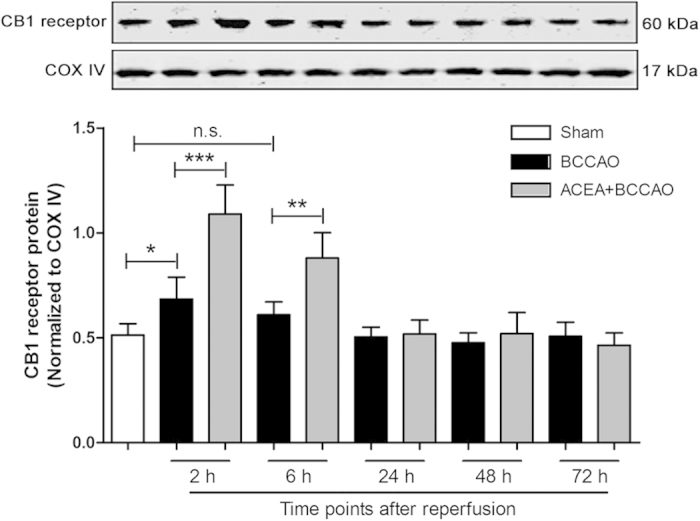
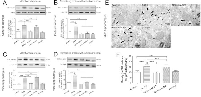
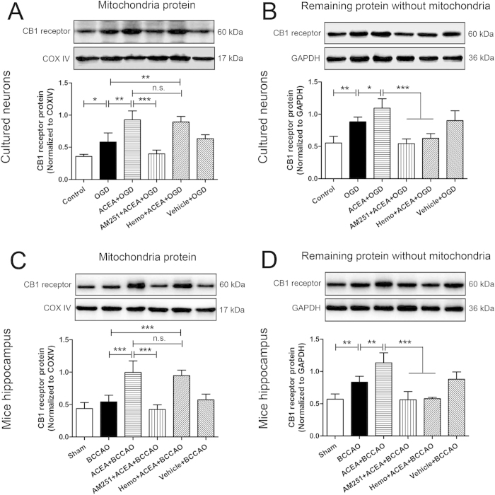
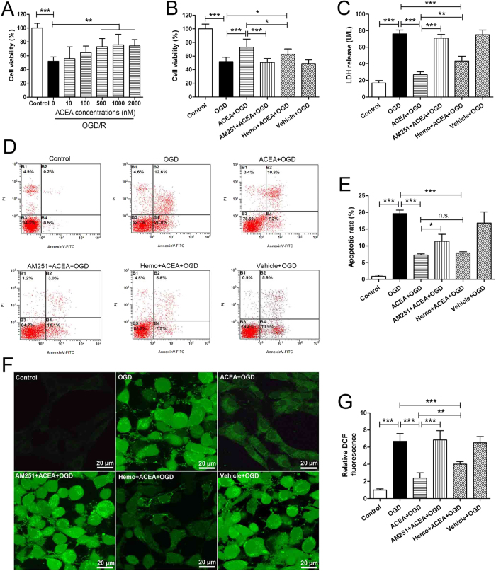
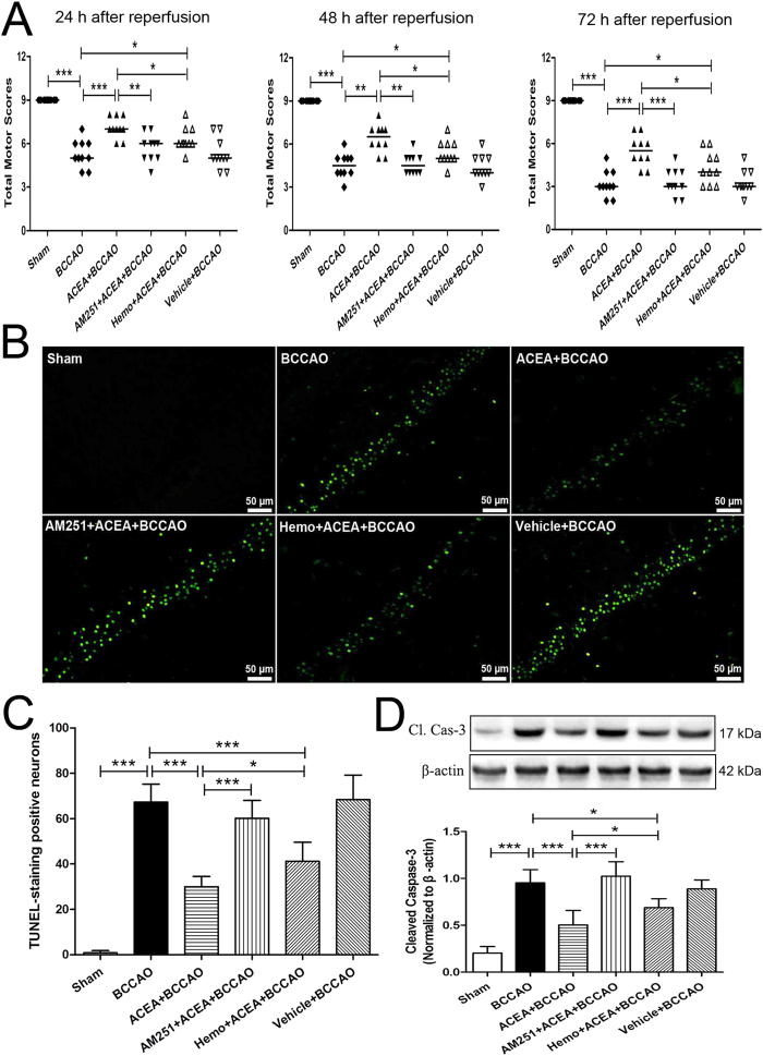
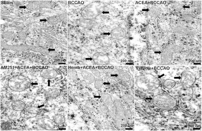
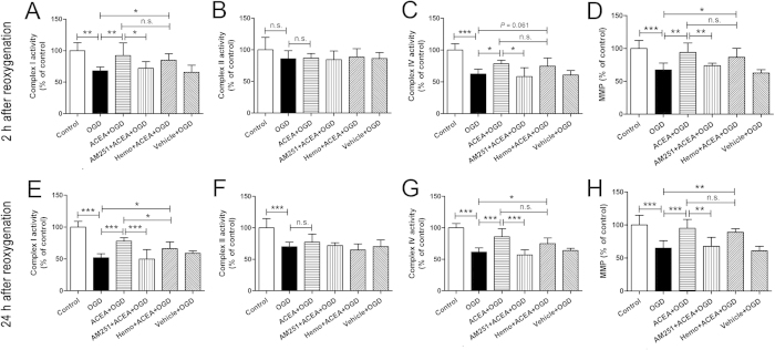
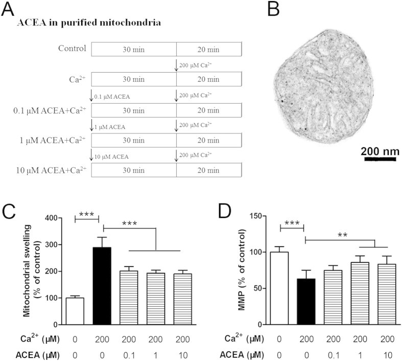
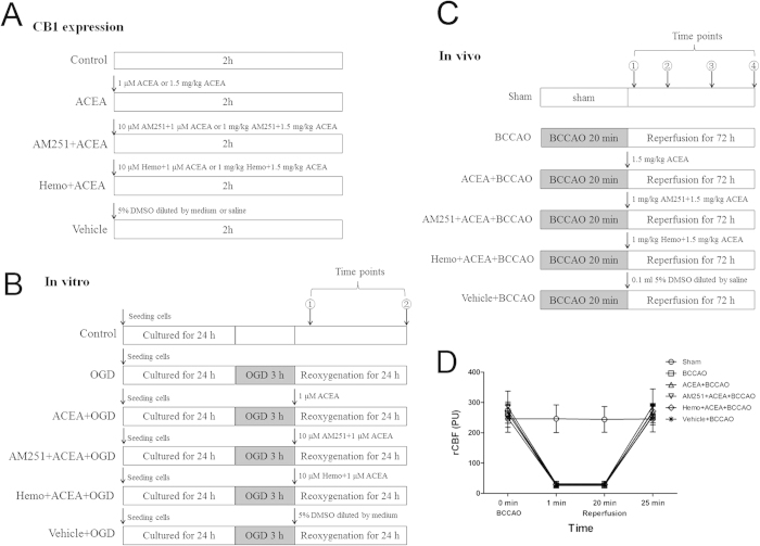
Mitochondrial dysfunction contributes to cell death after cerebral ischemia/reperfusion (I/R) injury. Cannabinoid CB1 receptor is expressed in neuronal mitochondrial membranes (mtCB1R) and involved in regulating mitochondrial functions under physiological conditions. However, whether mtCB1R affords neuroprotection against I/R injury remains unknown. We used mouse models of cerebral I/R, primary cultured hippocampal neurons exposed to oxygen-glucose deprivation/reoxygenation (OGD/R) and Ca(2+)-induced injury in purified neuronal mitochondria to investigate the role of mtCB1R in neuroprotection. Our results showed selective cell-permeant CB1 receptor agonist, arachidonyl-2-chloroethylamide (ACEA), significantly up-regulated the expression of mtCB1R protein in hippocampal neurons and tissue. In vitro, ACEA restored cell viability, inhibited generation of reactive oxygen species (ROS), decreased lactate dehydrogenase (LDH) release and reduced apoptosis, improved mitochondrial function. In vivo, ACEA ameliorated neurological scores, diminished the number of TUNEL-positive neurons and decreased the expression of cleaved caspase-3. However, ACEA-induced benefits were blocked by the selective cell-permeant CB1 receptor antagonist AM251, but just partially by the selective cell-impermeant CB1 receptor antagonist hemopressin. In purified neuronal mitochondria, mtCB1R activation attenuated Ca(2+)-induced mitochondrial injury. In conclusion, mtCB1R is involved in ACEA-induced protective effects on neurons and mitochondrial functions, suggesting mtCB1R may be a potential novel target for the treatment of brain ischemic injury.


Figures

Figure 1
Figure 1. Expression of mtCB1R protein in mouse hippocampus at different time points after reperfusion.
Western blot showing mtCB1R protein expression at 2 h, 6 h, 24 h, 48 h and 72 h after reperfusion respectively. Data represent mean ± SD. *P < 0.05; **P < 0.01; ***P < 0.001; n.s.: no significance. COX IV: cytochrome c oxidase; BCCAO: bilateral common carotid artery occlusion.
Figure 2
Figure 2. Expression of CB1 receptor protein in primary cultured hippocampal neurons and mouse hippocampus.
(A, C) Western blot showing mtCB1R protein expression at 2 h after administration in vitro and in vivo (n = 5). (B, D) Western blot showing CB1 protein expression in the remaining samples without mitochondria protein at 2 h after administration in vitro and in vivo (n = 5). (E) Immunogold electron microscopy showing mtCB1R protein expression. (F) Density of mtCB1R immunoparticles were calculated per area of mitochondria (n = 3). Scale bars = 200 nm. Data represent mean ± SD. **P < 0.01; ***P < 0.001; n.s.: no significance. COX IV: cytochrome c oxidase; Hemo: hemopressin.
Figure 3
Figure 3. Expression of CB1 receptor protein in primary cultured hippocampal neurons at 2 h after reoxygenation and mouse hippocampus at 2 h after reperfusion.
(A, C) Western blot showing mtCB1R protein expression in vitro and in vivo (n = 5). (B, D) Western blot showing CB1 protein expression in the remaining samples without mitochondria protein in vitro and in vivo (n = 5). Data represent mean ± SD. *P < 0.05; **P < 0.01; ***P < 0.001; n.s.: no significance. COX IV: cytochrome c oxidase; OGD: oxygen-glucose deprivation; Hemo: hemopressin; BCCAO: bilateral common carotid artery occlusion.
Figure 4
Figure 4. ACEA-induced cytoprotection against OGD/R injury in primary cultured hippocampal neurons.
(A) The effective dose of ACEA against OGD/R evaluated by WST-8 (n = 8). Cell injury was evaluated in terms of WST-8 cell viability (B), (n = 8), LDH release (C), (n = 6), apoptosis rate (D, E), (n = 5) and intracellular ROS level (F, G), (n = 6) at 24 h after reoxygenation. Scale bars  = 20 μm. Data represent mean ± SD. *P < 0.05; **P < 0.01; ***P < 0.001; n.s.: no significance. OGD/R: oxygen-glucose deprivation/reoxygenation; WST-8: 2-(2-methoxy-4-nitrophen​yl)-3-(4-nitrophenyl)-5-(​2,4-disulfophenyl)-2H-tet​razolium, monosodium salt; Hemo: hemopressin; LDH: lactate dehydrogenase; ROS: reactive oxygen species.
Figure 5
Figure 5. ACEA-induced neuroprotection against cerebral ischemia/reperfusion injury in mice.
(A) Neurological assessment at 24 h, 48 h and 72 h after reperfusion (n = 13). Data represent the median. (B) Representative photomicrographs of TUNEL staining in hippocampal CA1 region. Scale bars = 50 μm. (C) Quantitative analysis of the number of TUNEL-positive cells in hippocampal CA1 region at 72 h after reperfusion (n = 5). (D) Western blot showing representative the expression of cleaved caspase-3 (Cl. Cas-3) protein in the hippocampus at 72 h after reperfusion (n = 5). Data represent mean ± SD. *P < 0.05; **P < 0.01; ***P < 0.001. BCCAO: bilateral common carotid artery occlusion; Hemo: hemopressin.
Figure 6
Figure 6. Transmission electron micrographs showing mitochondrial ultrastructure.
Representative electron microphotographs show mitochondrial ultrastructure in mouse CA1 hippocampal neurons at 72 h after the reperfusion (n = 3). Mitochondrial ultrastructures are indicated by arrows. Scale bars = 200 nm. BCCAO: bilateral common carotid artery occlusion; Hemo: hemopressin.
Figure 7
Figure 7. Effects of ACEA on mitochondrial function at 2 and 24 h after reoxygenation.
Neuronal mitochondria were purified at 2 and 24 h after reoxygenation respectively, and the activities of complexes I (A, E), II (B, F) and IV (C, G) were measured spectrophotometrically; the mitochondrial membrane potential (MMP) (D, H) was evaluated by fluorescence spectrophotometry. Data represent mean ± SD (n = 6). *P < 0.05; **P < 0.01; ***P < 0.001; n.s.: no significance. OGD: oxygen-glucose deprivation; Hemo: hemopressin.
Figure 8
Figure 8. Effects of ACEA on Ca2 + -induced mitochondrial injury in vitro.
(A) Protocol. (B) Representative electron micrograph of purified intact mitochondria from normal hippocampal neurons. Scale bars = 200 nm. Mitochondrial swelling was measured spectrophotometrically (C) and mitochondrial membrane potential (MMP) was evaluated by a fluorescence spectrophotometer (D). Data represent mean ± SD (n = 6). **P < 0.01; ***P < 0.001.
Figure 9
Figure 9. Experimental protocol and regional cerebral blood flow (rCBF).
(A) Protocol 1: Detection of CB1 receptor expression in vitro and in vivo. (B) Protocol 2: ACEA treatment in vitro. ①: at 2 h after reoxygenation, the neuronal mitochondria were purified for the detection of CB1 receptor expression and mitochondrial function assay; ②: at 24 h after reoxygenation, cell viability was measured and the neuronal mitochondria were purified for mitochondrial function assay. (C) Protocol 3: ACEA treatment in vivo. ①: at 2 h after reperfusion, mitochondria were isolated for the measurement of CB1 receptor expression; At 24 h (②) and 48 h (③) after reperfusion respectively, neurological tests were conducted; at 72 h (④) after reperfusion, neurological tests were performed, and neuronal apoptosis and apoptosis markers were assessed. (D) Changes of rCBF in C57BL/6 mice. The rCBF was reduced to <10% of the pre-ischemic baseline value immediately after ischemia and was maintained for 20 min. The rCBF returned to the pre-ischemic value at 5 min after reperfusion. OGD: oxygen-glucose deprivation; BCCAO: bilateral common carotid artery occlusion; Hemo: hemopressin.

References

    1. Laver S., Farrow C., Turner D. & Nolan J. Mode of death after admission to an intensive care unit following cardiac arrest. Intensive Care Med 30, 2126–2128 (2004). - PubMed
    1. Nichol G. et al. Regional systems of care for out-of-hospital cardiac arrest: A policy statement from the American Heart Association. Circulation 121, 709–729 (2010). - PubMed
    1. Roine R. O., Kajaste S. & Kaste M. Neuropsychological sequelae of cardiac arrest. JAMA 269, 237–242 (1993). - PubMed
    1. MacAskill A. F. & Kittler J. T. Control of mitochondrial transport and localization in neurons. Trends Cell Biol 20, 102–112 (2010). - PubMed
    1. Mattson M. P., Gleichmann M. & Cheng A. Mitochondria in neuroplasticity and neurological disorders. Neuron 60, 748–766 (2008). - PMC - PubMed

Publication types

Substances