. 2015 Jul 28:6:7823.
doi: 10.1038/ncomms8823.

Aversive learning shapes neuronal orientation tuning in human visual cortex

Affiliations

Aversive learning shapes neuronal orientation tuning in human visual cortex

Lisa M McTeague et al. Nat Commun. .

Abstract

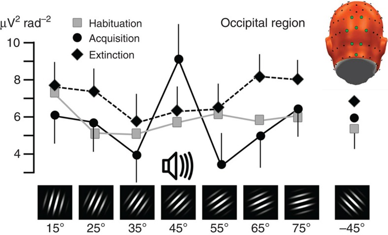
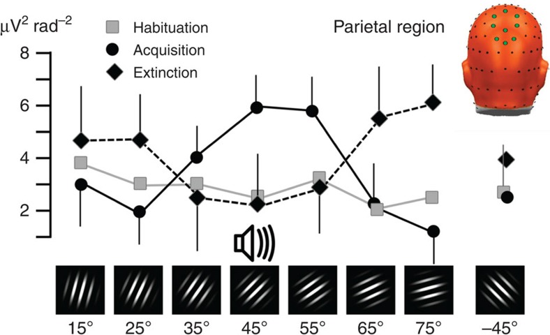
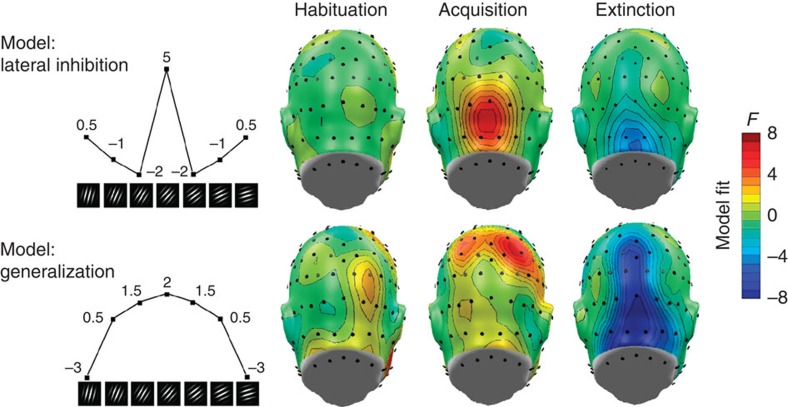
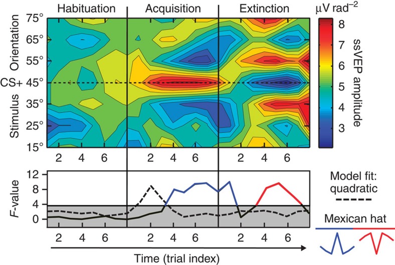
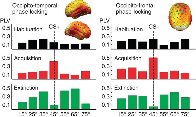
The responses of sensory cortical neurons are shaped by experience. As a result perceptual biases evolve, selectively facilitating the detection and identification of sensory events that are relevant for adaptive behaviour. Here we examine the involvement of human visual cortex in the formation of learned perceptual biases. We use classical aversive conditioning to associate one out of a series of oriented gratings with a noxious sound stimulus. After as few as two grating-sound pairings, visual cortical responses to the sound-paired grating show selective amplification. Furthermore, as learning progresses, responses to the orientations with greatest similarity to the sound-paired grating are increasingly suppressed, suggesting inhibitory interactions between orientation-selective neuronal populations. Changes in cortical connectivity between occipital and fronto-temporal regions mirror the changes in visuo-cortical response amplitudes. These findings suggest that short-term behaviourally driven retuning of human visual cortical neurons involves distal top-down projections as well as local inhibitory interactions.


Figures

Figure 1
Figure 1. Occipital retuning during aversive conditioning.
Changes in grand mean (N=15) visual electrocortical activity is shown for each phase of the experiment (habituation, acquisition and extinction) and for each orientation. Regional means of ssVEP amplitudes in CSD (Laplacian) space, averaged across occipital midline sensor locations, were used to estimate the occipital cortex surface potential. The inset shows a back view of the electrode array used, with sensor locations used for averaging highlighted in green. A pronounced Mexican-hat pattern is visible during the acquisition phase, consistent with lateral inhibitory interactions: Amplification of the 45° orientation, which was paired with a noxious sound, is accompanied by relative suppression of proximal orientations (55° and 35°). Error bars (s.e.m.) are shown for the acquisition and extinction phases.
Figure 2
Figure 2. Parietal retuning during aversive conditioning.
Grand mean (N=15) pooled visual electrocortical activity over parietal cortical sites is shown for each phase of the experiment (habituation, acquisition and extinction) and for each grating orientation. Spectral power of ssVEP current source density is shown, averaged across parietal midline sensor locations, shown as green circles in the top right inset. Note the generalization (quadratic) pattern with amplification of the 45° grating that was paired with noise, and a monotonic decline to distal orientations, specifically during fear acquisition. Error bars (s.e.m.) are shown for the acquisition and extinction phases.
Figure 3
Figure 3. Reflex physiology changes during aversive conditioning.
Grand mean (N=15) startle reflex magnitude (expressed in standardized T-scores) for each orientation, during the acquisition phase of the experiment. Paralleling parietal cortical engagement, a quadratic fear generalization pattern is visible, with amplification of the 45° noise-paired stimulus and monotonic decline to distal orientations. Error bars reflect the s.e.m.
Figure 4
Figure 4. Self-report changes during aversive conditioning.
Grand mean (N=15) self-reported emotional arousal (black lines, left scale) and pleasure (grey lines, right scale), comparing post-habituation (squares) and post-acquisition ratings (circles) for each orientation, on the SAM nine-point scale.
Figure 5
Figure 5. Cortical regions sensitive to aversive learning.
Topographical distribution (back views are shown) of planned contrast models testing the competing hypotheses of lateral inhibition (top, Mexican-hat contrast) versus fear generalization (bottom, quadratic contrast) of electrocortical responses across orientations, during the three experimental phases. Weights used for the planned contrasts are displayed for each orientation (left panel). F-values exceeding ±4.27 indicate reliable model fits at a given sensor location. Fits that matched the opposite pattern are shown in blue, indicating negative fit. Note the focal lateral inhibition (Mexican hat) pattern evident during acquisition over occipital areas, accompanied by parietal generalization indicated by a quadratic contrast. The tendency to reverse amplification of the noise-paired cue (that is, the 45° orientation) is reflected in the inverted patterns seen during the extinction phase, across parietal and occipital sites.
Figure 6
Figure 6. Temporal evolution of orientation tuning during aversive learning.
Top: color-coded single-trial amplitude of the visual electrocortical response, across the phases of the experiment. Learning dynamics in visual cortex are shown over eight trials per phase for seven neighbouring orientations, centred around the 45° orientation (the threat cue), which was paired with the noxious sound. Bottom: model fits (planned contrasts) for the competing hypotheses of fear generalization (top) and lateral inhibition (bottom, Mexican hat), calculated for each trial. Note that initial amplification of the threat cue (45°) is followed by increasing relative amplitude reduction for the proximal orientations. Complete reversal of these changes is visible during fear extinction, resulting in fitting an inverse Mexican-hat model, indicated in the lower panel by F-values shown in red.
Figure 7
Figure 7. Connectivity changes during visual retuning.
Pooled cortico-cortical connectivity relative to a reference site at the occipital pole across the three learning phases, shown for each orientation and for two regions of interest. Both occipitotemporal (left panel, pooled sensor locations shown as green circles) and occipitofrontal (right panel) connectivity show amplification of the CS+ orientation (45°) during acquisition and its suppression during extinction. PLV, phase-locking value.
Figure 8
Figure 8. Steady-state evoked potential time series.
Left panel: representative ssVEP time series evoked by the pattern-reversing grating stimuli used in this study, shown across all habituation trials for the CS+ orientation (45°), averaged across observers in the 15-Hz group (N=8). The right panel shows the topographical distribution of the spectral power of the ssVEP source density, with electrode location Oz used as a reference point for spatial pooling, highlighted by a green circle.

Similar articles

Cited by

References

    1. Bradley M. M., Keil A. & Lang P. J. Orienting and emotional perception: facilitation, attenuation, and interference. Front. Psychol. 3, 493 (2012). - PMC - PubMed
    1. Headley D. B. & Weinberger N. M. Gamma-band activation predicts both associative memory and cortical plasticity. J. Neurosci. 31, 12748–12758 (2011). - PMC - PubMed
    1. Miskovic V. & Keil A. Acquired fears reflected in cortical sensory processing: a review of electrophysiological studies of human classical conditioning. Psychophysiology 49, 1230–1241 (2012). - PMC - PubMed
    1. Miskovic V. & Keil A. Perceiving threat in the face of safety: excitation and inhibition of conditioned fear in human visual cortex. J. Neurosci. 33, 72–78 (2013). - PMC - PubMed
    1. Weinberger N. M. Specific long-term memory traces in primary auditory cortex. Nat. Rev. Neurosci. 5, 279–290 (2004). - PMC - PubMed

Publication types