. 2015 Aug 1;29(15):1605-17.
doi: 10.1101/gad.263574.115. Epub 2015 Jul 27.

miR-431 promotes differentiation and regeneration of old skeletal muscle by targeting Smad4

Affiliations

miR-431 promotes differentiation and regeneration of old skeletal muscle by targeting Smad4

Kwang-Pyo Lee et al. Genes Dev. .

Abstract

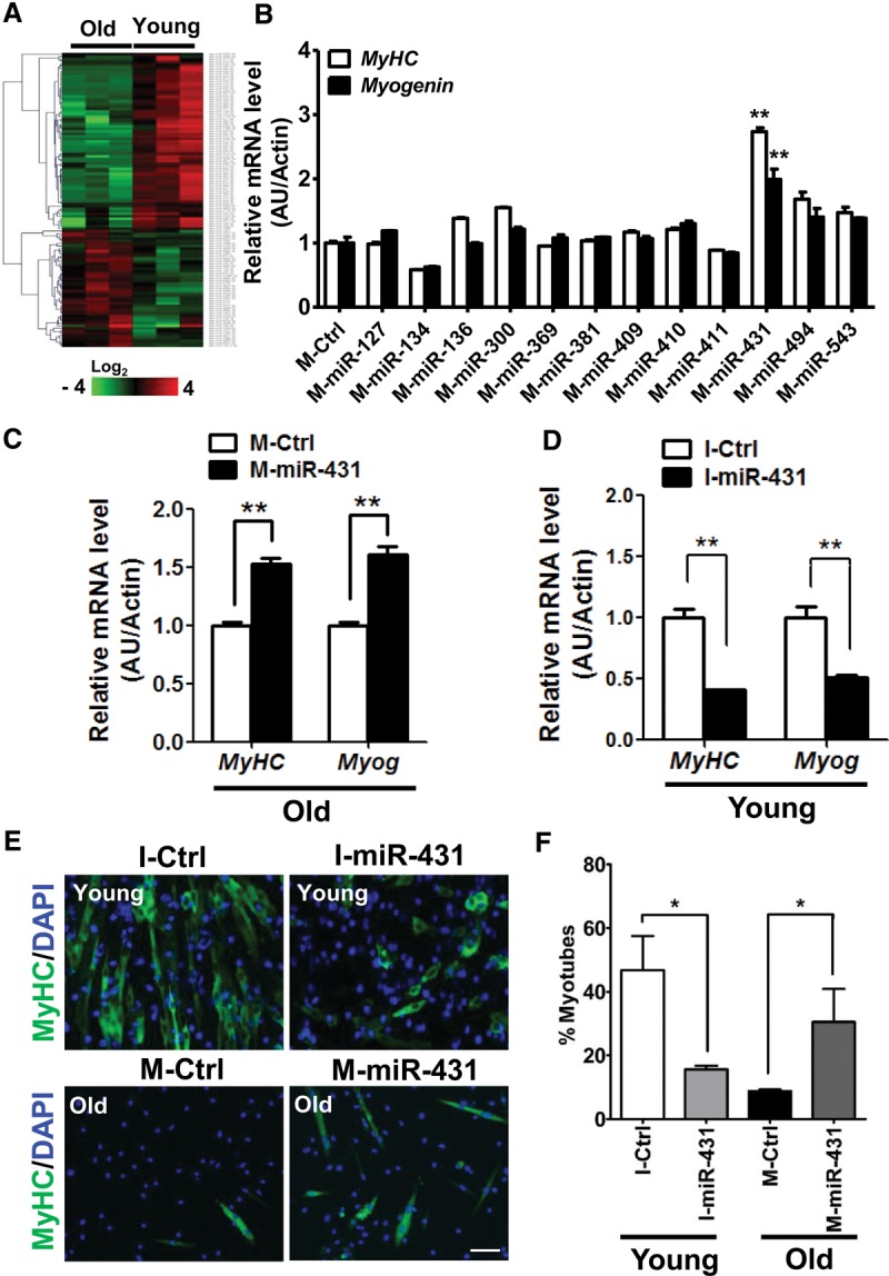
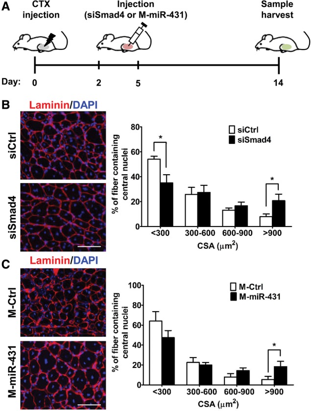
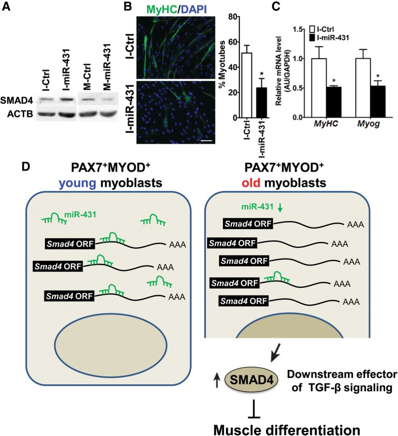
The myogenic capacity of myoblasts decreases in skeletal muscle with age. In addition to environmental factors, intrinsic factors are important for maintaining the regenerative potential of muscle progenitor cells, but their identities are largely unknown. Here, comparative analysis of microRNA (miRNA) expression profiles in young and old myoblasts uncovered miR-431 as a novel miRNA showing markedly reduced abundance in aged myoblasts. Importantly, elevating miR-431 improved the myogenic capacity of old myoblasts, while inhibiting endogenous miR-431 lowered myogenesis. Bioinformatic and biochemical analyses revealed that miR-431 directly interacted with the 3' untranslated region (UTR) of Smad4 mRNA, which encodes one of the downstream effectors of TGF-β signaling. In keeping with the low levels of miR-431 in old myoblasts, SMAD4 levels increased in this myoblast population. Interestingly, in an in vivo model of muscle regeneration following cardiotoxin injury, ectopic miR-431 injection greatly improved muscle regeneration and reduced SMAD4 levels. Consistent with the finding that the mouse miR-431 seed sequence in the Smad4 3' UTR is conserved in the human SMAD4 3' UTR, inhibition of miR-431 also repressed the myogenic capacity of human skeletal myoblasts. Taken together, our results suggest that the age-associated miR-431 plays a key role in maintaining the myogenic ability of skeletal muscle with age.

Keywords: SMAD4; differentiation; miR-431; muscle aging; myoblast; regeneration.


Figures

Figure 1.
Figure 1.
miR-431 promotes differentiation of old myoblasts. (A) Unsupervised hierarchical clustering of 118 differentially regulated miRNAs with aging; 47 miRNAs were up-regulated, and 71 miRNAs were down-regulated. Each column represents miRNA levels in young (n = 3) and old (n = 3) myoblasts isolated from 3-mo-old and 27-mo-old mice each. The intensity represents the magnitude of the difference. Red and green denote high and low expression, respectively. (B) After transfection with the miRNA mimics indicated, old myoblasts were induced to differentiate in differentiation medium. The relative expression of differentiation marker genes such as MyHC (white bar; n = 3 for each group) and Myog (black bar; n = 3 for each group) were quantified by RT-qPCR. The results were normalized to the amount of Actb (β-actin) mRNA. The data are presented as the means ± SD. (**) P < 0.01. (C) After transfection with Ctrl-mimic (M-Ctrl) or miR-431 mimic (M-miR-431), old myoblasts were induced to differentiate in differentiation medium and analyzed for the levels of differentiation markers such as MyHC and Myog mRNAs. (D) Young myoblasts transfected with the miR-431 inhibitor (I-miR-431) showed decreased levels of the differentiation markers tested (MyHC and Myog mRNAs). The results were normalized by the average of Actb mRNA. Data are presented as the means ± SD. (**) P < 0.01. (E) Representative images of differentiated myotubes in I-miR-431 transfected young myoblasts (top panels), M-miR-431 transfected old myoblasts (bottom panels), and the corresponding control transfections; immunofluorescence staining was used to detect MyHC (green) and DAPI (blue). Bar, 200 µm. (F) The fusion index was assessed by calculating the percentage of MyHC-positive cells that contained two or more nuclei versus total MyHC-positive cells. Six different views were randomly selected for measurement of the fusion index. (*) P < 0.05.
Figure 2.
Figure 2.
miR-431 regulates SMAD4 expression by directly interacting with the Smad4 3′ UTR. (A) Schematic depicting the sites of putative interaction of down-regulated miRNAs within the 3′ UTR of the Smad4 mRNA. (B) The effect of M-miR-431 on the activity of a luciferase-Smad4 3′ UTR reporter construct. (**) P < 0.01. (C) Effect of miR-431 on the activity of luciferase reporters bearing either a wild-type Smad4 3′ UTR or a mutant (MT) Smad4 3′ UTR with a deletion of the miR-431 site. (D) Lysates (300 µg) from young or old muscle tissues were incubated with biotinylated (Bi)-miR431-5p or Bi-cel-miR-67 (Ctrl miR) at 4°C with rotation. Four hours later, the RNA was isolated from pull-down material using streptavidin beads and was analyzed by RT-qPCR for Smad4 mRNA enrichment; normalization was carried out using Gapdh mRNA for young or old muscle tissue. The data represent the means ± SEM of three independent experiments. (*) P < 0.05. (E) Lysates from young myoblasts were incubated with antisense oligomers (ASOs) complementary to Smad4 mRNA or a negative control RNA (Gm14635 lincRNA). The RNA isolated after pull-down using streptavidin beads and Smad4 mRNA (normalized to Gapdh mRNA) (left) or for miR431-5p (normalized to RNU6B) (right) were measured by RT-qPCR analysis. (F) Levels of SMAD4 in I-miR-431 transfected young myoblasts (left) and in M-miR-431 transfected old myoblasts (right), each relative to control cells.
Figure 3.
Figure 3.
Knockdown of SMAD4 promotes myogenesis of old myoblasts. (A, top) Lysates of four independently isolated young and old myoblasts were subjected to immunoblot analysis with antibodies to detect the indicated proteins, and ACTB was used as a loading control. (Bottom) Quantification of Western blotting signals is presented as the means ± SD. (*) P < 0.05. (B, top) Twenty-four hours after old myoblasts were transfected with siCtrl and siSmad3 or siSmad4, myogenesis was induced using differentiation medium. Differentiation markers MyHC mRNA and Myog mRNA were identified by RT-qPCR analysis. (Bottom) The effect of SMAD4 knockdown by assessing the same parameters. (*) P < 0.05; (**) P < 0.01; (***) P < 0.001. (C) Immunofluorescence staining for MyHC (green) and DAPI (blue) to detect differentiated myotubes in old myoblasts expressing normal or silenced levels of SMAD4. Bar, 200 µm. (Right) The fusion index was measured by the percentage of MyHC-positive cells that contained two or more nuclei versus total MyHC-positive cells. Six different views were randomly selected for measurement of the fusion index. (*) P < 0.05. (D) Expression levels of the indicated proteins, as determined by Western blot analysis in young and old myoblasts cultured in differentiation medium for up to 3 d (DM3). ACTB was used as a loading control. (E) Relative expression of miR-431 in the cells described in D. The data were normalized to the amount of U6 snoRNA and were expressed relative to the normalized value for young myoblasts in growth medium. (GM) Growth medium; (DM) differentiation medium. (F) The effect of miR-431 on the repression of myogenesis due to TGF-β treatment. After transfection with M-Ctrl or M-miR-431, old myoblasts were incubated in differentiation medium with 5 ng/mL TGF-β1 for 2 d. The levels of MyHC mRNA and Myog mRNA were measured by RT-qPCR analysis. The results were normalized to Actb mRNA levels in each sample, as measured by RT-qPCR analysis. Data are presented as the means ± SD. (**) P < 0.01. (G) The effect of miR-431 on the SMAD signaling induced by the treatment with TGF-β1. C2C12 cells were transfected with M-miR-431 or I-miR-431 together with the 4xSBE-luc reporter construct. Cells were treated with 5 ng/mL TGF-β1 for 16 h before luciferase analysis. The values represent fold induction relative to the basal activity of 4xSBE-luc in a dual-luciferase assay. (*) P < 0.05.
Figure 4.
Figure 4.
Levels of SMAD4 and miR-431 during regeneration of injured muscles. (A) SMAD4 expression patterns were determined using Western blot analysis in CTX-injured young and old TA muscle tissues for up to 14 d post-injury (DPI). (Top) GAPDH was included as a loading control. RT-qPCR analysis of miR-431 in CTX-injured young and old TA muscle tissues for up to 14 d. (Bottom) Expression levels were measured in triplicate, and the data were normalized to the amount of U6 snoRNA and expressed relative to the normalized value for young TA muscle tissues at day 0. (B) At day 14 after CTX injury, young and old TA muscle tissue sections were stained with antibodies that recognized PAX7 (green) and SMAD4 (red), revealing the colocalization of both antigens in myoblasts localized in old muscle tissues. Bar, 20 µm. (White dotted lines) Muscle fibers. (***) P < 0.001.
Figure 5.
Figure 5.
siSmad4-treated or miR-431-treated old muscles showed enhanced regenerative capacity. (A) Scheme of the experimental procedures. CTX (20 µM) was injected into TA muscle tissues of old mice at day 0. Two days and 5 d later, injured muscle tissues were injected with siSmad4 or miR-431 mimic, and contralateral muscle tissues were injected with control siRNA or miRNA mimic. At 14 d after injury, muscle tissues were analyzed by immunohistochemistry. (B,C) Representative immunohistochemistry images (left panels) of laminin (red) and DAPI (blue) staining of siSmad4-treated (B) and miR-431 mimic-treated (C) regenerating fibers (n = 4) on day 14 after injury. Each cross-section area (CSA) was measured by ImageJ software, and six different views were randomly selected for measurement of CSA; (*) P < 0.05. Bar, 100 µm.
Figure 6.
Figure 6.
The effect of miR-431 inhibition on the differentiation of HSMMs. (A) Levels of SMAD4 protein after transfection of HSMMs in growth medium (GM) with I-miR-431 or M-miR-431. Protein levels were detected by Western blot analysis, and β-actin (ACTB) was used as a loading control. (B) After transfection with I-Ctrl or I-miR-431, HSMMs were placed in differentiation medium (DM), and the numbers of differentiated myotubes were compared with those in control cells after immunofluorescence staining for MyHC (green) and DAPI (blue). Bar, 200 µm. (C) Levels of differentiation markers (MyHC and Myog mRNAs) in I-miR-431 transfected HSMMs. Data represent the means ± SD. (*) P < 0.05. (D) Proposed model for the regulation of myoblast differentiation by miR-431 in different ages. Briefly, decreased miR-431 in old PAX7+MYOD+ myoblasts results in increased levels of SMAD4, a downstream effector of signaling by the myogenesis repressor TGF-β, thereby interrupting normal muscle differentiation.

Similar articles

Cited by

References

    1. Ambros V. 2004. The functions of animal microRNAs. Nature 431: 350–355. - PubMed
    1. Bartel DP. 2004. MicroRNAs: genomics, biogenesis, mechanism, and function. Cell 116: 281–297. - PubMed
    1. Bernet JD, Doles JD, Hall JK, Kelly Tanaka K, Carter TA, Olwin BB. 2014. p38 MAPK signaling underlies a cell-autonomous loss of stem cell self-renewal in skeletal muscle of aged mice. Nat Med 20: 265–271. - PMC - PubMed
    1. Brack AS, Bildsoe H, Hughes SM. 2005. Evidence that satellite cell decrement contributes to preferential decline in nuclear number from large fibres during murine age-related muscle atrophy. J Cell Sci 118: 4813–4821. - PubMed
    1. Callis TE, Chen JF, Wang DZ. 2007. MicroRNAs in skeletal and cardiac muscle development. DNA Cell Biol 26: 219–225. - PubMed

Publication types