. 2015 Sep;25(9):1025-42.
doi: 10.1038/cr.2015.90. Epub 2015 Jul 28.

RHOBTB3 promotes proteasomal degradation of HIFα through facilitating hydroxylation and suppresses the Warburg effect

Affiliations

RHOBTB3 promotes proteasomal degradation of HIFα through facilitating hydroxylation and suppresses the Warburg effect

Chen-Song Zhang et al. Cell Res. 2015 Sep.

Abstract

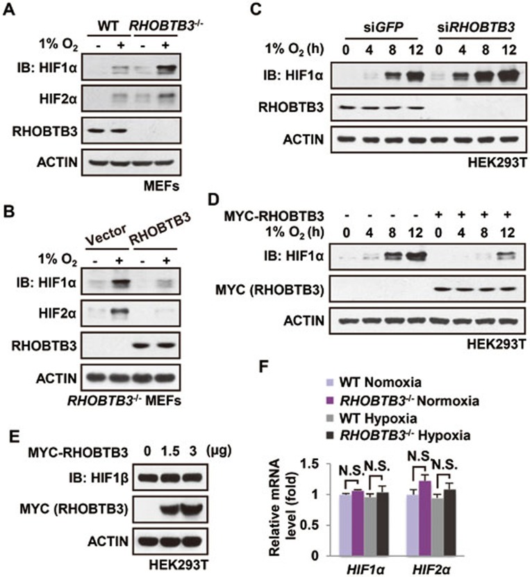
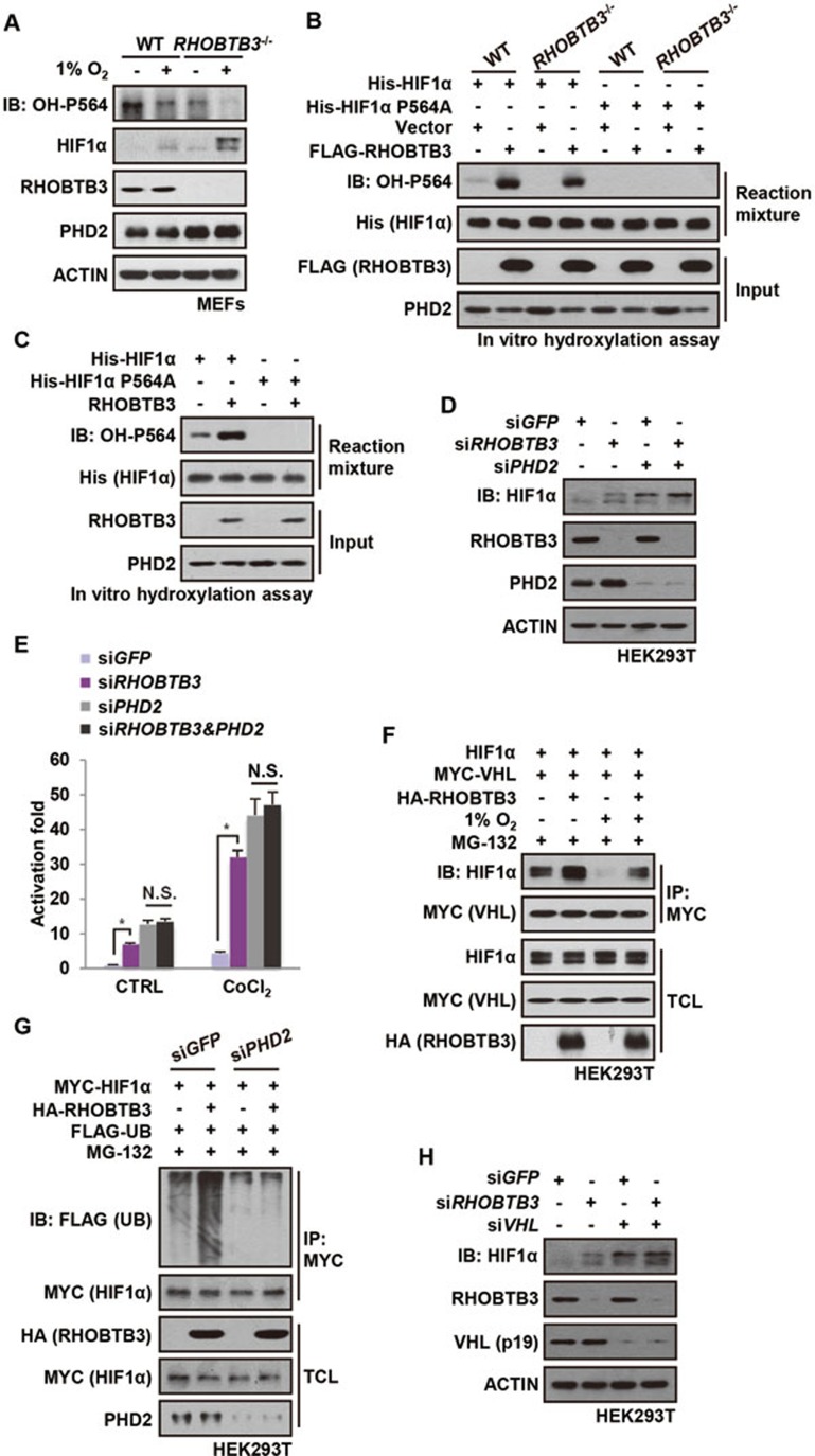
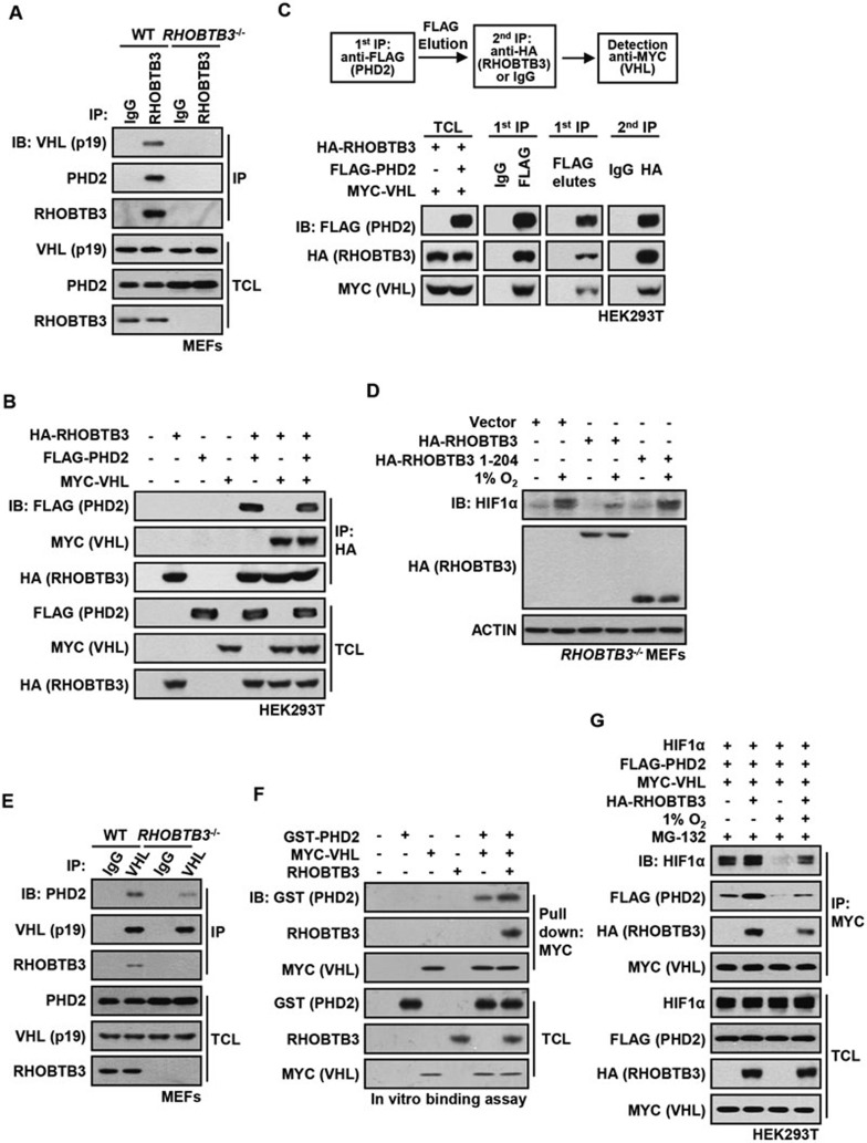
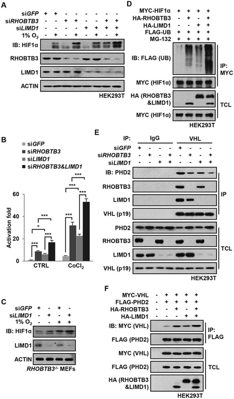
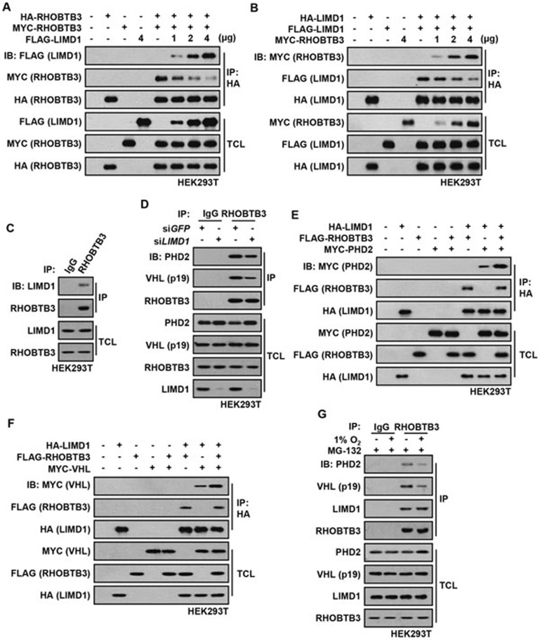
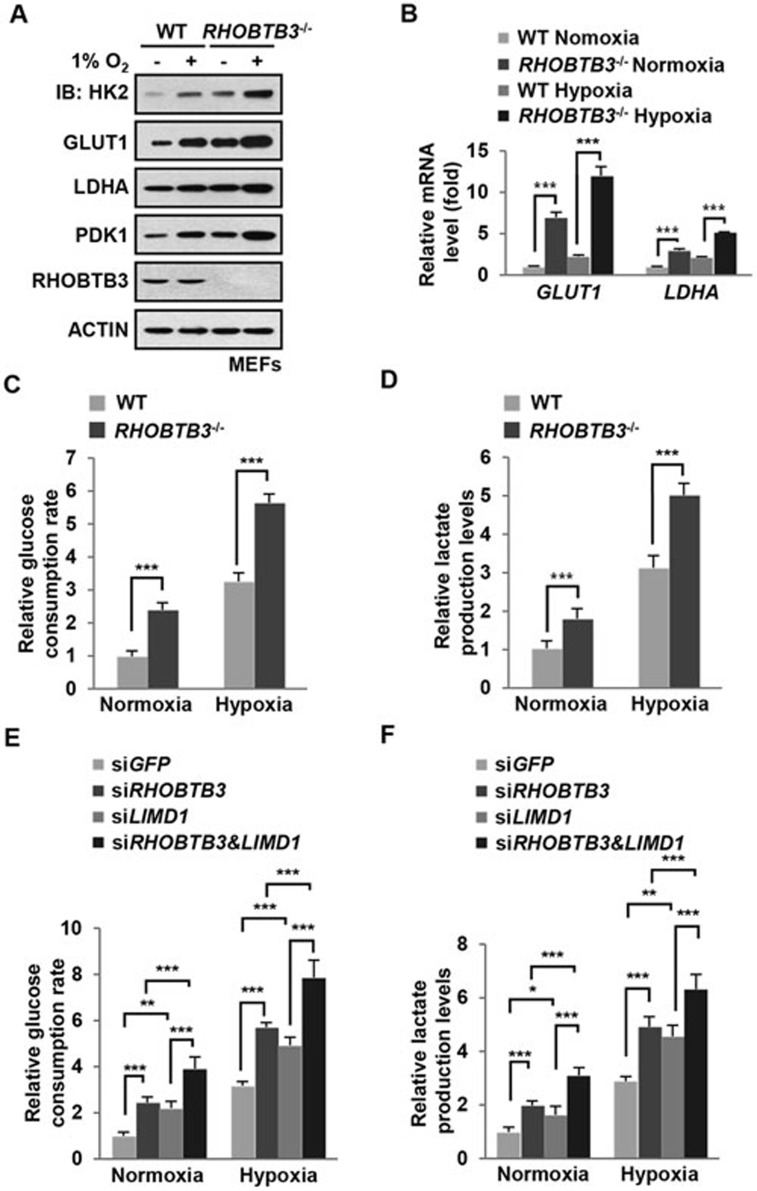
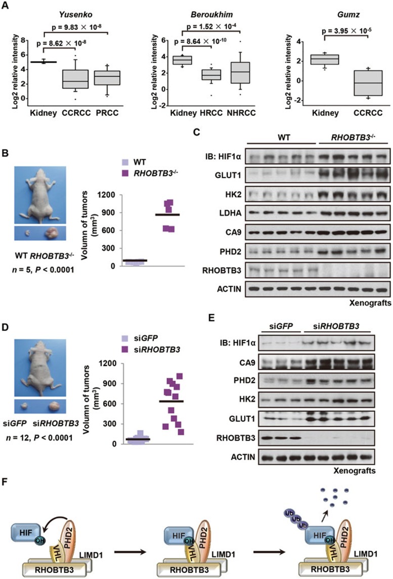
Hypoxia-inducible factors (HIFs) are master regulators of adaptive responses to low oxygen, and their α-subunits are rapidly degraded through the ubiquitination-dependent proteasomal pathway after hydroxylation. Aberrant accumulation or activation of HIFs is closely linked to many types of cancer. However, how hydroxylation of HIFα and its delivery to the ubiquitination machinery are regulated remains unclear. Here we show that Rho-related BTB domain-containing protein 3 (RHOBTB3) directly interacts with the hydroxylase PHD2 to promote HIFα hydroxylation. RHOBTB3 also directly interacts with the von Hippel-Lindau (VHL) protein, a component of the E3 ubiquitin ligase complex, facilitating ubiquitination of HIFα. Remarkably, RHOBTB3 dimerizes with LIMD1, and constructs a RHOBTB3/LIMD1-PHD2-VHL-HIFα complex to effect the maximal degradation of HIFα. Hypoxia reduces the RHOBTB3-centered complex formation, resulting in an accumulation of HIFα. Importantly, the expression level of RHOBTB3 is greatly reduced in human renal carcinomas, and RHOBTB3 deficiency significantly elevates the Warburg effect and accelerates xenograft growth. Our work thus reveals that RHOBTB3 serves as a scaffold to organize a multi-subunit complex that promotes the hydroxylation, ubiquitination and degradation of HIFα.


Figures

Figure 1
Figure 1
RHOBTB3 downregulates HIF1α expression. (A) Protein levels of HIF1α and HIF2α are elevated in RHOBTB3−/− MEFs under both normoxic and hypoxic conditions. RHOBTB3−/− MEFs and control (WT) MEFs were maintained in normoxia or exposed to hypoxia (1% O2) for 8 h. Cells were then lysed and analyzed by immunoblotting with antibodies indicated. (B) Re-introduction of RHOBTB3 into RHOBTB3−/− MEFs reduces the HIFα levels. RHOBTB3−/− MEFs stably expressing GFP or RHOBTB3 were maintained in normoxia or exposed to hypoxia for 8 h, and were then lysed and analyzed as described in A. (C) Knockdown of RHOBTB3 increases the protein levels of HIF1α. HEK293T cells were infected with lentiviruses expressing siRNA targeting either GFP (control) or RHOBTB3. At 16 h post-infection, cells were exposed to hypoxia for different periods of time as indicated, and were then lysed and analyzed by immunoblotting with antibodies indicated. (D) Ectopic expression of RHOBTB3 in HEK293T cells downregulates HIF1α. HEK293T cells were transfected with pcDNA3.3-MYC-RHOBTB3 or pcDNA3.3-MYC vector as a control. At 16 h post-transfection, cells were exposed to hypoxia for the indicated periods of time and were then lysed, and the protein levels of HIF1α were analyzed. (E) RHOBTB3 does not affect the protein levels of HIF1β/ARNT. HEK293T cells were transfected with RHOBTB3. At 16 h post-transfection, cells were lysed and analyzed by immunoblotting with antibodies indicated. (F) RHOBTB3 has no effect on the mRNA levels of HIF1α or HIF2α under both normoxic and hypoxic conditions. RHOBTB3−/− MEFs and WT MEFs, maintained in normoxia or hypoxia, were homogenized in Trizol reagent, and total RNAs were purified, and were subjected to real-time PCR analysis for mRNA levels of HIF1α and HIF2α. Values are presented as mean ± SEM; n = 3 for each group; three replicate experiments. N.S., not significant. Statistical analysis was carried out by ANOVA followed by Tukey.
Figure 2
Figure 2
RHOBTB3 promotes HIFα hydroxylation and ubiquitination in a PHD2- and VHL-dependent manner. (A) RHOBTB3 promotes hydroxylation of HIF1α in MEFs. RHOBTB3−/− MEFs and WT MEFs were maintained in normoxia or exposed to hypoxia for 8 h. Cells were then lysed and the hydroxylation on proline-564 of HIF1α (OH-P564) was analyzed by immunoblotting. As a consequence of sustained accumulation of HIF1α in RHOBTB3-null MEFs, the protein levels of PHD2 were increased. In contrast, the relatively short-term, 8-h hypoxic exposure did not change the protein levels of PHD2. (B) Ectopically expressed RHOBTB3 promotes hydroxylation of HIF1α in vitro. RHOBTB3−/− MEFs and WT MEFs were infected with blank lentiviruses or lentiviruses expressing FLAG-RHOBTB3. Following lysis, the cell lysates were incubated with nickel affinity resin-bound bacterially expressed His-HIF1α (aa 401-603) or the P564A mutant for 90 min at 30 °C. The mixtures were diluted twofold in a 2× SDS buffer, and analyzed by western blotting using antibodies indicated. (C) In vitro translated RHOBTB3 promotes hydroxylation of HIF1α. In vitro translated RHOBTB3 and His-HIF1α (aa 401-603) or the P564A mutant were separately added to cell lysates of RHOBTB3−/− MEFs, and the mixtures were incubated at 30 °C for 90 min. The mixtures were then analyzed for levels of HIF1α hydroxylation as in B. (D) Knockdown of PHD2 impairs RHOBTB3-induced degradation of HIF1α. HEK293T cells were infected by lentiviruses expressing control siRNA (GFP), or siRNA targeting RHOBTB3 or PHD2 or both. At 16 h post-infection, cells were exposed to hypoxia for 4 h, then lysed and analyzed by immunoblotting with antibodies indicated. (E) Double knockdown of RHOBTB3 and PHD2 does not significantly increase transcriptional activity of HIF1α in single knockdown of PHD2. HEK293T cells were infected with lentiviruses expressing different siRNAs as indicated. After 12 h, cells were treated with 200 μM CoCl2 for another 8 h and then lysed. The firefly luciferase reporter carrying HRE was measured and normalized against the Renilla luciferase activity in a dual luciferase assay system. Data are presented as mean ± SEM; n = 3 for each group; *P< 0.05 (ANOVA followed by Tukey); N.S., not significant. (F) RHOBTB3 promotes the interaction between HIF1α and VHL. HEK293T cells were transfected with different combinations of HIF1α, MYC-VHL and HA-RHOBTB3. At 16 h post-transfection, cells were treated with 10 μM MG-132 and maintained in normoxia or exposed to hypoxia for another 10 h, and were lysed. The protein extracts were immunoprecipitated with antibody against MYC for VHL, and were subjected to western blot analysis. TCL, total cell lysate. (G) Knockdown of PHD2 attenuates RHOBTB3-induced ubiquitination of HIF1α. HEK293T cells infected with lentiviruses expressing siRNA targeting GFP (control) or PHD2 were transfected with different combinations of MYC-HIF1α, HA-RHOBTB3 and FLAG-UB. At 16 h post-transfection, cells were treated with 10 μM MG-132 for another 10 h, and were then lysed with RIPA buffer containing 1% SDS and boiled. The protein extracts were diluted in RIPA buffer without SDS to a final concentration of 0.2% SDS, and were subjected to IP with antibody against MYC for HIF1α. The IP product was analyzed by immunoblotting. (H) Knockdown of VHL impairs RHOBTB3 deficiency-induced HIF1α accumulation. HEK293T cells were infected by lentiviruses expressing siRNA targeting GFP (control), RHOBTB3 and/or VHL. At 16 h post-infection, cells were exposed to hypoxia for 4 h, and analyzed by immunoblotting to determine HIF1α protein levels. Probably owing to the low expression levels of its 24 kDa isoform in HEK293T as described previously and the preferential affinity of antibody, only the 19 kDa isoform of VHL (VHL (p19)) could be detected and is shown here.
Figure 3
Figure 3
RHOBTB3, PHD2 and VHL form a complex. (A) RHOBTB3 interacts with endogenous PHD2 and VHL. Protein extracts of WT MEFs and RHOBTB3−/− MEFs (control) were immunoprecipitated with antibody against RHOBTB3 or control IgG, and analyzed by immunoblotting with antibodies indicated. (B) Ectopically expressed RHOBTB3 interacts simultaneously with PHD2 and VHL. HEK293T cells were transfected with different combinations of HA-RHOBTB3, MYC-VHL and FLAG-PHD2. At 16 h post-infection, cells were lysed and the protein extracts were immunoprecipitated with antibody against HA, and the IP product was analyzed by western blotting. (C) RHOBTB3, VHL and PHD2 form a complex. HEK293T cells were transfected with HA-RHOBTB3, FLAG-PHD2 and MYC-VHL. After 16 h, cells were harvested. Two-step co-IP was performed by first using anti-FLAG antibody, followed by elution with the FLAG peptide. The eluates were subjected to a second round of IP with anti-HA or control IgG, and the final precipitated proteins were analyzed by immunoblotting. (D) The N-terminal region of RHOBTB3 (aa 1-204) does not have a role in the degradation of HIF1α. RHOBTB3−/− MEFs were infected with lentiviruses expressing HA-RHOBTB3 or HA-RHOBTB3 (aa 1-201). At 36 h post-infection, cells were maintained in normoxia or exposed to hypoxia for 8 h, and analyzed after lysis by immunoblotting. (E) RHOBTB3 strengthens the interaction between PHD2 and VHL. RHOBTB3−/− MEFs and WT MEFs were lysed and the endogenous VHL was immunoprecipitated. The IP product was analyzed by immunoblotting. (F) RHOBTB3 promotes the PHD2-VHL interaction in vitro. In vitro translated RHOBTB3 and bacterially expressed GST-PHD2 were incubated with anti-MYC-conjugated resin-bound MYC-tagged VHL. The mixtures were then pulled down by centrifugation, and analyzed by immunoblotting. (G) RHOBTB3 promotes the interaction between PHD2 and HIF1α. HEK293T cells were transfected with different combinations of HIF1α, FLAG-PHD2, MYC-VHL and HA-RHOBTB3. At 8 h post-transfection, cells were treated with 10 μM MG-132 and maintained in normoxia or exposed to hypoxia for 10 h. The protein extracts were immunoprecipitated with antibody against MYC (for VHL), and precipitated proteins were analyzed by immunoblotting.
Figure 4
Figure 4
RHOBTB3 and LIMD1 cooperatively regulate HIF1α. (A) RHOBTB3 and LIMD1 cooperatively suppress the protein level of HIF1α. HEK293T cells were infected with lentiviruses expressing siRNA targeting GFP, RHOBTB3 and/or LIMD1. At 16 h post-infection, cells were maintained in normoxia or exposed to hypoxia for 8 h before lysis and immunoblotting with antibodies indicated. (B) RHOBTB3 and LIMD1 cooperatively suppress the transcriptional activities of HIF1α. HEK293T cells were infected with different combinations of lentiviruses as indicated. Transcriptional activities of HIF1α were measured using a dual luciferase assay system as described in Figure 2E. Data are presented as mean ± SEM, n = 3 for each group, *P < 0.05, ***P < 0.001 (ANOVA followed by Tukey). (C) Knockdown of LIMD1 in RHOBTB3−/− MEFs further increases the protein levels of HIF1α. RHOBTB3−/− MEFs were infected with lentiviruses expressing siRNA targeting GFP or LIMD1. At 36 h post-infection, cells were maintained in normoxia or exposed to hypoxia for 8 h, before the western blot analysis. (D) RHOBTB3 and LIMD1 cooperatively promote the ubiquitination of HIF1α. HEK293T cells were transfected with different combinations of MYC-HIF1α, HA-RHOBTB3, HA-LIMD1 and FLAG-UB (ubiquitin). After treatment with 10 μM MG-132 for 10 h, the cells were lysed, and the lysates were subjected to IP with antibody against MYC (for HIF1α). The IP product was analyzed by western blotting to determine the ubiquitination levels of HIF1α. (E) Knockdown of RHOBTB3 and/or LIMD1 decreases PHD2-VHL interaction. HEK293T cells were infected with lentiviruses expressing siRNA targeting GFP, RHOBTB3 and/or LIMD1. At 16 h post-infection, cells were lysed and the endogenous VHL was immunoprecipitated, and the IP product was analyzed by immunoblotting. (F) Ectopically expressed RHOBTB3 and LIMD1 cooperatively promote PHD2-VHL interaction. HEK293T cells were transfected with different combinations of MYC-VHL, HA-RHOBTB3, HA-LIMD1 and FLAG-PHD2. Protein extracts from the transfected cells were subjected to IP with antibody against FLAG and analyzed by immunoblotting with antibodies indicated.
Figure 5
Figure 5
Dimerization of RHOBTB3 and LIMD1. (A) Ectopically expressed HA-tagged RHOBTB3 interacts with MYC-tagged RHOBTB3 or FLAG-tagged LIMD1. HEK293T cells were transfected with different combinations of HA-RHOBTB3, MYC-RHOBTB3 and FLAG-LIMD1. Cells were then lysed and the protein extracts were immunoprecipitated with antibody against HA. The IP product was analyzed by immunoblotting. (B) Ectopically expressed HA-tagged LIMD1 interacts with FLAG-tagged LIMD1 or MYC-tagged RHOBTB3. Lysates from transfected cells were subjected to IP with antibody against HA (for LIMD1), and analyzed by immunoblotting as in A. (C) RHOBTB3 interacts with endogenous LIMD1. Lysates of HEK293T cells were immunoprecipitated with antibody against RHOBTB3 or IgG (control), and analyzed by immunoblotting using antibodies indicated. (D) Knockdown of LIMD1 attenuates the interaction between RHOBTB3 and PHD2/VHL. HEK293T cells were infected with lentivirus expressing siRNA targeting GFP or LIMD1. At 16 h post-infection, cells were lysed and the endogenous RHOBTB3 was then immunoprecipitated, and analyzed by immunoblotting with antibodies indicated. (E, F) Ectopically expressed RHOBTB3 promotes the interaction between LIMD1 and PHD2 (E), and the interaction between LIMD1 and VHL (F). HEK293T cells were transfected with different combinations of HA-LIMD1, FLAG-RHOBTB3, MYC-PHD2 (E) and MYC-VHL (F). Protein extracts were immunoprecipitated and analyzed by immunoblotting. (G) Hypoxia attenuates the interaction between endogenous RHOBTB3/LIMD1 and PHD2/VHL. HEK293T cells were maintained in normoxia or exposed to hypoxia for 8 h in presence of 10 μM MG-132 to prevent the degradation of VHL under hypoxic condition as described previously. Endogenous RHOBTB3 was then immunoprecipitated, and the IP product was analyzed by immunoblotting with antibodies indicated.
Figure 6
Figure 6
RHOBTB3 is a negative regulator of the Warburg effect. (A) Knockout of RHOBTB3 elevates the expression of HK2, LDHA GLUT1 and PDK1. RHOBTB3−/− MEFs and WT MEFs were maintained in normoxia or exposed to hypoxia for 16 h. Cells were then lysed and the protein extracts were analyzed by immunoblotting with antibodies indicated. (B) RHOBTB3 deficiency leads to increased mRNA levels of GLUT1 and LDHA. Total RNAs from RHOBTB3−/− MEFs and WT MEFs, maintained in normoxia or exposed to hypoxia for 16 h, were purified, and analyzed by real-time PCR analysis for the expression levels of GLUT1 and LDHA. Values are presented as mean ± SEM, n = 3 for each group, three replicate experiments. ***P< 0.001 (ANOVA followed by Tukey). (C, D) RHOBTB3 decreases rates of glucose consumption (C) and lactate production (D). RHOBTB3−/− and WT MEFs were maintained in normoxia or exposed to hypoxia for 8 h, and glucose consumption rates (C) and lactate production rates (D) were measured. Values are presented as mean ± SEM, n = 3 for each group, ***P< 0.001 (ANOVA followed by Tukey). (E, F) RHOBTB3 and LIMD1 cooperatively decrease glucose consumption (E) and lactate production (F). HEK293T cells were infected with lentiviruses expressing siRNAs targeting GFP, RHOBTB3 and/or LIMD1. At 16 h post-infection, cells were maintained in normoxia or exposed to hypoxia for 8 h and glucose consumption rates (E) and lactate production rates (F) were measured. Values are presented as mean ± SEM, n = 3 for each group, *P< 0.05, **P< 0.01, ***P< 0.001 (ANOVA followed by Tukey).
Figure 7
Figure 7
RHOBTB3 suppresses tumorigenesis. (A) Expression of RHOBTB3 in human renal cancer samples in public data sets summarized by the Oncomine Platform. (B) Xenografts derived from RHOBTB3−/− MEFs are significantly larger compared with those derived from control MEFs. Ras V12/E1A H133-transformed RHOBTB3−/− MEFs and control WT MEFs (1 × 10⁶) were injected intradermally into each flank of nude mice. Tumors were allowed to develop for 35 days. Tumor volumes were then determined by direct measurement using a caliper and calculated by the formula: (widest diameter × smallest diameter2)/2. Data were presented as mean ± SEM, n = 5 for each group, P < 0.0001 (Student's t-test). (C) The protein levels of HIF1α and its targets are upregulated in xenografts derived from RHOBTB3−/− MEFs. Xenografts derived from RHOBTB3−/− and WT MEFs as described in B were homogenized and analyzed by immunoblotting with the indicated antibodies. (D) Xenografts derived from RHOBTB3 knocked down HeLa cells are significantly larger compared with those derived from control cells. Suspensions of HeLa cells expressing siGFP or siRHOBTB3 (1 × 10⁶ each) were injected intradermally into each flank of nude mice. Tumors were allowed to develop for 37 days and their volumes were calculated as described in Figure 7B. The values of tumor volumes are presented as mean ± SEM, n = 12 for each group, P < 0.0001 (Student's t-test). (E) Protein levels of HIF1α and its target genes are upregulated in xenografts of RHOBTB3 knocked down HeLa cells. Xenografts derived from HeLa-siGFP or HeLa-siRHOBTB3 cells were homogenized and analyzed by immunoblotting with antibodies indicated. (F) Simplified model depicting that RHOBTB3 and LIMD1 promote the formation of the HIF1α degradation complex. In this scheme, RHOBTB3 and LIMD1 form a heterodimer, which interacts with PHD2 and VHL, and recruits HIFα, forming a RHOBTB3/LIMD1-PHD2-VHL-HIFα complex that promotes the hydroxylation, ubiquitination and degradation of HIFα. Hypoxia loosens the interaction between RHOBTB3-LIMD1 and HIFα-VHL-PHD2, allowing for the accumulation of HIFα in cells under hypoxia. Notably, RHOBTB3 can directly promote PHD2-mediated hydroxylation of HIFα, whereas LIMD1 cannot.

References

    1. Majmundar AJ, Wong WJ, Simon MC. Hypoxia-inducible factors and the response to hypoxic stress. Mol Cell. 2010;40:294–309. - PMC - PubMed
    1. Kaelin WG, Jr, Ratcliffe PJ. Oxygen sensing by metazoans: the central role of the HIF hydroxylase pathway. Mol Cell. 2008;30:393–402. - PubMed
    1. Semenza GL. Hypoxia-inducible factors in physiology and medicine. Cell. 2012;148:399–408. - PMC - PubMed
    1. Semenza GL. Oxygen sensing, homeostasis, and disease. N Engl J Med. 2011;365:537–547. - PubMed
    1. Semenza GL, Jiang BH, Leung SW, et al. Hypoxia response elements in the aldolase A, enolase 1, and lactate dehydrogenase A gene promoters contain essential binding sites for hypoxia-inducible factor 1. J Biol Chem. 1996;271:32529–32537. - PubMed

Publication types

MeSH terms

Substances