. 2015 Jul 29:14:140.
doi: 10.1186/s12943-015-0418-x.

Bevacizumab promotes venous thromboembolism through the induction of PAI-1 in a mouse xenograft model of human lung carcinoma

Affiliations

Bevacizumab promotes venous thromboembolism through the induction of PAI-1 in a mouse xenograft model of human lung carcinoma

Ni Chen et al. Mol Cancer. .

Abstract

Background: An increased incidence of venous thromboembolism (VTE) is associated with anti-vascular endothelial growth factor (VEGF) treatment in cancer. However, the mechanism underlying this effect remains elusive. In this study, we examined the effect of bevacizumab, a humanized monoclonal antibody against VEGF-A, on VTE in a murine xenograft A549 cell tumor model.

Methods: Inferior vena cava stenosis model and FeCl3-induced saphenous vein thrombosis model were performed in a mouse xenograft models of human lung adenocarcinoma.

Results: We found that treatment with bevacizumab significantly increased the thrombotic response to inferior vena cava obstruction and femoral vein injury. Plasminogen activator inhibitor (PAI-1) expression in tumors, plasma, and thrombi was significantly increased by bevacizumab. However, bevacizumab did not enhance VTE in PAI-1-deficient mice, suggesting that PAI-1 is a major mediator of bevacizumab's prothrombotic effect. VEGF inhibited expression of PAI-1 by A549 cells, and this effect was neutralized by bevacizumab, suggesting that bevacizumab increases PAI-1 expression in vivo by blocking the inhibitory effect of VEGF on PAI-1 expression by tumor cells. Pharmacological inhibition of PAI-1 with PAI-039 blocked bevacizumab-induced venous thrombosis.

Conclusion: Collectively, these findings indicate that PAI-1 plays a role in VTE associated with antiangiogenic therapy and the inhibition of PAI-1 shows efficacy as a therapeutic strategy for the prevention of bevacizumab-associated VTE.


Figures
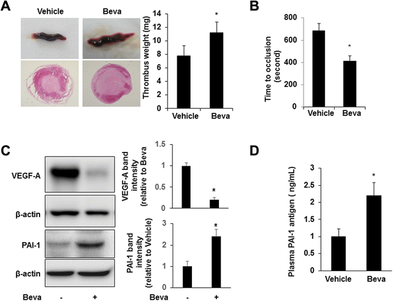
Fig. 1
Fig. 1
Bevacizumab promotes venous thrombosis. a. IVC stenosis was induced in tumor-bearing mice (n = 6/group). Three hours after ligation, mice were euthanized and the weight of the thrombus was determined. Representative whole thrombi and cross-sections of thrombi retrieved from mice treated with bevacizumab (Beva) or vehicle control are shown. *P < 0.05 vs. vehicle control. b. Saphenous vein thrombosis was induced using 10 % FeCl3 in control (n = 6) and A549 tumor-bearing mice (n = 6). Occlusion times were measured and are shown as the mean ± SEM. *P < 0.05 vs. the vehicle group. c. Tumors were excised and lysates were prepared and subjected to Western blotting to detect VEGF-A, PAI-1, and β-actin. Representative blots and densitometric analyses of 3 independent experiments are shown. *P < 0.05 vs. control. d. Plasma PAI-1 antigen was measured (n = 6/group); *P < 0.05 vs. the vehicle group. Beva: bevacizumab
Fig. 2
Fig. 2
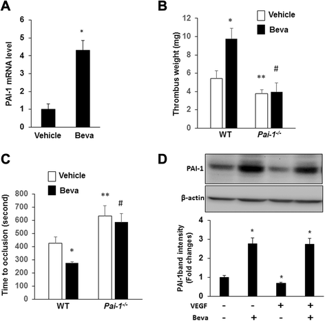
Bevacizumab promotes venous thrombosis in a PAI-1-dependent manner. a. The intrathrombotic gene expression of PAI-1 in the bevacizumab and vehicle groups was determined via real-time RT-PCR. All values represent mean ± SEM (n = 6/group). *P < 0.01 vs. the vehicle group. b. IVC stenosis was induced in WT and Pai-1 −/− mice (n = 6/group). Ten days after ligation, all mice were euthanized, and the weight of the thrombus was determined. *P < 0.05 vs. the vehicle group in WT mice; **P < 0.05 vs. the vehicle group in WT mice; #P < 0.05 vs. the bevacizumab group in WT mice, and #P = 0.74 vs. the vehicle group in Pai-1 −/− mice (n = 6/group). c. Saphenous vein thrombosis was induced using 10 % FeCl3 in WT and Pai-1 −/− mice (n = 6/group). Occlusion times were measured and are shown as the mean ± SEM. *P < 0.05 vs. the vehicle group in WT mice; **P < 0.05 vs. the vehicle group in WT mice; #P < 0.05 vs. the bevacizumab group in WT mice. d. A549 cells were cultured for 24 hrs in the presence and absence of VEGF (50 ng/mL) and bevacizumab (250 μg/mL), as indicated. Cell lysates were prepared and subjected to Western blotting to detect PAI-1and β-actin. Representative blot and densitometric analyses of 3 independent experiments are shown. *P < 0.05 vs. control (i.e. no VEGF or Beva)
Fig. 3
Fig. 3
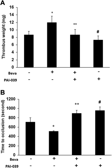
Pharmacological inhibition of PAI-1 blocks the prothrombotic effect of bevacizumab. a. IVC stenosis was induced in tumor-bearing mice. The mean weight of the thrombus was determined. *P < 0.05 vs. the vehicle group; **P < 0.05 vs. the beva group; #P > 0.05 vs. beva + PAI-039 group (n = 6/group). b. Mean occlusion times were measured in a saphenous vein thrombosis model and are shown as the mean ± SEM. *P < 0.01 vs. the vehicle group; **P < 0.05 vs. the beva group; #P > 0.05 vs. beva + PAI-039 group (n = 6/group). Beva: bevacizumab
Fig. 4
Fig. 4
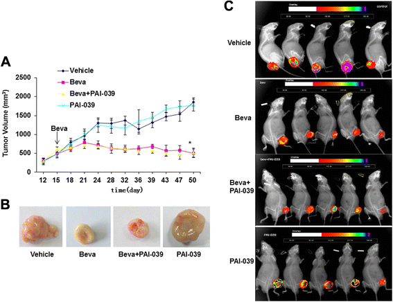
Tumor growth inhibition by bevacizumab in orthotopic A549 xenografts. a. A549 tumor growth curves of nude mice receiving bevacizumab (Beva; 200 μg/mouse; once weekly), PAI-039 (2 mg/kg/day), both, or vehicle control (n = 6/group). Arrow denotes first bevacizumab injection. Tumor sizes of Beva and Beva + PAI-039 groups were significantly less than those of vehicle group (P < 0.05) at 24 days and all subsequent time points, whereas differences between PAI-039 and vehicle groups were not statistically significant at any time point. b. Representative images of tumors retrieved at completion of treatment protocol. c. Bioluminescent imaging of A549-luc tumors after completion of treatment protocol. Note smaller tumor sizes in bevacizumab-treated mice

Similar articles

Cited by

References

    1. Zangari M, Fink LM, Elice F, Zhan F, Adcock DM, Tricot GJ. Thrombotic events in patients with cancer receiving antiangiogenesis agents. J Clin Oncol. 2009;27:4865–4873. doi: 10.1200/JCO.2009.22.3875. - DOI - PubMed
    1. Eremina V, Jefferson JA, Kowalewska J, Hochster H, Haas M, Weisstuch J, et al. VEGF inhibition and renal thrombotic microangiopathy. N Engl J Med. 2008;358:1129–1136. doi: 10.1056/NEJMoa0707330. - DOI - PMC - PubMed
    1. Rini BI, Escudier B, Tomczak P, Kaprin A, Szczylik C, Hutson TE, et al. Comparative effectiveness of axitinib versus sorafenib in advanced renal cell carcinoma (AXIS): a randomized phase 3 trial. Lancet. 2011;378:1931–1939. doi: 10.1016/S0140-6736(11)61613-9. - DOI - PubMed
    1. Evans CE, Grover SP, Humphries J, Saha P, Patel AP, Patel AS, et al. Antiangiogenic therapy inhibits venous thrombus resolution. Arterioscler Thromb Vasc Biol. 2014;34:565. doi: 10.1161/ATVBAHA.113.302998. - DOI - PubMed
    1. Nalluri SR, Chu D, Keresztes R, Zhu X, Wu S. Risk of venous thromboembolism with the angiogenesis inhibitor bevacizumab in cancer patients: a meta-analysis. JAMA. 2008;300:2277–2285. doi: 10.1001/jama.2008.656. - DOI - PubMed

Publication types

MeSH terms

Substances