. 2015 Oct;1848(10 Pt A):2422-9.
doi: 10.1016/j.bbamem.2015.07.014. Epub 2015 Jul 26.

Membrane-mediated amyloid formation of PrP 106-126: A kinetic study

Affiliations

Membrane-mediated amyloid formation of PrP 106-126: A kinetic study

Yen Sun et al. Biochim Biophys Acta. 2015 Oct.

Abstract

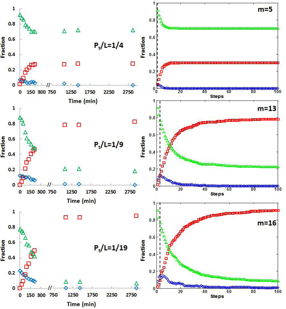
PrP 106-126 conserves the pathogenic and physicochemical properties of the Scrapie isoform of the prion protein. PrP 106-126 and other amyloidal proteins are capable of inducing ion permeability through cell membranes, and this property may represent the common primary mechanism of pathogenesis in the amyloid-related degenerative diseases. However, for many amyloidal proteins, despite numerous phenomenological observations of their interactions with membranes, it has been difficult to determine the molecular mechanisms by which the proteins cause ion permeability. One approach that has not been undertaken is the kinetic study of protein-membrane interactions. We found that the reaction time constant of the interaction between PrP 106-126 and membranes is suitable for such studies. The kinetic experiment with giant lipid vesicles showed that the membrane area first increased by peptide binding but then decreased. The membrane area decrease was coincidental with appearance of extramembranous aggregates including lipid molecules. Sometimes, the membrane area would increase again followed by another decrease. The kinetic experiment with small vesicles was monitored by circular dichroism for peptide conformation changes. The results are consistent with a molecular simulation following a simple set of well-defined rules. We deduced that at the molecular level the formation of peptide amyloids incorporated lipid molecules as part of the aggregates. Most importantly the amyloid aggregates desorbed from the lipid bilayer, consistent with the macroscopic phenomena observed with giant vesicles. Thus we conclude that the main effect of membrane-mediated amyloid formation is extraction of lipid molecules from the membrane. We discuss the likelihood of this effect on membrane ion permeability.

Keywords: Amyloidal peptides; Circular dichroism; Giant unilamellar vesicles; Lipid extracting effect; Neurodegenerative diseases; Prion protein.


Figures

Fig. 1
Fig. 1
Four representative runs of an aspirated GUV (containing 1% Rh-DOPE) exposed to 10 µM PrP 106-126 (out of 26 individual runs). The fractional change of the membrane area of the GUV, ΔA/A, was calculated from the change of the protrusion length as shown in the fluorescence images at the top, taken at 0, 15, 95, 318, 598 s from left to right for the run labeled by red squares. Scale bar = 10 µm. Note that extramembranous aggregates containing dye lipid began to appear as the protrusion length decreased for the first time. The plot includes two representative runs showing the membrane area increased and decreased twice in 10 mins, and two other representative runs showing membrane area increased and decreased once in 10 mins.
Fig. 2
Fig. 2
(A) CD spectra of PrP 106-126 in three different configurations: α-helix (blue), β-sheet (red), and random coil (green) in an unit expressed in mdeg for the same concentration 100 µM of peptide. (B) Time series of CD spectra, from bottom to top, for 100 µM PrP peptide in 0.7 mg/ml lipid vesicles (Pt/L = 1/9). (C) Time series of CD spectra, from bottom to top, for 100 µM PrP peptide in 4 mg/ml lipid vesicles (Pt/L = 1/50). In both (B) and (C), the data are shown by the symbols indicated by the time of measurement, and the red dots are the fit to a linear combination of three spectra shown in (A). All measurements were made with the same amount of peptide, so all CD spectra are normalized relatively to each other.
Fig. 3
Fig. 3
Kinetics of PrP peptide conformation changes at high total peptide to lipid ratios (Pt/L). The sample conditions were: 100 µM PrP in 0.35 mg/ml lipid (Pt/L = 1/4), in 0.7 mg/ml lipid (Pt/L = 1/9), and in 1.5 mg/ml lipid (Pt/L = 1/19). Data are on the left column (note the initial time scale expansion), and the corresponding simulations on the right, where m is the number of lipid vesicles in simulations (see text or Supporting Information). Each simulation result was the average of five repeated simulations. Green represents the fraction of the peptide in random coils, blue α-helices and red β-sheets. The black dashed lines in simulations are the “starting time” to be compared with the first measurements in the kinetic experiments.
Fig. 4
Fig. 4
Kinetics of PrP peptide conformation changes at low total peptide to lipid ratios (Pt/L). The sample conditions were: 100 µM PrP in 2 mg/ml lipid (Pt/L = 1/25), and in 4 mg/ml lipid (Pt/L =1/50); 50 µM PrP in 8 mg/ml lipid (Pt/L = 1/200). Data are on the left column (note the initial time scale expansion for Pt/L = 1/25), and the corresponding simulations on the right, where m is the number of lipid vesicles in simulations (see text or Supporting Information). Each simulation result was the average of five repeated simulations. Green represents the fraction of the peptide in random coils, blue α-helices and red β-sheets. The black dashed lines in simulations are the “starting time” to be compared with the first measurements in the kinetic experiments.
Fig. 5
Fig. 5
The fluorescence images of 100 µM PrP peptide mixed with 0.5 mg/ml lipid vesicles made of 7:3 DOPC/DOPG and 1% of Rh-DOPE (Pt/L = 1/9), at the time indicated. Initially the aggregates were too small to be detected, but the aggregates grew to macroscopic size as time went on. An additional confocal image was shown for day 5. These images show that the lipid was part of the aggregates formed by PrP peptides. Scale bar = 50 µm.

Similar articles

Cited by

References

    1. Perutz MF, Windle AH. Cause of neural death in neurodegenerative diseases attributable to expansion of glutamine repeats. Nature. 2001;412:143–144. - PubMed
    1. Lashuel HA, Lansbury PT., Jr Are amyloid diseases caused by protein aggregates that mimic bacterial pore-forming toxins? Q Rev Biophys. 2006;39:167–201. - PubMed
    1. Caughey B, Lansbury PT. Protofibrils, pores, fibrils, and neurodegeneration: separating the responsible protein aggregates from the innocent bystanders. Annu Rev Neurosci. 2003;26:267–298. - PubMed
    1. Xue WF, Hellewell AL, Gosal WS, Homans SW, Hewitt EW, Radford SE. Fibril fragmentation enhances amyloid cytotoxicity. J Biol Chem. 2009;284:34272–34282. - PMC - PubMed
    1. Kodali R, Wetzel R. Polymorphism in the intermediates and products of amyloid assembly. Curr Opin Struct Biol. 2007;17:48–57. - PubMed

Publication types

MeSH terms

LinkOut - more resources