. 2015 Dec;3(1):52.
doi: 10.1186/s40635-015-0052-3. Epub 2015 Jun 21.

Critical illness-induced bone loss is related to deficient autophagy and histone hypomethylation

Affiliations

Critical illness-induced bone loss is related to deficient autophagy and histone hypomethylation

Helen C Owen et al. Intensive Care Med Exp. 2015 Dec.

Abstract

Background: Survivors of critical illness are at increased risk of fractures. This may be due to increased osteoclast formation during critical illness, leading to trabecular bone loss. Such bone loss has also been observed in Paget's disease, and has been related to deficient autophagy. Deficient autophagy has also been documented in vital organs and skeletal muscle of critically ill patients. The objective of this study was to investigate whether deficient autophagy can be linked to critical illness-induced bone loss.

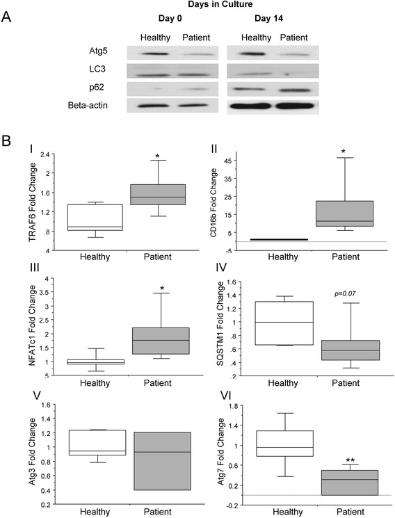
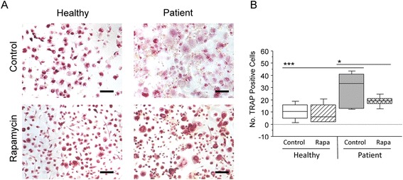
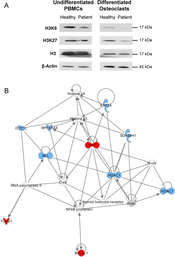
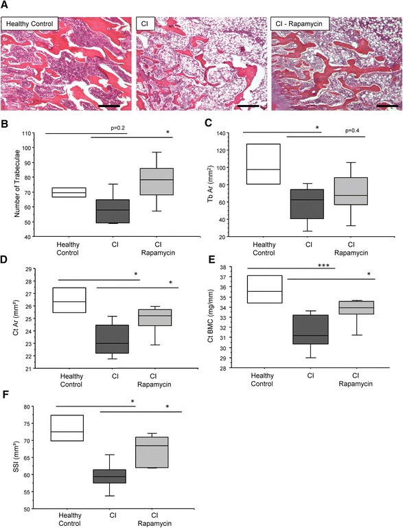
Methods: Osteoclasts grown in vitro and their precursor cells isolated from peripheral blood of critically ill patients and from matched healthy volunteers were analysed for the expression of autophagy genes (SQSTM1, Atg3 and Atg7), and proteins (p62, Atg-5, and microtubule-associated protein light chain 3-II (LC3-II)) and for autophagy and epigenetic signalling factors via PCR arrays and were treated with the autophagy inducer rapamycin. The effect of rapamycin was also investigated at the tissue level in an in vivo rabbit model of critical illness.

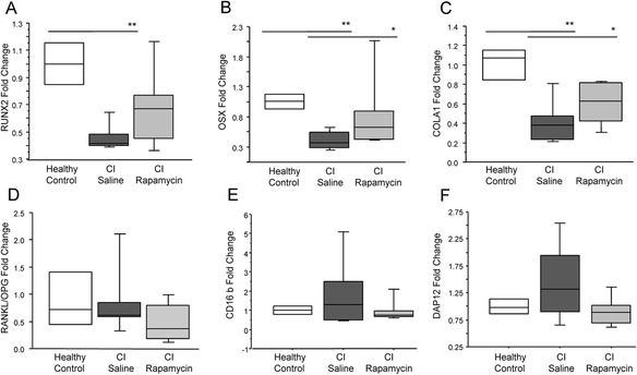
Results: Many more osteoclasts formed in vitro from the blood precursor cells isolated from critically ill patients, which accumulated p62, and displayed reduced expression of Atg5, Atg7, and LC3-II compared to healthy controls, suggesting deficient autophagy, whilst addition of rapamycin reduced osteoclast formation. PCR arrays revealed a down-regulation of histone methyltransferases coupled with an up-regulation of negative regulators of autophagy. Critically ill rabbits displayed a reduction in trabecular and cortical bone, which was rescued with rapamycin.

Conclusions: Deficient autophagy in osteoclasts and their blood precursor cells at least partially explained aberrant osteoclast formation during critical illness and was linked to global histone hypomethylation. Treatment with the autophagy activator Rapamycin reduced patient osteoclast formation in vitro and reduced the amount of bone loss in critically ill rabbits in vivo. These findings may help to develop novel therapeutic targets to prevent critical illness-induced bone loss.


Figures

Fig. 1
Fig. 1
Deficient autophagy in critically ill patient PBMCs. (a) Undifferentiated PBMCs from critically ill patients displayed decreased levels of Atg5 and increased levels of p62 protein, suggesting autophagy was already deficient in these cells. In PBMCs grown for 14 days in culture, protein levels of both Atg5 and LC3–II were decreased, in combination with an accumulation in p62. (b) At the level of gene expression, markers of mature osteoclast differentiation (TRAF6, CD16B, NFATc1) were significantly increased in osteoclasts from critically ill patients compared to healthy controls (1.6-fold, 17.5–fold, and 1.9–fold, respectively; p < 0.05). Expression of autophagy markers SQSTM1, the gene encoding p62, and Atg3 were not significantly different between cell populations, however, Atg7 was significantly decreased by 3.5–fold in patient cells compared to healthy controls (p < 0.01) (n = 3; *p < 0.05; **p < 0.01)
Fig. 2
Fig. 2
Induction of autophagy in vitro leads to decreased osteoclast formation in critically ill patient PBMCs. (a) The formation of mature, multi-nuclear (≥3 nuclei, tartrate-resistant acid phosphatase (TRAP) positive) osteoclasts was quantified visually using ImageJ software. (b) After 14 days in culture, osteoclast formation increased by 65 % in PBMC cultures from critically ill patients than those from healthy controls. The addition of rapamycin did not alter osteoclast formation in healthy control cultures, however, in patient cultures, osteoclast formation was reduced by 36 % (n = 6; *p < 0.05; ***p < 0.001)
Fig. 3
Fig. 3
Aberrant epigenetic modifications are linked to alterations in the autophagy pathway in critically ill patient osteoclasts. (a) Global histone methylation at histone H3 methylated Lys9 (H3K9) and histone H3 methylated Lys27 (H3K27) was decreased in freshly isolated PBMCs and mature osteoclasts from critically ill patients compared to healthy controls, whilst levels of unmethylated histone H3 were unchanged. (b) Using the Ingenuity Pathway Analysis site, a number of down-regulated chromatin modification enzymes (namely DOT1L, RPS6KA5, MLL, KAT2A, SUV39H1, HDAC1, and HDAC7) could be linked to up-regulated genes related to inhibition of autophagy (HTT and BC2L1), suggesting that histone hypomethylation may be associated with deficient autophagy in critically ill patient osteoclasts (red network hubs = autophagy-related genes; blue network hubs = chromatin modification-related genes)
Fig. 4
Fig. 4
Induction of autophagy restores bone loss in critically ill rabbits. (a) After 3 days, there was a trend (p = 0.2) for a reduction in number of trabeculae (b) and a significant reduction in trabecular area (c) in critically ill rabbits compared to controls. Similarly, the cortical area (d) cortical bone mineral content (BMC) (e) and strength strain index (SSI) (f) was significantly reduced. Critically ill rabbits treated with rapamycin displayed a significantly higher number of trabeculae, cortical area, and SSI compared to untreated sick rabbits (p < 0.05; n = 6 per group)
Fig. 5
Fig. 5
Induction of autophagy restores the expression of osteoblast differentiation markers in trabecular bone of critically ill rabbits. The expression of RUNX2 (a), OSX (b), and COLA1 (c) was significantly decreased in the critically ill rabbits as compared to healthy controls (p < 0.05; n = 6 per group). (d) No change in the RANKL/OPG ratio was observed critically ill rabbits, however a trend for increased CD16b (e) and DAP12 (f) expression was observed (n = 6 per group)

Similar articles

Cited by

References

    1. Van den Berghe G, Van Roosbroeck D, Vanhove P, Wouters PJ, De Pourcq L, Bouillon R. Bone turnover in prolonged critical illness: effect of vitamin D. J Clin Endocrinol Metab. 2003;88:4623–32. doi: 10.1210/jc.2003-030358. - DOI - PubMed
    1. Orford NR, Saunders K, Merriman E, Henry M, Pasco J, Stow P, Kotowicz M. Skeletal morbidity among survivors of critical illness. Crit Care Med. 2011;39:1295–300. doi: 10.1097/CCM.0b013e318211ff3d. - DOI - PubMed
    1. Vanhees I, Gunst J, Janssens T, Wauters A, Van Herck E, Van Cromphaut S, Van den Berghe G, Owen HC. Enhanced immunoreceptor tyrosine-based activation motif signaling is related to pathological bone resorption during critical illness. Horm Metab Res. 2013;45:862–9. doi: 10.1055/s-0033-1351290. - DOI - PubMed
    1. Owen HC, Vanhees I, Solie L, Roberts SJ, Wauters A, Luyten FP, Van Cromphaut S, Van den Berghe G. Critical illness-related bone loss is associated with osteoclastic and angiogenic abnormalities. J Bone Miner Res. 2012;27:1541–52. doi: 10.1002/jbmr.1612. - DOI - PubMed
    1. Hocking LJ, Lucas GJ, Daroszewska A, Mangion J, Olavesen M, Cundy T, Nicholson GC, Ward L, Bennett ST, Wuyts W, Van Hul W, Ralston SH. Domain-specific mutations in sequestosome 1 (SQSTM1) cause familial and sporadic Paget’s disease. Hum Mol Genet. 2002;11:2735–9. doi: 10.1093/hmg/11.22.2735. - DOI - PubMed

LinkOut - more resources