Review
. 2015 May;35(3):222-36.
doi: 10.1016/j.semnephrol.2015.04.008.

APOL1 Kidney Disease Risk Variants: An Evolving Landscape

Affiliations
Review

APOL1 Kidney Disease Risk Variants: An Evolving Landscape

Patrick D Dummer et al. Semin Nephrol. 2015 May.

Abstract

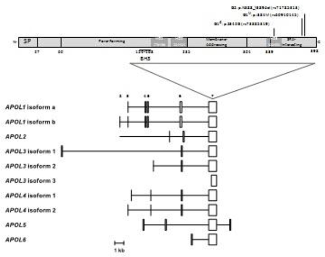
Apolipoprotein L1 (APOL1) genetic variants account for much of the excess risk of chronic and end-stage kidney disease, which results in a significant global health disparity for persons of African ancestry. We estimate the lifetime risk of kidney disease in APOL1 dual-risk allele individuals to be at least 15%. Experimental evidence suggests a direct role of APOL1 in pore formation, cellular injury, and programmed cell death in renal injury. The APOL1 BH3 motif, often associated with cell death, is unlikely to play a role in APOL1-induced cytotoxicity because it is not conserved within the APOL family and is dispensable for cell death in vitro. We discuss two models for APOL1 trypanolytic activity: one involving lysosome permeabilization and another involving colloid-osmotic swelling of the cell body, as well as their relevance to human pathophysiology. Experimental evidence from human cell culture models suggests that both mechanisms may be operative. A systems biology approach whereby APOL1-associated perturbations in gene and protein expression in affected individuals are correlated with molecular pathways may be productive to elucidate APOL1 function in vivo.

Keywords: APOL1; Health disparities; chronic kidney disease; focal segmental glomerulosclerosis; innate immunity.


Conflict of interest statement

conflict of interest: No conflicts of interest.

Figures

Figure 1
Figure 1. APOL1 predicted protein domains and human predicted APOL1 isoform transcripts
The upper panel shows APOL1 domain organization as proposed by Pays and colleagues, with residues numbered according to APOL1 isoform A. The BH3 domain proposed by Hu and colleagues is also shown [10]. MB, predicted membrane binding domains; SP, signal peptide. The locations of mutations that comprise the G1 and G2 alleles are shown. The lower panel shows the RNA transcript structures for the six human APOL family members, limited to protein-coding exons or portions of exons. Exon numbering is according to APOL1. Exon 7 encodes amino acid residues 106 to 398 of APOL1. APOL1 has three transcripts; only a and b are shown. [Permission pending from Smith and Malik, Genome Res. 2009. 19: 850–858 - PMID: 19299565]
Figure 2
Figure 2. APOL gene family in selected primate species and mouse
Genes, lost genes, and pseudogenes are represented by plain black box, empty box, and light gray box with Ψ, respectively. The hashed boxes represent mouse homologous, but not orthologous, genes. (1) Macaque also exhibits an APOL2.1 gene and an APOL7 pseudogene. (2) Baboon also exhibits two APOL2.1 pseudogenes and an APOL7 pseudogene. The trypanolytic potential against T. brucei is also depicted. [Permission pending from Capewell et al., Parasitology, 2015 - PMID:25656360]
Figure 3
Figure 3. APOL1 domain organization and homology of the BH3 domain
Panel A shows APOL1 domain organization as in figure 1. Also shown are the reported cleavage sites at amino acid residues 27 (signal peptidase) and 54 (unknown), and the Nglycosylation site (□) at N264. Panel B presents sequence analysis of residues that comprise the proposed BH3 domain of APOL family members, compared to the canonical BH3 domain motif. The middle panel shows an alignment of the amino residues for the six human APOL family members. The upper and lower panels show alignment logos generated with Skyligne software using a hidden Markov model (HMM), where the height of the amino acid code letter reflects residue frequency among the analyzed protein sequences. The upper panel shows an alignment logo for APOL family proteins found in the Pfam database, while the lower panel shows that of BH3 domain-containing proteins found in the InterPro database. For both HMM logos, sequences were filtered to remove redundancy. Each logo was generated from at least 300 aligned sequences, using sequences from all species included in the respective databases.
Figure 4
Figure 4. APOL1 domain organization and proposed mechanisms of APOL1 trypanolysis
Panel A shows the domain structure suggested by Pays and colleagues, identical that shown in figure 1. Panel B shows the model of APOL1 trypanolysis proposed by Pays and colleagues. In this model the HDL-bound APOL1 is endocytosed by the trypanosome and trafficked to the trypanosome lysosome. Once in the lysosome, the acidic environment leads to opening of the hairpin-like membrane-addressing domain, followed by dissociation of APOL1 from the HDL particle and insertion membrane-addressing domain into the lysosomal membrane. The pore-forming domain of APOL1 forms a chloride channel that depolarizes the lysosomal membrane, leading to irreversible swelling of the lysosome, followed by trypanolysis. SRA inhibits APOL1 within the lysosome by binding the SRA-interacting domain on APOL1, though the mechanism of inhibition is unclear. Panel C depicts the model of trypanolysis proposed by Thomson and Finkelstein. In this model, following endocytosis and trafficking to the lysosome, the acidic environment leads to irreversible insertion of APOL1 into the lysosomal membrane, a process mediated by the SRA-interacting domain. The APOL1 channel is inactive until recycled to the trypanosome cell surface, where neutral pH leads to APOL1 channel activation. Cation influx through APOL1 depolarizes the plasma membrane and is coupled to potassium efflux and anion and water influx, followed by loss of osmoregulation, cytoplasmic vacuolization, and lysis. SRA binding to APOL1 is speculated to inhibit insertion of the SRA-interacting domain into the lysosomal membrane, thereby preempting APOL1 channel formation and lysis. Panel D depicts a revised model for APOL1 domain organization. The pore-forming domain has been removed, as the residues responsible for channel formation remain to be identified. The membrane-addressing domain has been replaced by a proposed membrane insertion domain at the C terminus, within the SRA-interacting domain. PF, pore-forming domain. MA, membrane-addressing domain. SI,SRA-interacting domain. Other abbreviations as in Figure 1. [Permission pending from Pays et al., Nat Rev Microbiol. 2006, PMID: 16710327; Thomson and Finkelstein, Proc Natl Acad Sci U S A. 2015PMID: 25730870]

References

    1. United States Renal Data System. Bethesda, MD: National Institutes of Health, National Institute of Diabetes and Digestive and Kidney Diseases; 2014. Annual Data Report: Epidemiology of Kidney Disease in the United States.
    1. Kiberd BA. Cumulative Risk for Developing End-Stage Renal Disease in the US Population. Journal of the American Society of Nephrology. 2002;13:1635–1644. - PubMed
    1. Genovese G, Friedman DJ, Ross MD, Lecordier L, Uzureau P, et al. Association of trypanolytic ApoL1 variants with kidney disease in African Americans. Science. 2010;329:841–845. - PMC - PubMed
    1. Kopp JB, Nelson GW, Sampath K, Johnson RC, Genovese G, et al. APOL1 genetic variants in focal segmental glomerulosclerosis and HIV-associated nephropathy. J Am Soc Nephrol. 2011;22:2129–2137. - PMC - PubMed
    1. Thomson R, Genovese G, Canon C, Kovacsics D, Higgins MK, et al. Evolution of the primate trypanolytic factor APOL1. Proc Natl Acad Sci U S A. 2014;111:E2130–E2139. - PMC - PubMed

Publication types