. 2015 Jul 27;4(7):e002044.
doi: 10.1161/JAHA.115.002044.

Manganese-Enhanced Magnetic Resonance Imaging Enables In Vivo Confirmation of Peri-Infarct Restoration Following Stem Cell Therapy in a Porcine Ischemia-Reperfusion Model

Affiliations

Manganese-Enhanced Magnetic Resonance Imaging Enables In Vivo Confirmation of Peri-Infarct Restoration Following Stem Cell Therapy in a Porcine Ischemia-Reperfusion Model

Rajesh Dash et al. J Am Heart Assoc. .

Abstract

Background: The exact mechanism of stem cell therapy in augmenting the function of ischemic cardiomyopathy is unclear. In this study, we hypothesized that increased viability of the peri-infarct region (PIR) produces restorative benefits after stem cell engraftment. A novel multimodality imaging approach simultaneously assessed myocardial viability (manganese-enhanced magnetic resonance imaging [MEMRI]), myocardial scar (delayed gadolinium enhancement MRI), and transplanted stem cell engraftment (positron emission tomography reporter gene) in the injured porcine hearts.

Methods and results: Twelve adult swine underwent ischemia-reperfusion injury. Digital subtraction of MEMRI-negative myocardium (intrainfarct region) from delayed gadolinium enhancement MRI-positive myocardium (PIR and intrainfarct region) clearly delineated the PIR in which the MEMRI-positive signal reflected PIR viability. Human amniotic mesenchymal stem cells (hAMSCs) represent a unique population of immunomodulatory mesodermal stem cells that restored the murine PIR. Immediately following hAMSC delivery, MEMRI demonstrated an increased PIR viability signal compared with control. Direct PIR viability remained higher in hAMSC-treated hearts for >6 weeks. Increased PIR viability correlated with improved regional contractility, left ventricular ejection fraction, infarct size, and hAMSC engraftment, as confirmed by immunocytochemistry. Increased MEMRI and positron emission tomography reporter gene signal in the intrainfarct region and the PIR correlated with sustained functional augmentation (global and regional) within the hAMSC group (mean change, left ventricular ejection fraction: hAMSC 85±60%, control 8±10%; P<0.05) and reduced chamber dilatation (left ventricular end-diastole volume increase: hAMSC 24±8%, control 110±30%; P<0.05).

Conclusions: The positron emission tomography reporter gene signal of hAMSC engraftment correlates with the improved MEMRI signal in the PIR. The increased MEMRI signal represents PIR viability and the restorative potential of the injured heart. This in vivo multimodality imaging platform represents a novel, real-time method of tracking PIR viability and stem cell engraftment while providing a mechanistic explanation of the therapeutic efficacy of cardiovascular stem cells.

Keywords: ischemia–reperfusion injury; magnetic resonance imaging; manganese‐enhanced magnetic resonance imaging; peri‐infarct region imaging; stem cell imaging.


Figures

Figure 1
Figure 1
Lentiviral HSV-tk reporter gene construct. Viral vector map of HSV-tk positron emission tomography reporter gene construct used to transfect a subpopulation (≈15 million of 80 million total) hAMSCs prior to cell transplantation. This construct contains dual reporter genes Fluc and HSVtk. HSVtk confers the ability to metabolize and retain the radioisotope 18F-FHBG. AmpR indicates ampicillin resistance; bp, base pairs; Fluc, firefly luciferase; hAMSCs, human amniotic mesenchymal stem cells; HSV-tk, herpes simplex virus thymidine kinase; LVLTR, lentiviral long terminal repeat; LV-TF, triple fusion lentiviral vector; SINLTR, self-inactivating long terminal repeat; WPRE, woodchuck hepatitis virus post-transciptional response element.
Figure 2
Figure 2
Fluoroscopically guided catheter cell delivery. Cell and control injections are targeted to the mid- to apical segments in each quadrant using (A) delayed gadolinium enhancement reference to the infarct zone and (B) biplane fluoroscopic mapping. Manual tracings documenting the locations of the injections are shown from (C and D) 2 different fluoroscopic projections. The sternum is used as an anatomical landmark, and the locations of injection are tracked numerically as marked.
Figure 3
Figure 3
Regions of interest for DEMRI and MEMRI. Representative (A and B) short-axis DEMRI and (C) MEMRI images showing semiautomatic tracing of ROIs in a pig heart 6 weeks after human amniotic mesenchymal stem cell delivery. The signal threshold standard deviation method was used for DEMRI and MEMRI quantification with manual correction. Note the overall smaller MEMRI-defect area (blue ROI) compared with the yellow DEMRI-positive region. (D) Superimposed DEMRI and MEMRI infarct ROIs show the extent of PIR on either edge of the positive DEMRI regions still labeled yellow. DEMRI indicates delayed gadolinium enhancement magnetic resonance imaging; MEMRI, manganese-enhanced magnetic resonance imaging; PIR, peri-infarct region; ROI, region of interest.
Figure 4
Figure 4
Timeline of cell-delivery experiments. Timeline outlining the ischemia–reperfusion injury and subsequent cardiac injections of hAMSC at both early (1 week, blue) and late (4 week, red) time points, in addition to the CMR, PET, harvest, and IHC performed for each group. CMR indicates cardiac magnetic resonance imaging; hAMSC, human amniotic mesenchymal stem cell; IHC, immunohistochemistry; PET, positron emission tomography.
Figure 5
Figure 5
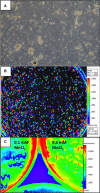
Isolation, culture, and reporter gene transduction of hAMSCs. A, A subset of hAMSCs was cultured for 6 weeks after transduction with the herpes simplex virus thymidine kinase positron emission tomography reporter gene and showed expansion properties similar to nontransduced hAMSCs. B, Robust in vitro BLI signal from hAMSCs 6 weeks after transduction with the HSV-tk and firefly luciferase double-fusion reporter gene. C, BLI of plated hAMSCs 5 days after Mn2+ labeling. Note the preserved BLI signal at 0.1 and 0.5 mmol/L MnCl2, indicating no impairment of cell survival with Mn2+ labeling. BLI indicates bioluminescence; hAMSCs, human amniotic mesenchymal stem cells.
Figure 6
Figure 6
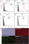
Flow cytometry and immunohistological characterization of hAMSCs. A, Flow cytometry analysis indicated 0% HLA-DR–positive hAMSCs. PE, phycoeryhthrin. B, Flow cytometry analyses showed a high proportion of CD59 positive cells, with smaller proportions of (C) HLA-G; (D) c-kit–positive hAMSCs; and (E) immunohistological evidence of hAMSC cell surface markers, including uAMC, Thy-1, SSEA-4, and c-kit. hAMSCs indicates human amniotic mesenchymal stem cells.
Figure 7
Figure 7
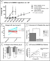
LVEF and remodeling improvements in the hAMSC-treated hearts. hAMSC treatment led to sustained improvement in cardiac function. A, Mean percentage of LVEF increases in early and late-hAMSC hearts vs control hearts compared with post-IR, preinjection LVEF. The hAMSC-treated swine (light green) exhibited a significant LVEF increase immediately after hAMSC delivery (day 7) that was sustained for 21 days after delivery (day 28 after IR, day 21 after injection). NS-injected and lysed hAMSC controls showed no improvement in LVEF over the same time period. Late-hAMSC hearts, which received hAMSCs 28 days after IR injury, exhibited a predictable, significant, and steady rise in LVEF after cell delivery. B, Absolute LVEF changes before and after injection showed a consistent increase in the LVEF in hAMSC-treated swine and a consistent decrease in LVEF in the control group. C, LVEDV in hAMSC hearts exhibited 86% less chamber dilatation (percentage change in LVEDV) than the control group, indicating improved LV remodeling due to hAMSC delivery. D, MEMRI/DEMRI infarct size improvement in hAMSC-treated hearts, which exhibit a significantly lower MEMRI-defect volume and positive DEMRI scar volume compared with control hearts at 21 days after hAMSC therapy. E, A strong linear correlation was observed between the percentage change in LVEF and the percentage of infarct size reduction by MEMRI scar volume in the early hAMSC IR hearts (values expressed as percentage of total LV myocardium). Notably, the degree of scar reduction in late-hAMSC hearts is not as pronounced as in the early hAMSC hearts, despite similar LVEF increases. DEMRI indicates delayed gadolinium enhancement MRI; EF, ejection fraction; hAMSC, human amniotic mesenchymal stem cell; IR, ischemia-reperfusion; LV, left ventricular; LVEDV, left ventricular end-diastolic volume; MEMRI, manganese-enhanced magnetic resonance imaging; NS, normal saline.
Figure 8
Figure 8
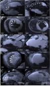
DEMRI and MEMRI for hAMSC- and control-injected hearts. Representative MEMRI images shown in (A) 2-chamber and (B) short-axis views from the same animals demonstrate positive MEMRI signals (white arrows) throughout the infarct zone in the hAMSC group, resulting in a reduced MEMRI-defect region and increased contrast/noise ratio compared with the control group MEMRI-defect area (white arrowheads). Corresponding DEMRI images from the hAMSC and control groups 21 days after injection in (C) 3-chamber, (D) short-axis, and (E) 2-chamber views. Smaller regions of positive delayed enhancement in the anteroseptal walls of the hAMSC group are noted. Blue brackets delineate the region of positive DEMRI. hAMSC-treated hearts also exhibit increased viability in the expected infarct zone, with viable myocardium mixed with scar in the DEMRI and MEMRI images (white arrows, D and E). The region of transmural DEMRI-positive myocardium is substantially reduced (C through E, blue brackets). 2CH indicates 2-chamber; 3CH, 3-chamber; DEMRI, delayed gadolinium enhancement magnetic resonance imaging; hAMSC, human amniotic mesenchymal stem cell; MEMRI, manganese-enhanced magnetic resonance imaging; SAX, short-axis.
Figure 9
Figure 9
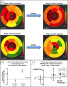
Segmental improvement in hAMSC-treated hearts by percentages of both MEMRI and radial thickening. A, Polar maps depicting representative percentages of radial thickening at days 0 to 21 after injection from the mid- to apical segments (segments 7 to 16 of American Heart Association 16-segment model) of the control and hAMSC-treated hearts. Note the significant improvement (brighter color map in mid- to apical segments) in the hAMSC-treated heart compared with control (darker color map in day 21 mid- to apical segments). B, Percentage of mid- to apical segmental radial strain measurements in control vs hAMSC-treated hearts. Note the significant increase in mean percentage of segmental radial strain in hAMSC hearts. C, Scatterplot of change in segmental MEMRI signal (y-axis) vs change in percentage of radial thickening (x-axis) from both control (open symbols) and hAMSC-treated (closed symbols) hearts. Note the distribution of hAMSC-treated hearts in the upper right quadrant, reflecting an association of improved MEMRI signal of myocardial viability and improved radial thickening. Conversely, the control hearts exhibited worsened MEMRI signal and lower percentage of radial thickening, consistent with the lack of functional recovery in the control hearts. hAMSC indicates human amniotic mesenchymal stem cell; MEMRI, manganese-enhanced magnetic resonance imaging; SNR, signal/noise ratio.
Figure 10
Figure 10
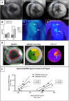
MEMRI and PET colocalization of the hAMSC-injected hearts. In vivo confirmation of MEMRI signal for live hAMSCs within the myocardium of a hAMSC-injected swine is shown. hAMSCs were delivered 1 week after ischemia–reperfusion injury into the peri-infarct zones of the mid- to apical infero- and anteroseptum. A, SAX MEMRI image on day 2 with a characteristic MEMRI defect in the infarct zone (blue region of interest). Note area of inferoseptum with increased signal (star). Serial MEMRI shows increasing signal intensity in inferoseptum on days 21 and 38 after cell delivery, with overall increased signal within the infarct zone. B, Absolute increase in MEMRI CNR of the infarct zone in the hAMSC-treated vs control (normal saline–injected) hearts on day 21 after hAMSC delivery. C and D, Axial PET reporter gene signals from hAMSC populations in the apical septum, confirming specific live hAMSC activity on days 21 and 38 (IVS, LV). E, SAX MEMRI images (left) from an early hAMSC animal with traced endo- and epicardial contours; corresponding polar maps (middle) of MEMRI signal from the mid- to apical slices of the LV (the regions of hAMSC injection), with yellow and tan colors indicating increasing signal intensity; polar maps (right) from corresponding PET images. Note the similar patterns between MEMRI and PET polar maps. Arrows denote the focal signal in both MEMRI and PET images. F, Significant linear correlation of MEMRI signals (y-axis, signal intensity units) and PET signals (x-axis, signal intensity units) for both early hAMSC (left plot) and late-hAMSC (right plot) hearts. CNR indicates contrast-to-noise ratio; CT, computed tomography; hAMSCs, human amniotic mesenchymal stem cells; IVS, interventricular septum; LV, left ventricle; MEMRI, manganese-enhanced magnetic resonance imaging; PET, positron emission tomography; SAX, short-axis.
Figure 11
Figure 11
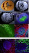
Immunostaining confirmed intact hAMSC populations in vivo. A, Gross SAX section of a hAMSC-injected heart (6 weeks after cell delivery) demonstrated prominent anterior and inferoseptal peri-infarct segments, which were hAMSC injection sites (note: inset white box corresponds to MEMRI, DEMRI, and PET images and to the immunostaining tissue specimen). B, A bright focus (white box) of MEMRI signal is detected within the inferoseptal peri-infarct region corresponding to cell injection site. C, Matched DEMRI SAX image shows preserved myocardium (null signal) throughout most of the septum. D, 3-dimensional coregistration of PET-RG and MEMRI images acquired on the same day, showing a colocalized focus of intense inferoseptal signal from the PET-RG–transduced hAMSCs and increased MEMRI signal (white box). E and F, Myocardial sections stain positive for (E) anti-human nuclear antigen Ab and (F) anti-human mitochondrial antigen Ab, confirming the presence of live hAMSC populations up to 6 weeks after cell transplantation in these matched MEMRI and PET-positive regions. G and H, These same cell clusters are negative for both anti-α-actinin and anti-troponin Ab staining. Ab indicates antibody; DEMRI, delayed gadolinium enhancement magnetic resonance imaging; hAMSC, human amniotic mesenchymal stem cell; IVS, interventricular septum; LV, left ventricle; MEMRI, manganese-enhanced magnetic resonance imaging; PET, positron emission tomography; PET-RG, positron emission tomography reporter gene; SAX, short-axis.

Similar articles

Cited by

References

    1. Lloyd-Jones D, Adams R, Carnethon M, De Simone G, Ferguson TB, Flegal K, Ford E, Furie K, Go A, Greenlund K, Haase N, Hailpern S, Ho M, Howard V, Kissela B, Kittner S, Lackland D, Lisabeth L, Marelli A, McDermott M, Meigs J, Mozaffarian D, Nichol G, O’Donnell C, Roger V, Rosamond W, Sacco R, Sorlie P, Stafford R, Steinberger J, Thom T, Wasserthiel-Smoller S, Wong N, Wylie-Rosett J, Hong Y. Heart disease and stroke statistics—2009 update: a report from the American Heart Association Statistics Committee and Stroke Statistics Subcommittee. Circulation. 2009;119:480–486. - PubMed
    1. Heidary S, Patel H, Chung J, Yokota H, Gupta SN, Bennett MV, Katikireddy C, Nguyen P, Pauly JM, Terashima M, McConnell MV, Yang PC. Quantitative tissue characterization of infarct core and border zone in patients with ischemic cardiomyopathy by magnetic resonance is associated with future cardiovascular events. J Am Coll Cardiol. 2010;55:2762–2768. - PubMed
    1. Tsukiji M, Nguyen P, Narayan G, Hellinger J, Chan F, Herfkens R, Pauly JM, McConnell MV, Yang PC. Peri-infarct ischemia determined by cardiovascular magnetic resonance evaluation of myocardial viability and stress perfusion predicts future cardiovascular events in patients with severe ischemic cardiomyopathy. J Cardiovasc Magn Reson. 2006;8:773–779. - PubMed
    1. Jaussaud J, Biais M, Calderon J, Chevaleyre J, Duchez P, Ivanovic Z, Couffinhal T, Barandon L. Hypoxia-preconditioned mesenchymal stromal cells improve cardiac function in a swine model of chronic myocardial ischaemia. Eur J Cardiothorac Surg. 2012;43:1050–1057. - PubMed
    1. Kawamura M, Miyagawa S, Miki K, Saito A, Fukushima S, Higuchi T, Kawamura T, Kuratani T, Daimon T, Shimizu T, Okano T, Sawa Y. Feasibility, safety, and therapeutic efficacy of human induced pluripotent stem cell-derived cardiomyocyte sheets in a porcine ischemic cardiomyopathy model. Circulation. 2012;126:S29–S37. - PubMed

Publication types