. 2015 Jul 28:8:399.
doi: 10.1186/s13071-015-1006-z.

ANISERP: a new serpin from the parasite Anisakis simplex

Affiliations

ANISERP: a new serpin from the parasite Anisakis simplex

Elizabeth Valdivieso et al. Parasit Vectors. .

Abstract

Background: Serine proteinase inhibitors (serpins) finely regulate serine proteinase activity via a suicide substrate-like inhibitory mechanism. In parasitic nematodes, some serpins interact with host physiological processes; however, little is known about these essential molecules in Anisakis. This article reports the gene sequencing, cloning, expression and preliminary biochemical and bioinformatically-based structural characterization of a new Anisakis serpin (ANISERP).

Methods: The full AniSerp gene was cloned by specific RACE-PCR after screening an Anisakis simplex (L3) cDNA library. For biochemical assays, the AniSerp gene was subcloned into both prokaryotic and eukaryotic vectors, and the recombinant proteins were purified. The inhibitory properties of the proteins were tested in classical biochemical assays using human serine peptidases and AMC substrates. Immunolocalization of ANISERP, theoretical structural analysis and bioinformatically-based structural modelling of the ANISERP protein were also conducted.

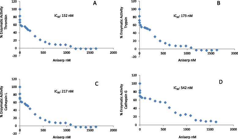
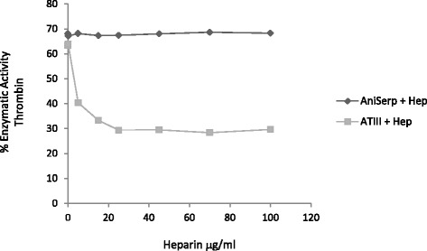
Results: The AniSerp gene was found to have 1194 nucleotides, coding for a protein of 397 amino acid residues plus a putative N-terminal signal peptide. It showed significant similarity to other nematode, arthropod and mammalian serpins. The recombinant ANISERP expressed in the prokaryotic and eukaryotic systems inhibited the human serine proteases thrombin, trypsin and cathepsin G in a concentration-dependent manner. No inhibitory activity against Factor Xa, Factor XIa, Factor XIIa, elastase, plasmin or chymotrypsin was observed. ANISERP also acted on the cysteine protease cathepsin L. ANISERP was mainly localized in the nematode pseudocoelomic fluid, somatic muscle cell bodies and intestinal cells. The findings of molecular dynamics studies suggest that ANISERP inhibits thrombin via a suicide substrate-like inhibitory mechanism, similar to the mechanism of action of mammalian coagulation inhibitors. In contrast to findings concerning human antithrombin III, heparin had no effect on ANISERP anticoagulant inhibitory activity.

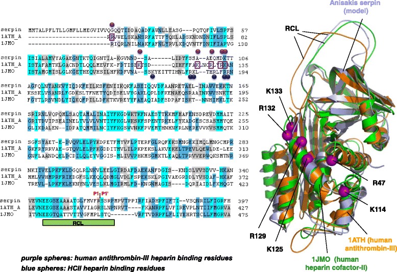
Conclusions: Our findings suggest that ANISERP is an internal Anisakis regulatory serpin and that the inhibitory activity against thrombin depends on a suicide substrate-like inhibitory mechanism, similar to that described for human antithrombin (AT)-III. The fact that heparin does not modulate the anticoagulant activity of ANISERP might be explained by the absence in the latter of five of the six positively charged residues usually seen at the AT-III-heparin binding site.


Figures

Fig. 1
Fig. 1
Effect of ANISERP on the proteolytic activity of human thrombin, trypsin, cathepsin L and cathepsin G. Inhibitory effect of different doses of recombinant ANISERP, expressed in a baculovirus system, on the enzymatic activity of a Thrombin (74 nM) on Boc-Val-Arg-AMC, b Trypsin (54 nM) on Boc-Gln-Ala-Arg-AMC, c Cathepsin L (0.83 nM) on Z-Phe-Arg-AMC, and d Cathepsin G (0.33 nM) on N-succinyl-Ala-Ala-Phe-AMC
Fig. 2
Fig. 2
Immunolocalization of ANISERP in sections of Anisakis simplex L3 with a rabbit hyperimmune serum. a Cross section at the level of the oesophagus showing strongly positive staining in SM and PF, moderate staining in SOM and slight staining of LC. No staining was observed in the EC. b The negative control incubated with the preimmune serum did not stain. c Cross section through the intestinal level showing in detail strongly positive immunostaining in the SM, which is restricted to the nSM; no staining was observed in the cSM, the C or EC, including the ca. Less intense staining was also observed in IC. d Section C was decolorized to remove the 4CN stain and subsequently stained with Wheatley’s trichrome. DOG, dorsal oesophageal gland; SOM, subventral muscular sectors of oesophagus; SM, somatic musculature; LC, lateral hypodermal cords; PF, pseudocoelomic fluid; EC, excretory cell; V, ventricle; IC, intestinal cells; nSM, non-contractile portion of somatic muscle cells (myocytons); cSM, contractile portion of somatic muscle cells; C, cuticle; ca, excretory canal; n, nucleus
Fig. 3
Fig. 3
Model for ANISERP-human thrombin binding. a General view of the model for ANISERP binding to human thrombin. The location of the ANISERP reactive centre loop (RCL [green]) in the structural groove of the thrombin is indicated. The thrombin surface is coloured according to its theoretical electrostatic properties (blue = positive, red = negative). b Residues in the heavy chain of human thrombin; binding of amino acids Met358 and Phe374 in ANISERP. c Position of residues in the catalytic triad of human thrombin (His43, Asp99 and Ser205) and their positions relative to ANISERP residues Arg361 and Ser362 (P1 and P1′ respectively). Distances between active residues and atoms in the peptide bond of the substrate, compatible with proteinase activity, are indicated
Fig. 4
Fig. 4
Effect of heparin on ANISERP inhibition of human thrombin proteolytic activity. Residual activity of thrombin after incubation with recombinant ANISERP (circles) or AT (squares) with increasing concentrations of heparin (0.1-100 μg/ ml). ANISERP was used at a fixed concentration of 2.17 nM (IC50: 152 nM)
Fig. 5
Fig. 5
Sequence and structural alignment of ANISERP, human antithrombin III (1 ATH_A), and human heparin cofactor-II (1JMO). The key residues involved in heparin binding described for human antithrombin III (Arg 47, Lys114, Lys125, Arg 129, Arg132 and Lys133) are shown as purple boxes and spheres. The key residues involved in heparin binding described for heparin cofactor-II (Lys173, Arg184, Lys185, Arg189, Arg192 and Arg193) are shown as blue spheres. The positions of the P1-P1’residues in the ANISERP sequence are indicated. The residues in the 1JMO and 1ATH_A sequences are numbered according to Baglin et al. [26]

References

    1. Audícana MT, del Pozo MD, Iglesias R, Ubeira FM. Anisakis simplex and Pseudoterranova decipiens. In: Miliotis M, Bier J, editors. International handbook on foodborne pathogens. New York: Marcell Dekker; 2003. pp. 613–36.
    1. Geraci JR, St Aubin DJ. Effects of parasites on marine mammals. Int J Parasitol. 1987;17:407–14. doi: 10.1016/0020-7519(87)90116-0. - DOI - PubMed
    1. Ishikura H, Kikuchi K, Nagasawa K, Ooiwa T, Takamiya H, Sato N, et al. Anisakidae and anisakidosis. Prog Clin Parasitol. 1993;3:43–102. doi: 10.1007/978-1-4612-2732-8_3. - DOI - PubMed
    1. Bier JW. Anisakiasis. Laboratory diagnosis of infectious diseases. In: Balows A, Hausler WJ Jr, Ohashi M, Turano A, editors. Bacterial, mycotic and parasitic diseases. New York: Springer; 1988. pp. 768–74.
    1. Valiñas B, Lorenzo S, Eiras A, Figueiras A, Sanmartín ML, Ubeira FM. Prevalence of and risk factors for IgE sensitization to Anisakis simplex in a Spanish population. Allergy. 2001;56:667–71. doi: 10.1034/j.1398-9995.2001.00987.x. - DOI - PubMed

Publication types

MeSH terms