. 2015 Dec 1;152(Pt B):329-39.
doi: 10.1016/j.physbeh.2015.07.025. Epub 2015 Jul 26.

Effects of high-fat diet and gastric bypass on neurons in the caudal solitary nucleus

Affiliations

Effects of high-fat diet and gastric bypass on neurons in the caudal solitary nucleus

A J Boxwell et al. Physiol Behav. .

Abstract

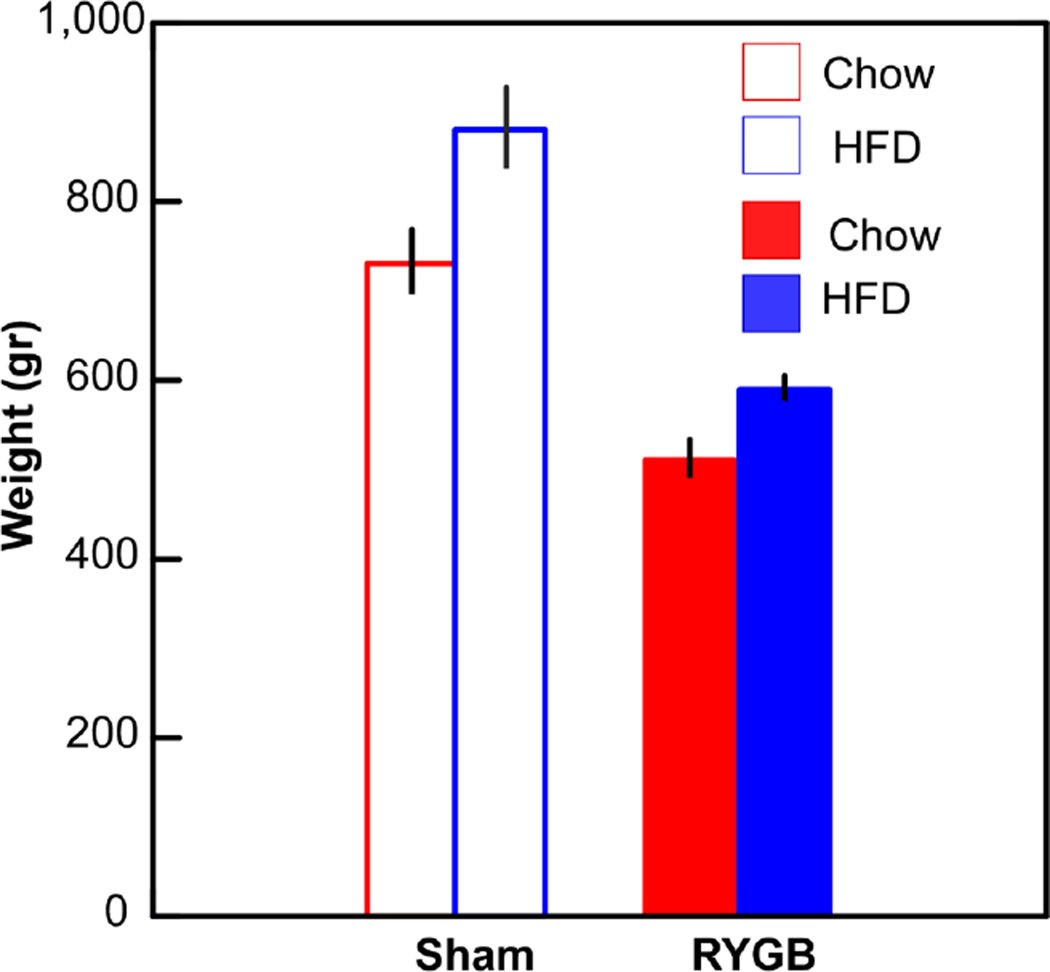
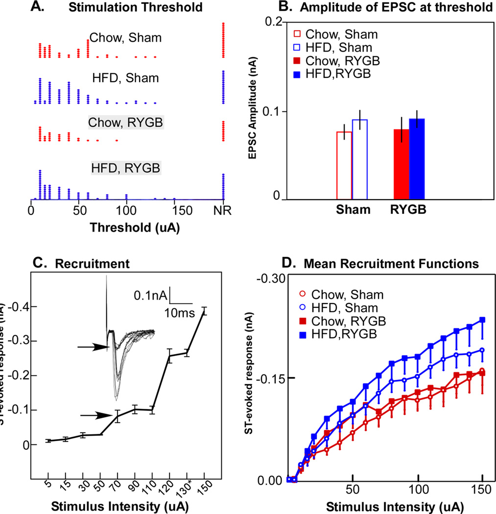
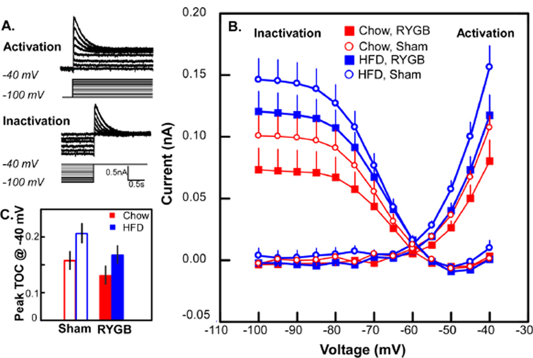
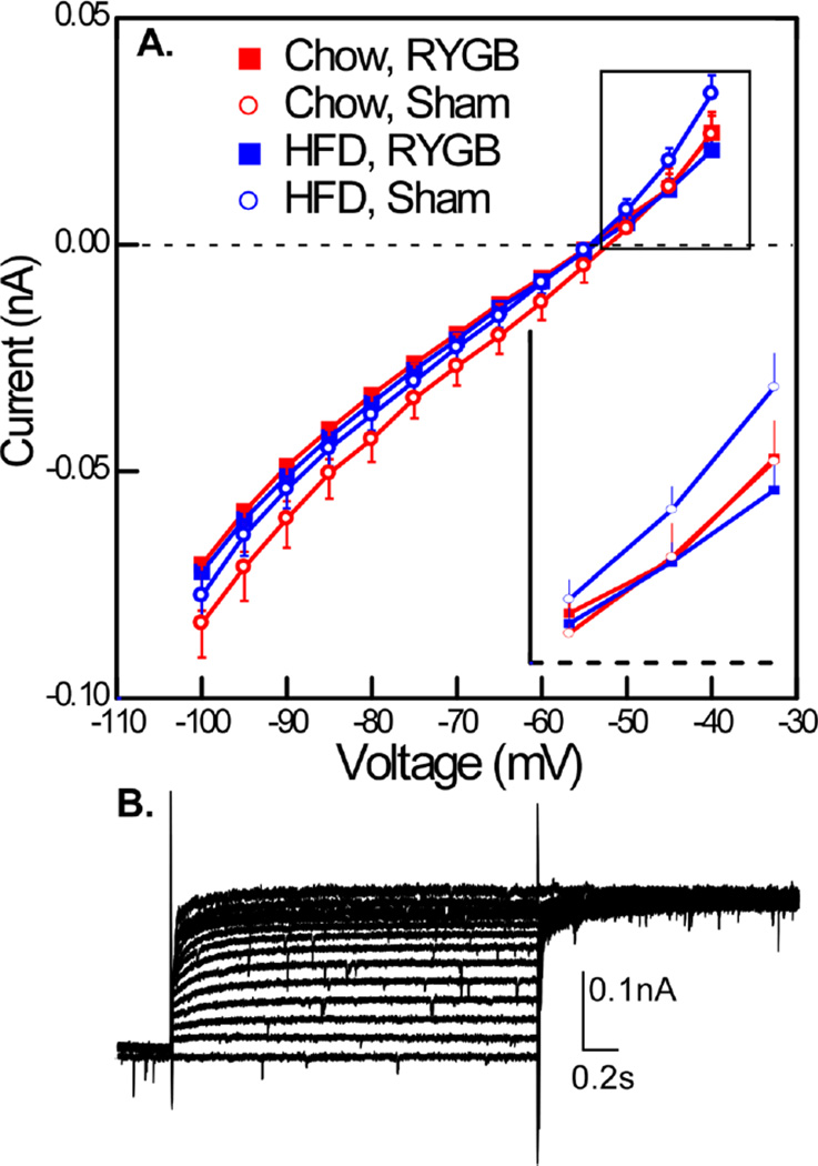
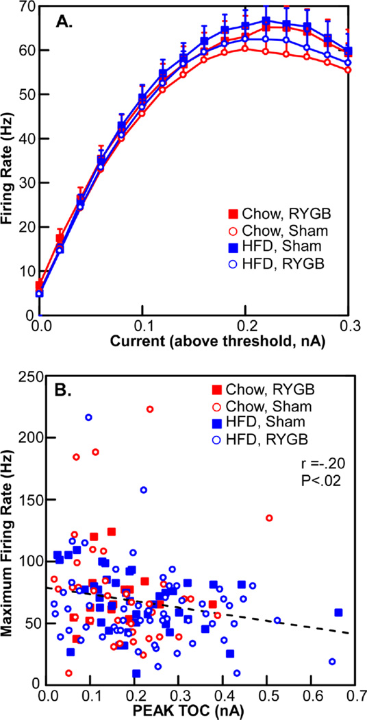
Bariatric surgery is an effective treatment for obesity that involves both peripheral and central mechanisms. To elucidate central pathways by which oral and visceral signals are influenced by high-fat diet (HFD) and Roux-en-Y gastric bypass (RYGB) surgery, we recorded from neurons in the caudal visceral nucleus of the solitary tract (cNST, N=287) and rostral gustatory NST (rNST,N=106) in rats maintained on a HFD and lab chow (CHOW) or CHOW alone, and subjected to either RYGB or sham surgery. Animals on the HFD weighed significantly more than CHOW rats and RYGB reversed and then blunted weight gain regardless of diet. Using whole-cell patch clamp recording in a brainstem slice, we determined the membrane properties of cNST and rNST neurons associated with diet and surgery. We could not detect differences in rNST neurons associated with these manipulations. In cNST neurons, neither the threshold for solitary tract stimulation nor the amplitude of evoked EPSCs at threshold varied by condition; however suprathreshold EPSCs were larger in HFD compared to chow-fed animals. In addition, a transient outward current, most likely an IA current, was increased with HFD and RYGB reduced this current as well as a sustained outward current. Interestingly, hypothalamic projecting cNST neurons preferentially express IA and modulate transmission of afferent signals (Bailey, '07). Thus, diet and RYGB have multiple effects on the cellular properties of neurons in the visceral regions of NST, with potential to influence inputs to forebrain feeding circuits.

Keywords: Gastric bypass; Potassium channels; Rat; Solitary nucleus.


Figures

Fig. 1
Fig. 1
Time-line indicating relationships between start of diet, time of surgery and transport of the animals to Ohio State University for the patch clamp experiments.
Fig. 2
Fig. 2
Weights of the animals at the time of the current study. ANOVA revealed main effects of diet (P < .009) and surgery (P < .001) but no interaction between these variables.
Fig. 3
Fig. 3
A and B. Photomicrographs showing a section from the cNST at the level of the area postrema (AP), the region where most cNST neurons were recorded. Panel A shows a photomicrograph of a fixed section taken after recording. Six neurons filled with Lucifer Yellow (pseudocolored magenta) are apparent. The arrowhead indicates the location of the neuron being recorded in panel B. Panel B shows a photomicrograph of the same section taken during recording. The arrow indicates the tip of the recording pipette. Note the position of the stimulating electrode on the solitary tract (ST). C. Photomicrograph of a fixed, dopamine-beta hydroxylase-(DBH) stained cNST section taken subsequent to recording. This section was caudal to the area postrema. The arrows indicate two Lucifer Yellow-filled (pseudocolored green) neurons that are double-labeled for DBH (magenta). D. Higher-power photomicrograph of the neurons indicated in C taken during the preparation. Scale bars: 100µm in A, B, & C; 10µm in D.
Fig. 4
Fig. 4
A. Distributions of the threshold for solitary tract (ST) stimulation necessary to elicit an excitatory postsynaptic current (EPSC) for the 4 experimental groups. NR, no reponse. B. Mean amplitude of the ESPCs at threshold; there were no significant differences across the 4 experimental groups. C. Example of recruitment of additional afferents with increases in the current amplitude of ST stimulation; 3 afferent inputs were identified for this neuron. The arrows indicate the response at threshold. D. Mean amplitudes of EPSCs at different levels of ST stimulation indicated that high fat diet (HFD) groups elicited significantly larger EPSCs, as shown by an ANOVA with a significant main effects of diet (P <.04), stimulus intensity (P < .001) an interaction between these variables (P < .001), but no interaction with surgery.
Fig. 5
Fig. 5
A. Example of the protocol for activation and inactivation for a transient outward current (TOC). For clarity, only a subset of recorded traces are shown; voltage steps for which recorded traces are not shown are designated in grey. B. Mean inactivation and activation curves for a TOC across the 4 experimental groups. ANOVA demonstrated significant main effects for both diet (P < .03) and surgery (P < .02) for the activation curve whereas only diet was significantly different for the inactivation curve (P < .02, see text). C. Bar graph for TOC observed at −40 mV exemplifies main effects: a high fat diet (HFD) is associated with a larger TOC and RYGB surgery suppresses the increased TOC. Note that RYGB in the HFD group reduced TOC to levels similar to sham operated chow animals.
Fig. 6
Fig. 6
A. Sustained current measured at the end of the TOC activation protocol also showed effects of surgery. An ANOVA conducted over the entire curve revealed an interaction between surgery and applied voltage (P < .001). At the two most depolarized levels (−40 and −45mv, see inset), the outward current in the HFD sham group was magnified, and an ANOVA conducted at these levels of depolarization demonstrated a significant interaction between applied voltage, surgery and diet (P < .05). B. Example of sustained current in a neuron without a TOC.
Fig. 7
Fig. 7
A. The number of action potentials in response to different levels of membrane depolarization did not change across the 4 experimental groups. B. A weak, albeit significant correlation between the magnitude of a transient outward current and maximum firing rate.

References

    1. Alhadeff AL, Rupprecht LE, et al. GLP-1 neurons in the nucleus of the solitary tract project directly to the ventral tegmental area and nucleus accumbens to control for food intake. Endocrinology. 2012;153(2):647–658. - PMC - PubMed
    1. Altschuler SM, Bao XM, et al. Viscerotopic representation of the upper alimentary tract in the rat: sensory ganglia and nuclei of the solitary and spinal trigeminal tracts. J Comp Neurol. 1989;283(2):248–268. - PubMed
    1. Amberg GC, Koh SD, et al. A-type potassium currents in smooth muscle. Am J Physiol Cell Physiol. 2003;284(3):C583–C595. - PubMed
    1. Andresen MC, Doyle MW, et al. Differentiation of autonomic reflex control begins with cellular mechanisms at the first synapse within the nucleus tractus solitarius. Braz J Med Biol Res. 2004;37(4):549–558. - PubMed
    1. Appleyard SM, Bailey TW, et al. Proopiomelanocortin neurons in nucleus tractus solitarius are activated by visceral afferents: regulation by cholecystokinin and opioids. J Neurosci. 2005;25(14):3578–3585. - PMC - PubMed

Publication types